Eureka AIR delivers breakthrough ideas for toughest innovation challenges, trusted by R&D personnel around the world.

Removing environment factors from signals generated from video images captured for biomedical measurements

a technology of biomedical measurements and environment factors, applied in image enhancement, television systems, instruments, etc., to achieve the effect of improving accuracy and reliability of biomedical measurements

Active Publication Date: 2013-08-22
XEROX CORP
View PDF2 Cites 64 Cited by
  • Summary
  • Abstract
  • Description
  • Claims
  • Application Information

AI Technical Summary

Benefits of technology

This patent describes a way to remove unwanted signals from video images captured by RGB or IR cameras. This helps to improve the accuracy and reliability of biomedical measurements.

Problems solved by technology

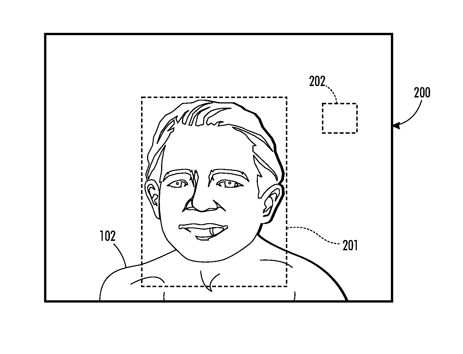
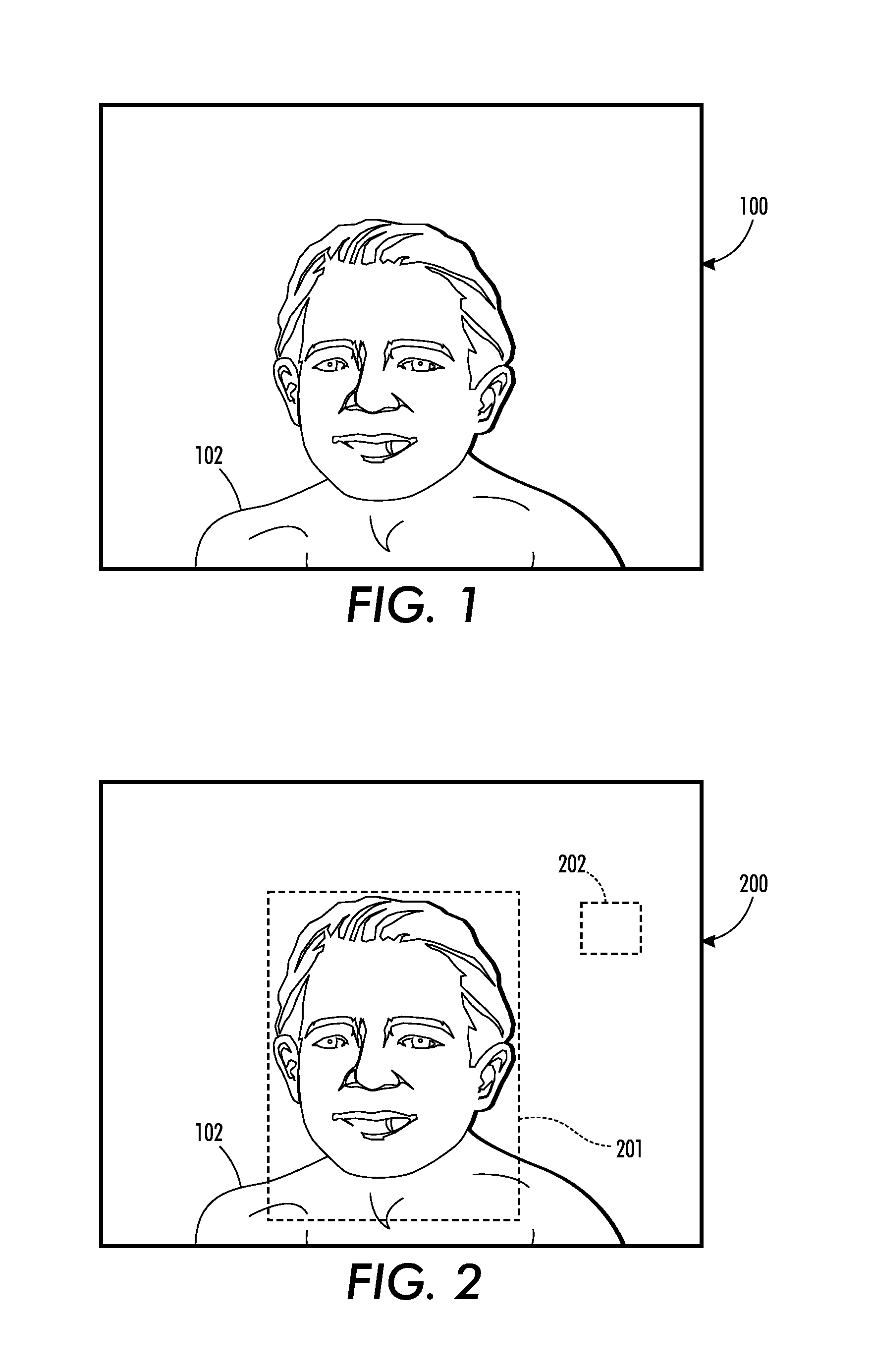
Both of these regions have been exposed to the same environmental factors containing undesirable environmental factors such as periodic signals and random background noise.

Method used


Image

  • Removing environment factors from signals generated from video images captured for biomedical measurements
  • Removing environment factors from signals generated from video images captured for biomedical measurements
  • Removing environment factors from signals generated from video images captured for biomedical measurements

Examples


example signal processing

System

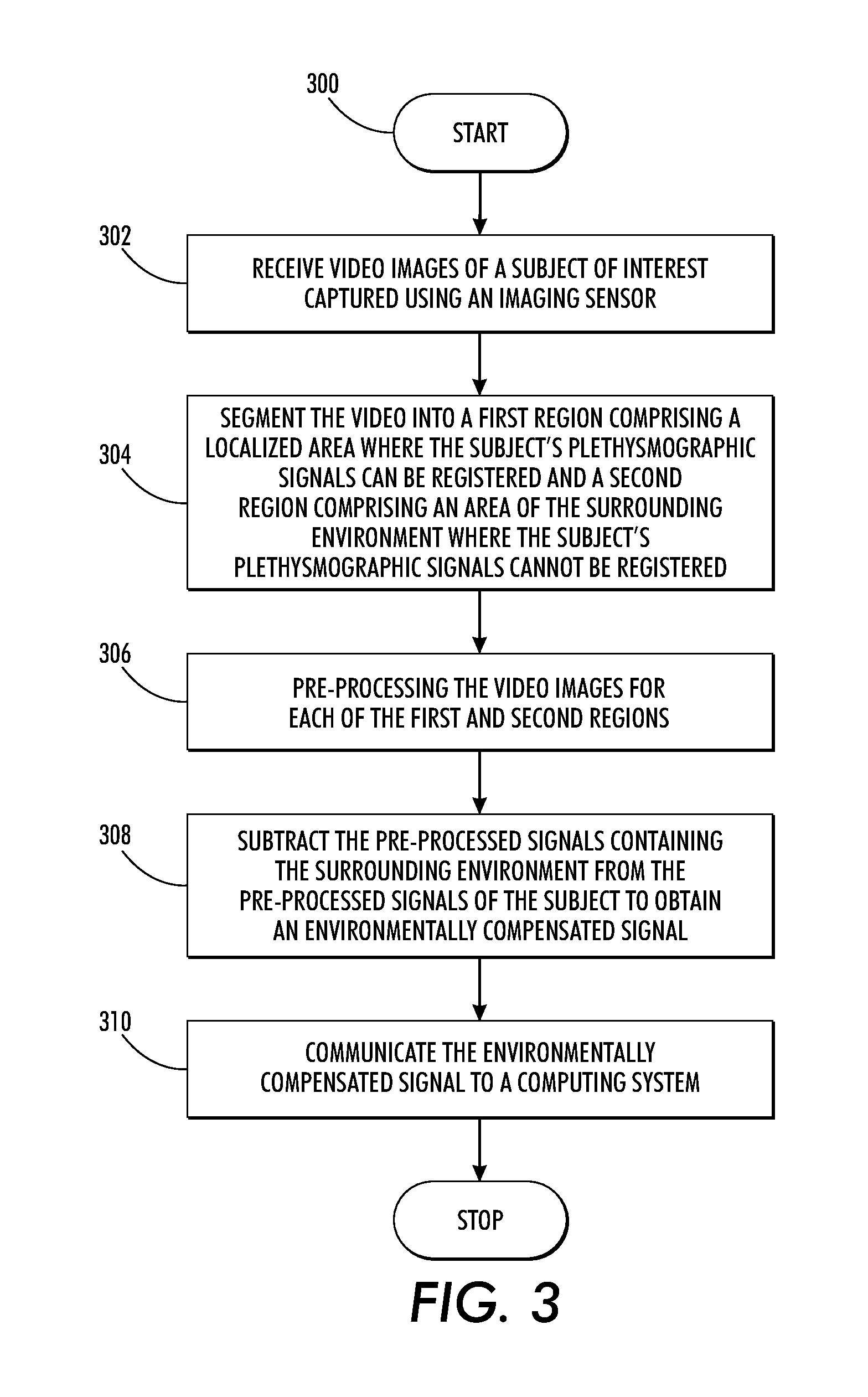
[0041]Reference is now being made to FIG. 4 which is a block diagram of an example networked video image processing system wherein various aspects of the present method as described with respect to the flow diagram of FIG. 3 are implemented.

[0042]In FIG. 4, imaging sensor 402 acquires source video images of a subject of interest in the sensor's field of view 403 over at least one imaging channel. The source video images are communicated to Video Image Processing System 404 wherein various aspects of the present method are performed. The example image processing system is shown comprising a Buffer 406 for buffering frames of the source video image for processing. Buffer 406 may further store data, formulas and other mathematical representations as are necessary to process the source video images in accordance with the teachings hereof. Image Stabilizer Module 408 is provided to process the images to compensate for motion induced blur, imaging blur, slow illuminant variation, an...


PUM


Abstract

What is disclosed is a system and method for automatically removing undesirable periodic or random background noise from heart rate measurement signals obtained from a video camera, ambient illuminator and other unknown electromagnetic sources to improve the overall reliability of biomedical measurements. In one embodiment, a time varying video image acquired over at least one imaging channel of a subject of interest is received. The video images are then segmented into a first region comprising a localized area where plethysmographic signals of the subject can be registered and a second region comprising a localized area of the environment where the plethysmographic signals cannot be registered. Both of the regions are exposed to the same environmental factors. The segmented video signals are pre-processed and the processed signals are subtracted from each other to generate an environmentally compensated signal. The environmentally compensated signal is then communicated to a computer system.

Description

TECHNICAL FIELD[0001]The present invention is directed to systems and methods for removing undesirable signals and background noise from signals generated from video images captured using a RGB camera or an Infrared (IR) camera for improved accuracy and reliability of biomedical measurements derived from those images.BACKGROUND[0002]Current Electro-Cardio Graphic (ECG) systems require the patient to be located in close proximity to the ECG machine obtaining measurements via electrodes attached to the skin. The adhesive electrodes can cause skin irritation, infection, discomfort, and other issues to the patient. This can especially be a problem to newborns with sensitive skin. Methods for non-contact cardiac pulse measurement based on imaging patients using RGB and / or multi-spectral infrared (IR) cameras have arisen in this art. By recording video images of the region of exposed skin where concentrations of blood vessels exist, small changes in pulsating inside blood vessels are regi...

Claims


Application Information

Patent Timeline
Patent Type & Authority Applications(United States)
IPC IPC(8): H04N7/18
CPCH04N7/18G06T5/002G06T5/003G06T2207/10016G06T2207/30088G06T2207/10048G06T2207/20056G06T2207/20076G06T2207/20144G06T2207/10024G06T7/194G06T5/73G06T5/70
Inventor MESTHA, LALIT KESHAVXU, BEILEI
Owner XEROX CORP
Why Eureka
Eureka Blog
Learn More