
Surgical lighting sources for use with fluophore-tagged monoclonal antibodies or fluorophore-tagged tumor avid compounds

a technology of fluorophore and fluorescent dye, applied in the field of medical devices, can solve the problems of difficult identification of tumors, inability to use fluorescent dyes in tumor diagnosis, and difficulty in identifying tumors, etc., and achieve the effect of easy identification and less cos

Inactive Publication Date: 2013-04-04
ONCOFLUOR
View PDF7 Cites 12 Cited by
  • Summary
  • Abstract
  • Description
  • Claims
  • Application Information

AI Technical Summary

Benefits of technology

The patent text describes a technology that allows for direct visualization of disease tissue without the need for image processing equipment. This is achieved by using fluorescein and its derivatives, which have high safety and easily distinguishable green emission colors. The technology can be used through endoscopic devices or by an operating physician, and does not require expensive charged couple device (CCD) equipment. The green fluorescence emanating from diseased tissue can be easily seen without the need for yellow filters, allowing for accurate removal of the diseased tissue. Overall, the technology provides a simple and cost-effective solution for visualizing disease sites.

Problems solved by technology

In medical applications, a similar difficulty arises because both tumors and normal healthy tissue fluoresce naturally, albeit at different wavelengths.
Consequently, when UV-activated fluorescence is used to detect tumors against a background of healthy tissue, identification of tumors is difficult.
Thus, the use of fluorescent dyes in diagnosis of tumors had not been wholly successful.
A still more complex method of visualizing images from an endoscopic device uses television scanning apparatus.
These endoscopic systems, which rely on photographic imaging of the area of interest (i.e. via a TV monitor), while effective, have historically relied on increasingly complex and expensive equipment and substitute image processing to construct a diagnostic image (i.e. indirect viewing) for direct viewing of the affected body part by the naked eye.
While effective for diagnosing and treating tumor, such methods have two major drawbacks.
Although these techniques have met with considerable success in determining the presence of tumor tissue, scintigraphic techniques are difficult to apply during a surgical procedure because of the equipment necessary for viewing the image provided by the radioisotope.

Method used


Image

  • Surgical lighting sources for use with fluophore-tagged monoclonal antibodies or fluorophore-tagged tumor avid compounds
  • Surgical lighting sources for use with fluophore-tagged monoclonal antibodies or fluorophore-tagged tumor avid compounds
  • Surgical lighting sources for use with fluophore-tagged monoclonal antibodies or fluorophore-tagged tumor avid compounds

Examples


Embodiment Construction

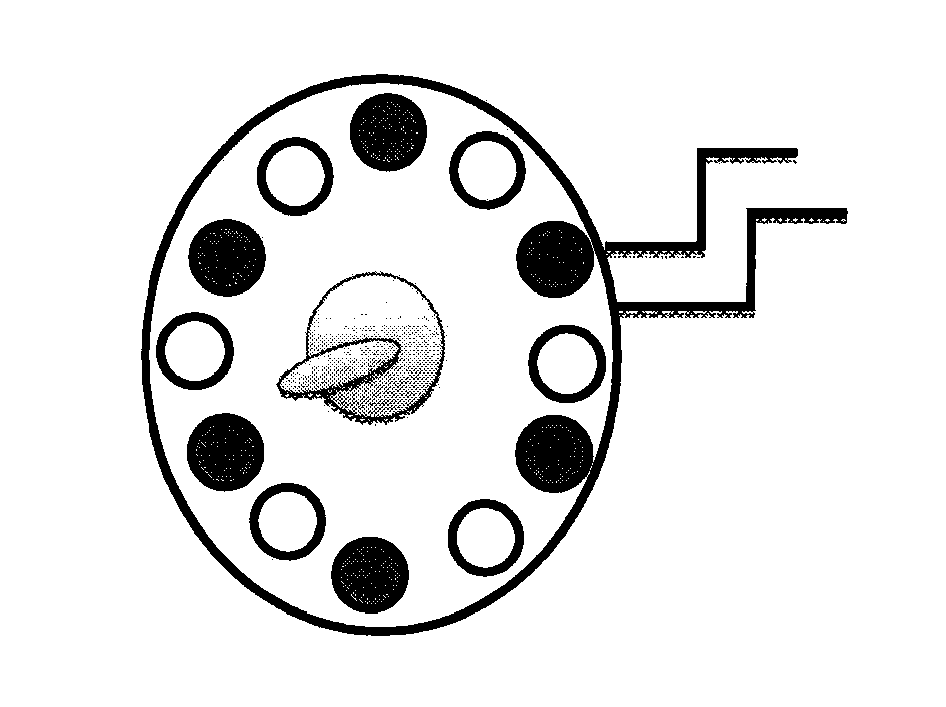
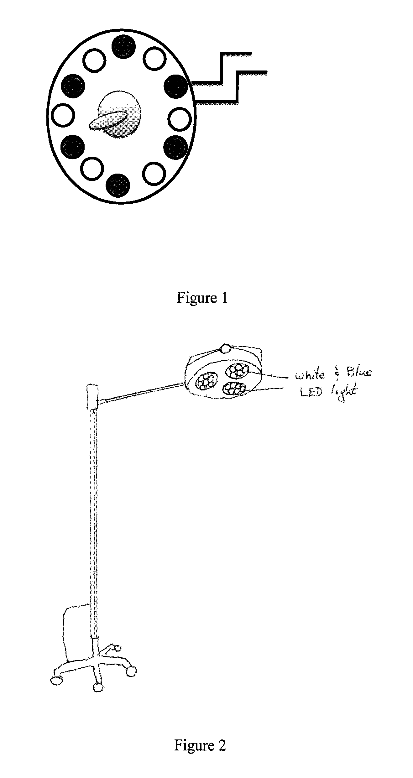
[0031]The present invention describes devices for in vivo identification of diseased tissue in a subject in need thereof. The invention includes a variety of light sources for irradiating an in vivo body part of the subject containing tumor tissue or other diseased tissue with light having at least one excitation wavelength in the range from about 401 nm to about 500 nm. Fluorescence emanating from a fluorescent targeting construct administered to the subject and which has specifically bound to and / or been taken up by the diseased tissue in the body part, in response to the at least one excitation wavelength (i.e., 470-495 nm range) is directly viewed to determine the location and / or surface area of the diseased tissue in the subject.

[0032]The devices and methods described herein relate to and are intended for use with fluorescence imaging technology as described in U.S. Pat. Nos. 6,652,836, 6,299,860 and 6,284,223, all titled “Method For Viewing Diseased Tissue Located Within A Bod...


PUM


Abstract

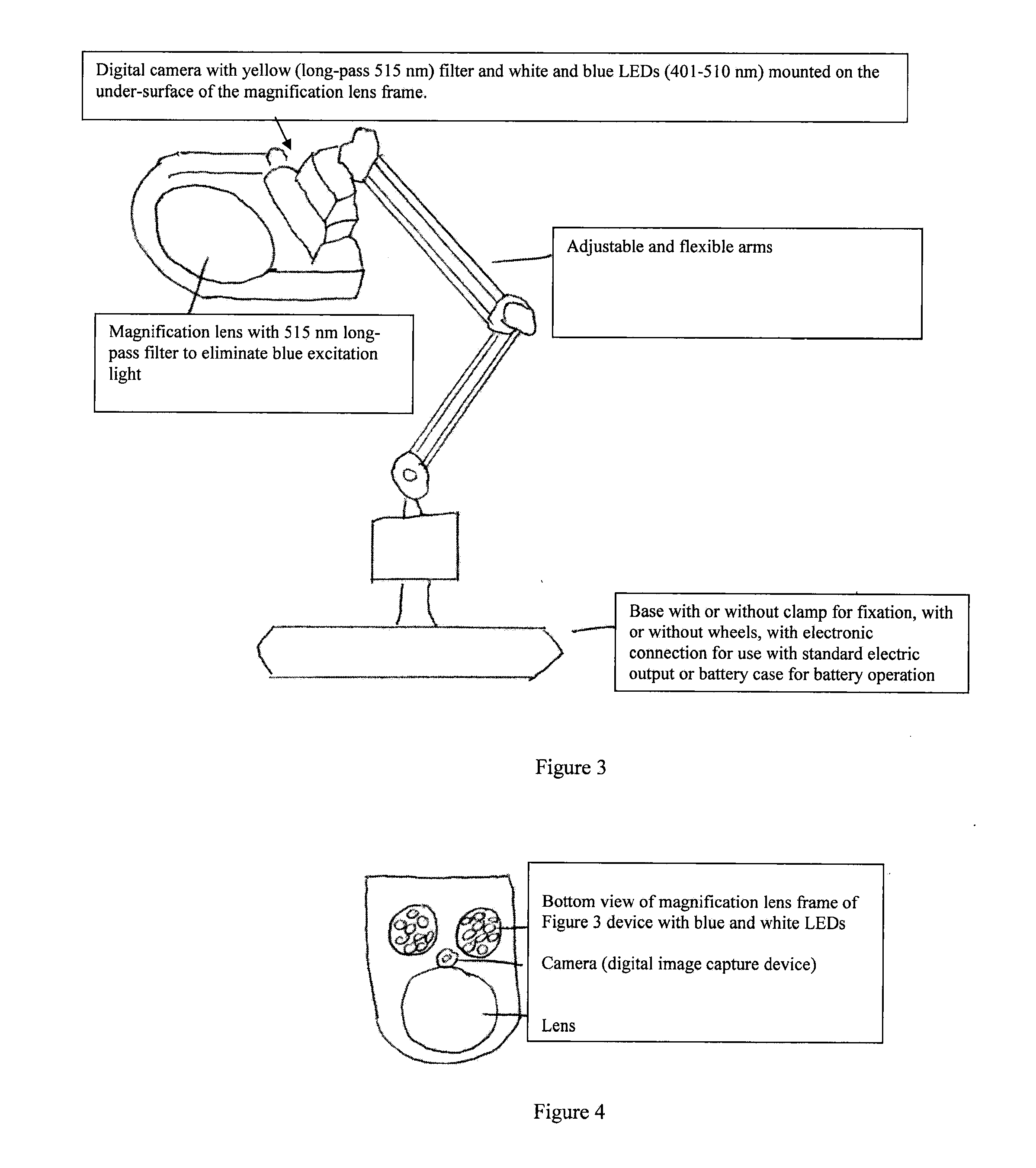
The present invention describes light source devices to provide white and blue (401-510 nm) light for the in vivo identification of diseased tissue using fluorescence based tissue targeting. The light source devices are configured with a variety of LED lights capable of emitting white and blue light with at least one excitation wavelength in the range from about 401 nm to about 500 nm (for example, 470 nm to 495 nm) to irradiate an in vivo body part of a subject containing tumor or diseased tissue. The tumor or diseased tissue has fluorophore-tagged targeting constructs attached. The fluorophores used in the targeting constructs have emission spectra greater than 515 nm. The fluorescence emanating from the fluorescent targeting construct in response to the excitation wavelength is directly viewed with long-pass filtered (515 nm) lenses and is used to determine the location and / or surface area of the diseased tissue in the subject. Fluorescence based surgical identification provides more accurate disease resection.

Description

CROSS REFERENCE TO RELATED APPLICATION(S)[0001]This application claims the benefit of priority under 35 U.S.C. §119(e) of U.S. Ser. No. 61 / 489,158, filed May 23, 2011, the entire contents of which is incorporated herein by reference in its entirety.BACKGROUND[0002]1. Field of the Invention[0003]The present invention relates generally to the medical field and more specifically to methods and devices for viewing the state of a body cavity or an internal organ of a mammalian body. The present invention relates to methods and devices more particularly blue light emitting diode (LED) surgical lighting systems for viewing the state of a body cavity or an internal organ of a mammalian body. More particularly, the invention relates to the light sources and methods used for detecting diseased or tumor tissue at an exterior or interior body site using an intravenously administered fluorescent targeting construct that binds to diseased tissue and which targeting construct is excited by light i...

Claims


Application Information

Patent Timeline
Patent Type & Authority Applications(United States)
IPC IPC(8): A61B5/00A61B17/3205
CPCA61B5/0071A61B17/3205G01J3/10G01N21/6447A61B1/0692G01N2021/6439A61B1/00039A61B1/043A61B1/0684G01N21/6456
Inventor LUIKEN, GEORGE A.
Owner ONCOFLUOR
Learn More