
Functionalized chromophoric polymer dots and bioconjugates thereof

a technology bioconjugates, which is applied in the direction of luminescent compositions, instruments, chemistry apparatuses and processes, etc., can solve the problems of low absorption, poor photostability, and insufficient brightness and photostability of chromophoric polymer dots, so as to reduce the non-specific adsorption of biological molecules

Active Publication Date: 2012-11-08
UNIV OF WASHINGTON +1
View PDF4 Cites 80 Cited by
  • Summary
  • Abstract
  • Description
  • Claims
  • Application Information

AI Technical Summary

Benefits of technology

[0016]In another aspect, the present invention provides a method for conjugating a biological molecule to a functionalized chromophoric polymer dot, the method comprising incubating a functionalized chromophoric polymer dot with the biological molecule in a solution containing polyethylene glycol under conditions suitable for conjugating the biological molecule to the functionalized chromophoric polymer dot, wherein the presence of polyethylene glycol in the solution reduces non-specific adsorption of the biological molecule to the surface of the polymer dot.

Problems solved by technology

However, for many imaging tasks and ultrasensitive assays, their brightness and photostability cannot provide sufficient signal to overcome the background associated with various autofluorescence and scattering processes within the cells.
Other factors such as blinking and saturated emission rate may also pose difficulties in high-speed and high-throughout fluorescent assays.
But intrinsic limitations of the conventional dyes, such as low absorptivity and poor photostability, have posed great difficulties in further developments of high-sensitivity imaging techniques and high-throughout assays (Resch-Genger, U.; Grabolle, M.; Cavaliere-Jaricot, S.; Nitschke, R.; Nann, T. Nat. Methods 2008, 5, 763-775; Fernandez-Suarez, M.; Ting, A. Y. Nat. Rev. Mol. Cell Biol.
However, Qdots are not bright enough for many photon-starved applications because of their low emission rates, blinking, and a significant fraction of non-fluorescent dots (Yao, J.; Larson, D. R.; Vishwasrao, H. D.; Zipfel, W. R.; Webb, W. W. Proc. Natl. Acad. Sci. USA 2005, 102, 14284-14289).
There has been recent work to develop non-blinking Qdots (Wang, X. Y.; Ren, X. F.; Kahen, K.; Hahn, M. A.; Rajeswaran, M.; Maccagnano-Zacher, S.; Silcox, J.; Cragg, G. E.; Efros, A. L.; Krauss, T. D. Nature 2009, 459, 686-689), but their toxicity, caused by the leaching of heavy metal ions, is still a critical concern for in vivo applications.
Another critical issue with quantum dot probes is their toxicity due to the leaching of heavy metal Cd2+ ions.
Additionally, the low emission rates, blinking, and a significant fraction of nonfluorescent dots also raise potential problems, particularly for single molecule / particle imaging applications.
However, there are also a number of limitations with the dye-loaded beads such as limited dye-loading concentration (a few percent) due to self-quenching, and the relatively large particle size (>30 nm) that would preclude sensing schemes involving the use of energy transfer to report analyte concentrations.
However, for applying these probes in biological imaging or sensing applications, an important problem has yet to be solved, that is, the surface functionalization and bioconjugation.

Method used


Image

  • Functionalized chromophoric polymer dots and bioconjugates thereof
  • Functionalized chromophoric polymer dots and bioconjugates thereof
  • Functionalized chromophoric polymer dots and bioconjugates thereof

Examples


example 1

Method for Preparing Functionalized Chromophoric Polymer Dots

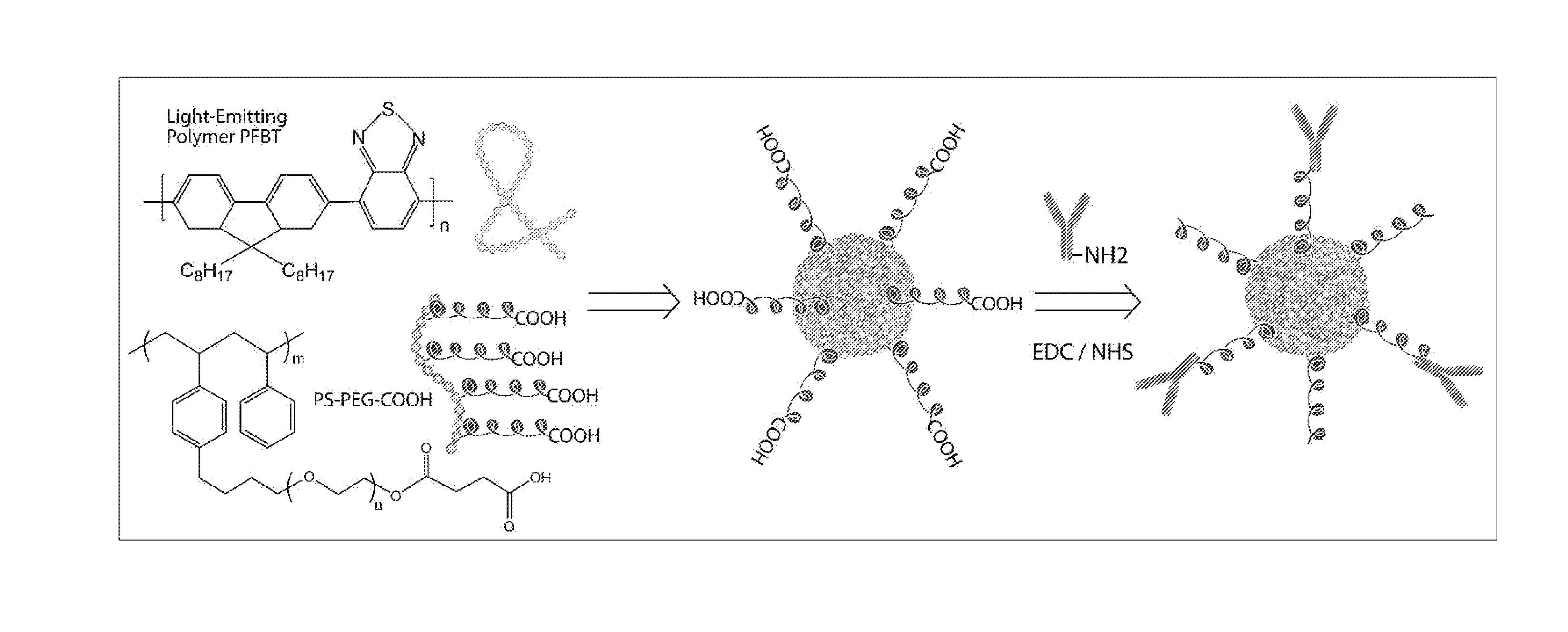
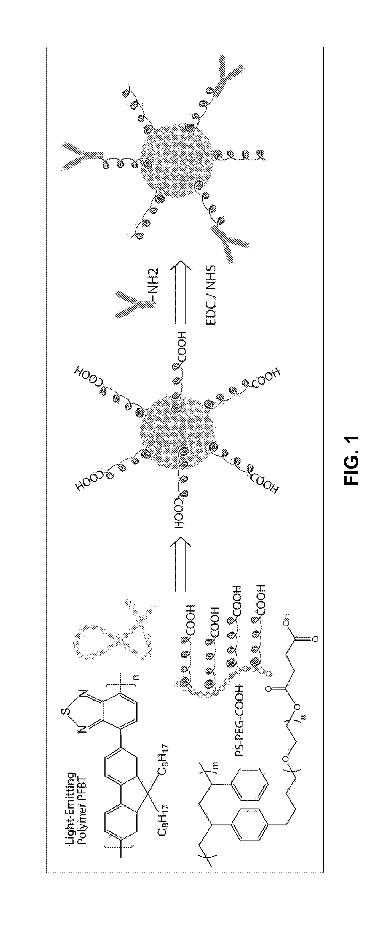
[0239]The present example provides a method for obtaining a quantity of functionalized chromophoric polymer dots for subsequent characterization and biomolecular conjugation. FIG. 1 shows a schematic diagram for preparing the functionalized chromophoric polymer dots and their biomolecular conjugates.

[0240]Functionalized chromophoric polymer dots in aqueous solution were prepared as follows. First, a chromophoric polymer, for example PFBT, was dissolved in tetrahydrofuran (THF) by stirring under inert atmosphere to make a stock solution with a concentration of 1 mg / mL. Certain amount of functionalization agent, for example PS-PEG-COOH in THF solution, was mixed with a diluted solution of PFBT to produce a solution mixture with a PFBT concentration of 40 μg / mL and a PS-PEG-COOH concentration of 4 μg / mL. The mixture was stirred to form homogeneous solutions. A 5 mL quantity of the solution mixture was added quickly to 10 mL o...

example 2

AFM Characterization of Functionalized Chromophoric Polymer Dots

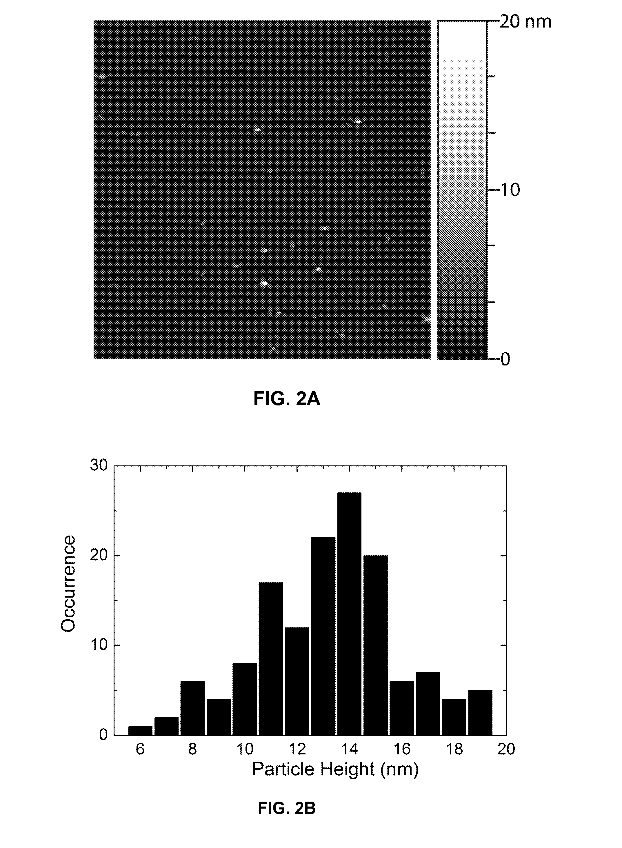
[0241]The functionalized chromophoric polymer dots prepared according to the method provided in Example 1 were assessed by AFM for their size, morphology and monodispersity. For the AFM measurements, one drop of the nanoparticle dispersion was placed on freshly cleaved mica substrate. After evaporation of the water, the surface was scanned with a Digital Instruments multimode AFM in tapping mode. FIG. 2A shows a representative AFM image of the functionalized chromophoric polymer dots. A particle height histogram taken from the AFM image indicates that most particles possess diameters in the range of 10-20 nm (FIG. 2B). The lateral dimensions are also in the range of 10-20 nm after the tip width is taken into account. The morphology and size are consistent with those of the polymer dots prepared without functionalization polymer, indicating the presence of a small amount of amphiphilic polymer has no apparent effect on p...

example 3

Optical Characterization of Functionalized Chromophoric Polymer Dots

[0242]The functionalized chromophoric polymer dots prepared according to the method provided in Example 1 were assessed for their fluorescence properties. UV-Vis absorption spectra were recorded with a DU 720 spectrophotometer using 1 cm quartz cuvettes. Fluorescence spectra were collected with a Fluorolog-3 fluorometer using a 1 cm quartz cuvette. The functionalized chromophoric polymer dots exhibit similar absorption and emission spectra to those of bare chromophoric polymer dots (FIG. 3), indicating the surface functionalization does not effect the optical properties of the polymer dots. Depending on the chromophoric polymer species, the chromophoric polymer dots exhibit absorption bands ranging from 350 nm to 550 nm, a wavelength range that is convenient for fluorescence microscopy and laser excitation. FIG. 3 shows the absorption and emission spectra of functionalized chromophoric polymer PFBT. Analysis of the ...


PUM

PropertyMeasurementUnit
Fractionaaaaaaaaaa
Fractionaaaaaaaaaa
Fractionaaaaaaaaaa

Abstract

The present invention provides, among other aspects, functionalized chromophoric polymer dots comprising a hydrophobic core and a hydrophilic cap, and bioconjugates thereof. Also provided are improved methods for preparing functionalized chromophoric polymer dots. Methods for in vivo imaging and molecular labeling are also disclosed.

Description

CROSS-REFERENCES TO RELATED APPLICATIONS[0001]The present application claims the benefit of U.S. Provisional Application No. 61 / 259,611 filed Nov. 9, 2009, which is expressly incorporated herein by reference in its entirety for all purposes.STATEMENT AS TO RIGHTS TO INVENTIONS MADE UNDER FEDERALLY SPONSORED RESEARCH OR DEVELOPMENT[0002]This invention was made with US Government support under grant numbers R21AG029574, R21CA147831, and R01NS062725, awarded by the NIH. The US Government has certain rights in this invention.REFERENCE TO A “SEQUENCE LISTING,” A TABLE, OR A COMPUTER PROGRAM LISTING APPENDIX SUBMITTED ON A COMPACT DISK[0003]NOT APPLICABLEBACKGROUND OF THE INVENTION[0004]Fluorescent probes have played a key role in modern cell biology and medical diagnostics. Organic small dye molecules are generally used in fluorescent based techniques such as fluorescence microscopy, flow cytometry, and versatile fluorescent assays and sensors. Historically, common fluorophores were deri...

Claims


Application Information

Patent Timeline
IPC IPC(8): C08L25/18G01N33/566C08L89/00G01N33/53
CPCG01N33/587G01N33/582C08G81/00C08L65/00C09K11/06C09K2211/1483C08L87/00
Inventor CHIU, DANIEL T.WU, CHANGFENGMCNEILL, JASONYU, JIANGBO
Owner UNIV OF WASHINGTON
Learn More