In vitro urogenital co-culture models

a co-culture model and urogenital technology, applied in the field of in vitro urogenital co-culture models, can solve the problems of limited anatomical replication of physiological tissue, loss of t and b cell viability and function, and epidemic proportions of sexually transmitted infections

Inactive Publication Date: 2012-06-21
SANOFI PASTEUR VAX DESIGN
View PDF4 Cites 3 Cited by
  • Summary
  • Abstract
  • Description
  • Claims
  • Application Information

AI Technical Summary

Benefits of technology

[0014]The present invention is directed to methods for assaying responses of cells found in the female reproductive mucosa to different test agents. The methods utilize a novel in vitro co-culture model, under static or non-static conditions, that comprises both cells of the reproductive mucosa and immune cells. One component of the co-culture of the present invention uses RWV-cultured epithelial or differentiated tissue attached to microcarrier beads that is subjected to chemical or biological (bacterial or viral), inter alia, insult under microgravity or non-mic

Problems solved by technology

In addition to direct explants or single-cell cultures, some of these methods include the use of cellular aggregates, which can also serve as microcarriers to maximize the cell loading capacity, with limited anatomical replication of physiological tissue.
However, explant culture of tonsilar lymphoid tissue (Nauman, 2007; Hammond, 2001) or lymphoid cells under microgravity conditions show loss of T and B cell viability and function over long-term culture.
Additionally, sexually transmitted infections (STI) have reached epidemic proportions.
Healthy intact vaginal epithelium provides a barrier to STI; however, external insults (e.g., pathogens or chemical) can disrupt this barrier and result in increased susceptibility t

Method used


Image

  • In vitro urogenital co-culture models
  • In vitro urogenital co-culture models
  • In vitro urogenital co-culture models

Examples


example 1

Summary of the Technique

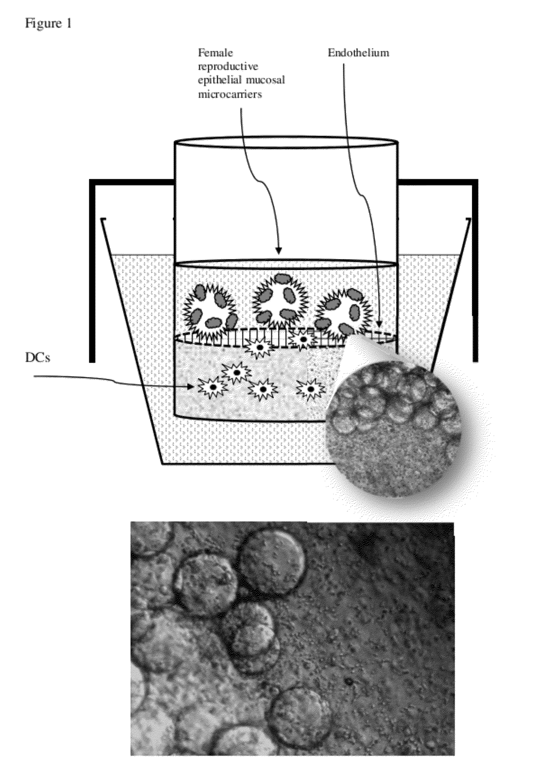
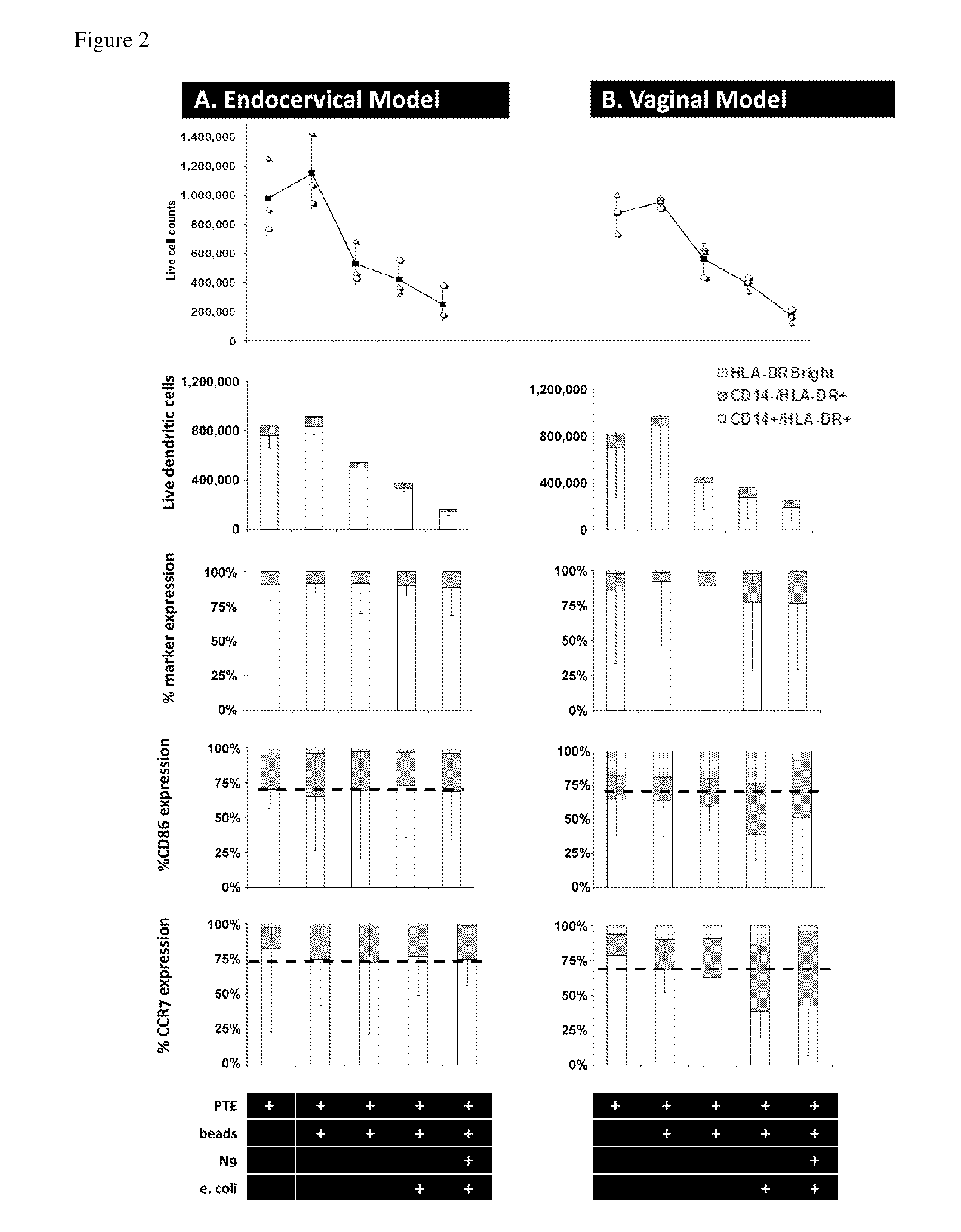
[0046]Our experimental design of the MIMIC® co-culture of RWV bioreactor-grown epithelial tissue from selected female reproductive tract tested the efficacy of the differentiated epithelia to pick up and present antigen (FIG. 1). RWV-cultured female reproductive microcarriers of differentiated vaginal and endocervical mucosa were exposed to microbicide / spermicide and bacterial antigen and then introduced to the APC-containing PTE module to study the innate immune responses to Nonoxynol-9 and E. coli bioparticulate treatments.

Production of Vaginal Tissue Microcarriers and Endocervical Tissue Microcarriers

[0047]Approximately 3.5-week cultured vaginal tissue aggregates and endocervical tissue aggregates were used to mimic the local mucosal tissue of the female reproductive vaginal and endocervical regions. Cell-bearing microcarrier beads were prepared as follows. Collagen-coated Cytodex beads (Sigma) composed of dextran and having an average size of ˜60-87 micro...


PUM


Abstract

The invention is directed to co-culture systems comprising (i) rotating wall vessel (RWV)-cultured epithelial or differentiated tissue attached to microcarrier beads and (ii) the peripheral tissue equivalent (PTE) module of the MIMIC® system, and to methods of using the co-culture systems for assessing chemical or biological (bacterial or viral) insults. The system models mucosal exposure to chemicals, pathogens or antigen at various sites in the human body. The microcarrier and MIMIC® co-culture approach provides an in vitro co-culture model that simultaneously demonstrates mucosa-mediated antigen presentation and immunogenic responses. Models of the present invention can be used, for example, in assessments of disease pathogenesis and in pharmaceutical development, reproductive physiology, and immunological and toxicological evaluations. Models of the present invention can generate patient-specific localized mucosal immunology using primary cells, resembling the human physiological situation.

Description

CROSS-REFERENCE TO RELATED APPLICATIONS[0001]This application claims benefit under 35 U.S.C. §119(e) of U.S. Patent Application No. 61 / 413,698, filed Nov. 15, 2010, which is hereby incorporated herein by reference in its entirety.BACKGROUND TO THE INVENTION[0002]Three-dimensional (3D) tissue models offer great potential in generating in vitro human functional tissue with similarities to the native in vivo environment. Methods for such systems generally include the use of extracellular matrix (ECM) materials, modified culture media nutrients, and static and / or flow conditions to mimic the 3D physiological environment for nutrient transport.[0003]In addition to direct explants or single-cell cultures, some of these methods include the use of cellular aggregates, which can also serve as microcarriers to maximize the cell loading capacity, with limited anatomical replication of physiological tissue. As an example, rotating-wall vessel (RWV) bioreactors have been used successfully for ce...

Claims


Application Information

Patent Timeline
IPC IPC(8): C12Q1/70C12Q1/02
CPCC12N5/0683C12N5/0697C12N2502/1121C12N2502/246C12N2502/28C12N2503/04G01N2500/10C12N2531/00C12N2533/54C12N2533/70G01N33/5044G01N33/54313C12N2525/00
Inventor MAHMOOD, AYESHAHERBST-KRALOVETZ, MELISSA M.WARREN, WILLIAM
Owner SANOFI PASTEUR VAX DESIGN
Why Eureka
Try Eureka