
Adhesion-Resistant Surgical Access, Reinforcement and Closure Prosthetic

a surgical access and prosthetic technology, applied in the field of medical implants, can solve the problems of affecting the healing process, and causing excessive or clinically unacceptable scar tissue or fibrosis, and achieves the effects of minimizing the proliferation of fibrosis, facilitating healing, and facilitating healing

Inactive Publication Date: 2012-01-12
MAST BIOSURGERY
View PDF2 Cites 4 Cited by
  • Summary
  • Abstract
  • Description
  • Claims
  • Application Information

AI Technical Summary

Benefits of technology

[0051]An adhesion-resistant surgical access, reinforcement and closure prosthetic (closure prosthetic) is provided for assisting the healing function of a human tissue disposed within a body and comprising a plurality of outer layers sandwiching an inner layer which may be porous and conductive to both chemical and cellular infiltrates. The sandwiching layers may resist tissue attachment, be comprised of a bio-resorbable polymer, such as poly-lactic acid or certain polyurethanes, and possess a maximum thickness suitable for insertion into the body via laparoscopic means. Optimally, there are two sandwiching layers defining proximal and distal surfaces that mimic the proximal and distal surfaces of the original tissue on which a surgical incision is intended. Between the sandwiching layers may be disposed a tissue scaffold which is oriented such that the edges of the incision made by a surgical procedure preferentially grow together in a normal anatomical aspect when the prosthetic is implanted to repair such a surgical incision. The sandwiching layers may overlap the tissue scaffold middle layer so as to provide marginal or peripheral regions between which living tissue may be engage and held in proximity to the tissue scaffold sandwiched therein. Preferably, the engagements thus made affect a fluidic seal able to withstand several mmHg of pressure. This is a beneficial feature since it is a common occurrence that such tissue layers enclose a lubricious fluid, for example, the dura and the parietal pericardium.
[0053]There is further provided a method for assisting the healing function of a human tissue disposed within a body, including outer walls and cell conductive medium, the method utilizing a first attachment means; an incision means; an inner wall unfolding means such that when the prosthetic is applied to a target tissue layer, this layer having proximal and distal sides, the prosthetic affixes to the distal surface of said tissue layer, an incision is made through the prosthetic and affixed tissue layer and the second inner layer of the prosthetic deploys onto the proximal side of said tissue layer, and a means for closing said formed incision.
[0055]The closure prosthetic described herein is advantageous because compared to other anti-adhesion devices, it utilizes tissue scaffolding to conduct the healing power of the body without allowing adjacent tissue to adhere thereof, or the cellular contents released by the incision to cause fibrosis away from the healing site. In a preferred embodiment the closure prosthetic uses at least one attachment means to reinforce the prosthetic-tissue prior to the placement of a surgical incision and a second attachment means capable of sandwiching the tissue edges of the incision at the moment the incision is executed permitting the incision to be spread open by a simple procedure that can be quickly performed without damaging said sandwiched tissue. In addition, the closure device localizes the wounded tissue so as to minimize proliferation of fibrosis and focuses the healing response to the wound site. The closure prosthetic mimics the functional performance of the tissue prior to incision once it is closed after the incision is made, which minimizes repeated re-injury of the healing site. Repeated re-injury of a wound site is an important etiology for chronic inflammation, excessive scarring, and adhesion to adjacent tissues. This ultimately generates a faster repair, and hastens the onset of tissue quiescence, or resumption of normal cytokine levels. As a result, a greater variety of surgical procedures can be provided with minimal post-operative pain under a greater variety of circumstances, including but not limited to, enhanced capacity for tissue healing, restoration of pre-incisional function, and enhanced or even permanent functionality because it utilizes a novel combination of wound healing stimulus in combination with sequestration of the wound heating process.

Problems solved by technology

This aspect of the tissue repair mechanism presents a special problem for surgical techniques that require access to tissue enclosed in living tissue membranes meant to separate dynamic tissue surfaces, such as the function of the pericardium in separating the beating heart from the lungs, surrounding vasculature and bone.
The healing process subsequent to surgery is non-specific in its locus of activity and tends to generate excessive or clinically unacceptable amounts of scar tissue, or fibrosis.
Scar tissue can in several instances block arteries, immobilize joints, damage internal organs, and can in some instances generally or partially impede a body's ability to maintain vital functions.
Too much scar tissue may cause physiological roadblocks that in severe cases can disfigure, cripple or even kill.
The number of patients undergoing cardiac surgery has been steadily increasing, and as a consequence, the number of cardiac complications has also increased.
Such conditions can persist often times with lower rates of treatment success as compared to similar complications of other tissues.
Despite the need for effective treatments, doctors typically have few optimal remedies to help them control this relatively prevalent condition.
That is, in the context of cardiac surgical procedures, scar tissue treatments tend to be less effective in preventing or attenuating fibrosis than with other procedures.
For example, thin-membrane implantation measures for attenuating adhesion following cardiac surgery, such as disclosed in U.S. application Ser. No. 10 / 660,461, tend to be less effective than similar procedures applied to other tissues and / or other parts of the body.
Wherever the cellular environment tends to remain in a transitional state, the region affected by fibrotic repair processes increases, and tissues with specialized functions tend to be compromised by the indiscriminate joining these processes precipitate.
Nevertheless, these approaches tend to be less effective in providing an optimum or even adequate attenuation of tissue adhesions in that vicinity, again potentially as a result of a relatively excessive amount, or nature, of movements of or near the affected tissue in need of treatment.
In particular, while such membranes provide temporary barriers to adhesion formation, they do not direct the healing process toward repair of the tissue, and consequently the non-specific scar tissue that forms becomes the source of continued irritation, remodeling, and release of cytokines that promote adhesion, sometimes long after the surgical barrier has been resorbed or encapsulated.
The nature of the device is that it tends not to allow the pericardium to expand further with time.
They are by their dimensional and mechanical aspects difficult to identify and repair, and repairs if achieved, compromise their barrier functionality.
They are particularly susceptible to stress, strain, and very slight abrasion.
Many of these tissues undergo contraction when incised, and the edges of the incised tissue cannot be brought back in apposition in a repair procedure in a tension-free manner.

Method used


Image

  • Adhesion-Resistant Surgical Access, Reinforcement and Closure Prosthetic
  • Adhesion-Resistant Surgical Access, Reinforcement and Closure Prosthetic
  • Adhesion-Resistant Surgical Access, Reinforcement and Closure Prosthetic

Examples


example 1

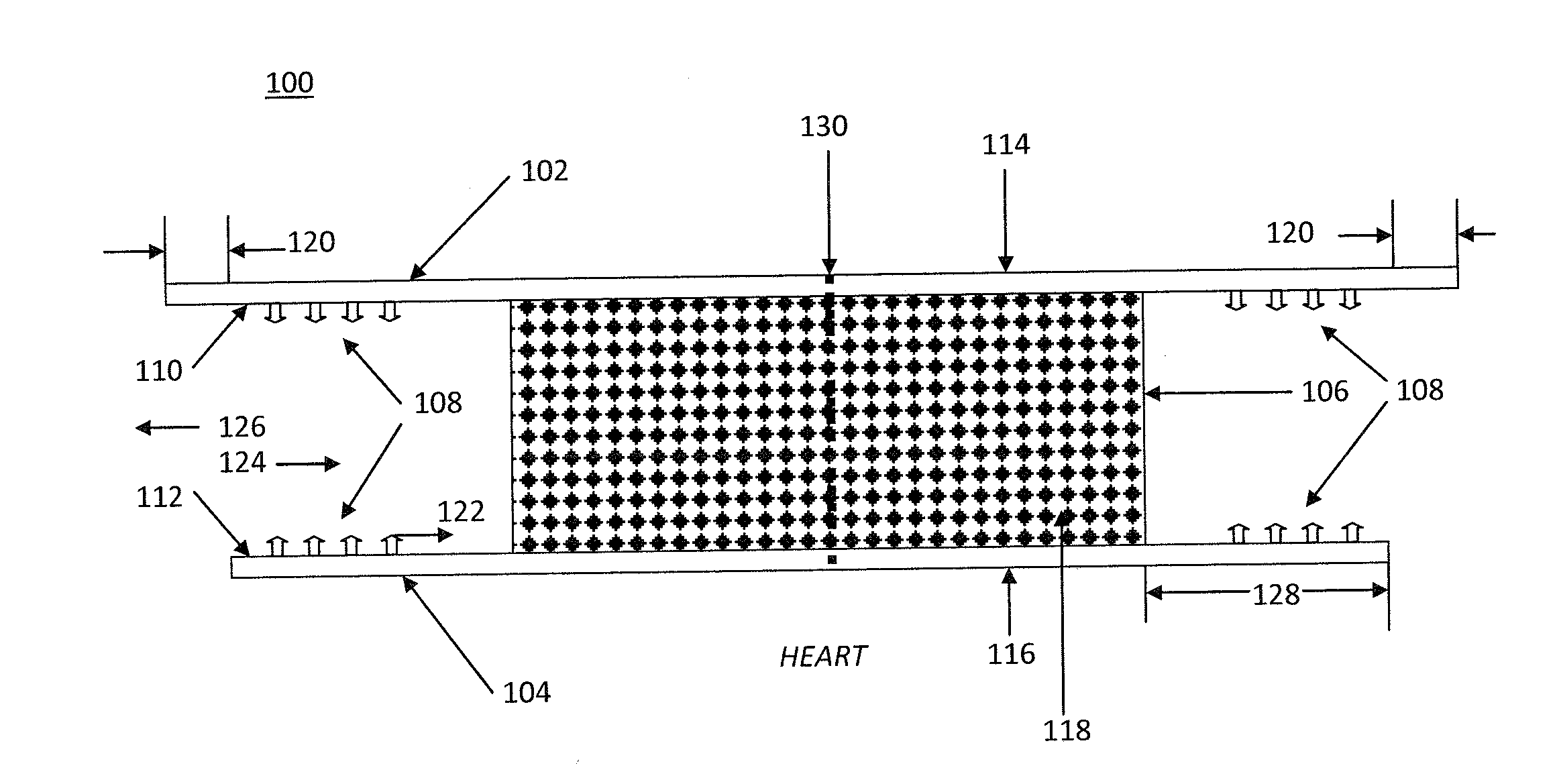
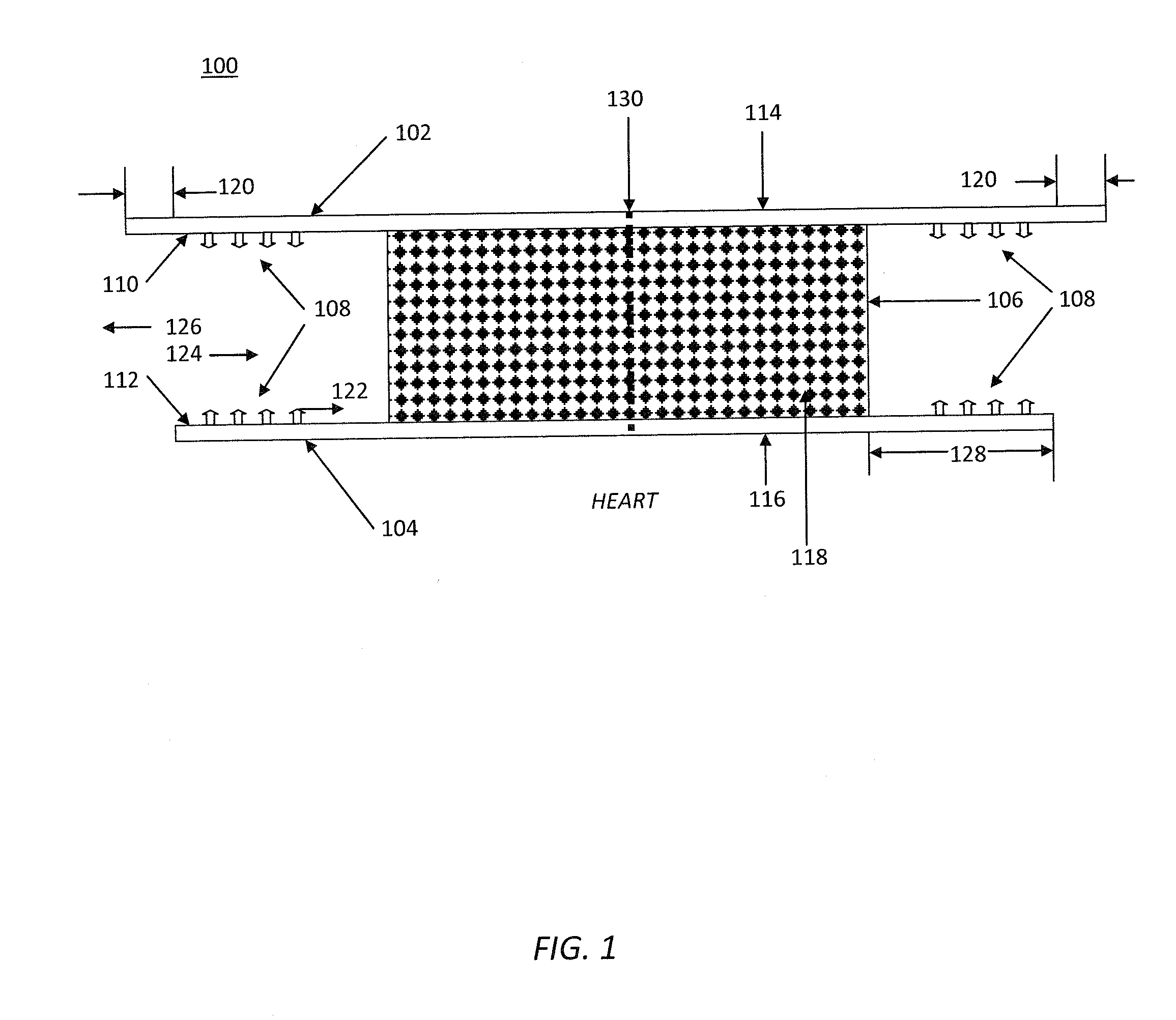
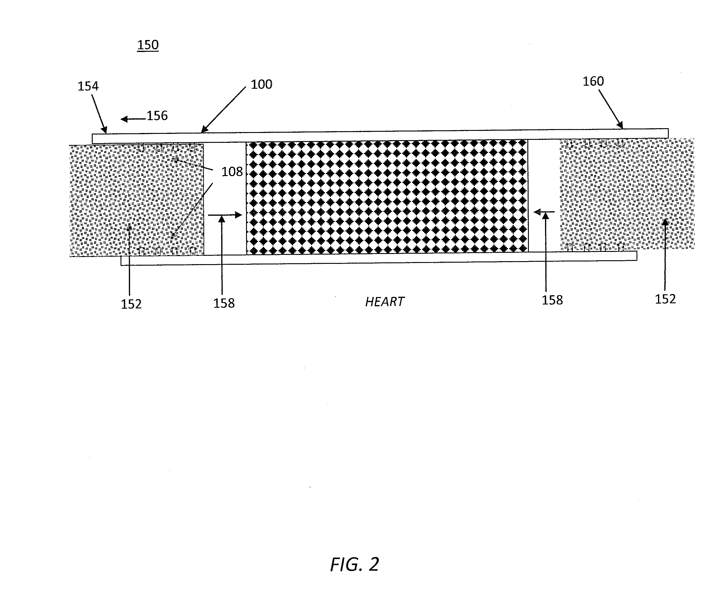
[0077]Referring to FIG. 1, a closure prosthetic 100 is comprised of an absorbable distal layer 102, an absorbable proximal layer 104, a sandwiched tissue scaffold layer 106, and attachment means 108. The attachment means 108 are located on distal interior side 110 of layer 102 and proximal interior side 112 of layer 104. Layers 102 and 104 have exterior distal sides 114 and exterior proximal sides 116, respectively. Sides 114 and 116 are smooth and resist tissue attachment. Layers 102 and 104 are bonded to opposite sides of tissue scaffold layer 106. Tissue scaffold layer 106 is a porous, flexible medium. The pores 118 are interconnected and sufficiently large and numerous to allow cells to infiltrate and tissue to grow into the tissue scaffold layer 106. Distal layer 102 is slightly larger than proximal layer 104, and both layers 102 and 104 are larger than tissue scaffold layer 106. The layers are bonded together in this arrangement, thereby defining a margin 120. Margin 120 allow...

example 2

[0081]Referring now to FIGS. 4A and 4B, a closure prosthetic 400 is shown with pre-folded proximal layer 104 in FIG. 4A. Edges 402 possess a proximally directed lip 404, as illustrated in FIG. 4B. Between edges 402 is defined the locus of incision such that when incision 406 is made, incision edges 408 naturally open and pass beyond lips 404 to position 410. Now when a dilator (not pictured) is placed in incision 406 proximal layers 104 unfolds. The fold in proximal layer 104 can be manufactured such that it is bistable, and can reside in either the fully folded position or the fully open position such that no matter the degree of dilation of incision 406 proximal layer 104 of closure prosthetic 400 becomes fully open.

[0082]Alternatively, the substance of proximal layer can be hydrophilic and induced to swell when in contact with bodily fluids such that shortly after incision 406 is made the swelling of proximal layer 104 causes it to assume the fully open position.

Closure Prostheti...

example 3

[0083]Referring now to FIG. 5, a closure prosthetic 500 is illustrated that includes inversion pulls 502. An incision 504 is made in tissue layer 506 and incision 504 naturally opens. Proximal layer 104 is naturally in the open position. To cause it to fold toward the incision opening, pulls 502 are pulled in direction 508. Proximal layer edges 508 engage incision edges 510 and slide proximally around them, causing tissue layer 506 to be sandwiched between distal layer 102 and proximal layer 104.

Closure Prosthetic


PUM


Abstract

Described are devices and methods for reinforcing a layer of living tissue, which when affixed to a tissue layer prior to surgical incision, reinforces the tissue to be incised, provides a fibro-conductive matrix to promote healing in a preferred plane, and provides for a subsequent closure and fluidic seal. A partially or entirely absorbable growth matrix is disclosed, comprising two adhesion-resistant layers enclosing a cellular conductive medium for promoting fibrosis in a preferred plane. The cellular conductive portion is partially or entirely sequestered from surrounding tissue. The device is constructed in a physiologic range of tensile strengths and elasticity suitable for closure of the pericardium, peritoneum, or other typically thin membranes enclosing organs in the body, whose function is to prevent adhesions between tissue surfaces normally in motion.

Description

CROSS-REFERENCE TO RELATED APPLICATIONS[0001]This application claims the benefit of U.S. Provisional Application No. 61 / 353,156, filed Jun. 9, 2010 and entitled Adhesion-Resistant Surgical Access, Reinforcement and Closure Prosthetic (Att. Docket MB8134CIPPR2) and U.S. Provisional Application No. 61 / 355,053, filed Jun. 15, 2010 and entitled Adhesion-Resistant Surgical Access, Reinforcement and Closure Prosthetic (Att. Docket MB811.34CIPPR3), and is a continuation-in-part of U.S. application Ser. No. 12 / 498,291, filed Jul. 6, 2009 and entitled Resorbable Membrane Implanting Methods for Reducing Adhesions (Att. Docket MB8134P), the entire contents all of which are expressly incorporated herein by reference. U.S. application Ser. No. 12 / 498,291 claims priority to U.S. Provisional Application No. 61 / 078,431, filed Jul. 6, 2008 and entitled Resorbable Membrane Implanting Methods for Reducing Adhesions (Att. Docket MB8134PR) and is a continuation-in-part of U.S. application Ser. No. 10 / 66...

Claims


Application Information

Patent Timeline
Patent Type & Authority Applications(United States)
IPC IPC(8): A61F2/02
CPCA61B17/0057A61B17/085A61B2017/00004A61B2017/00243A61B2017/00597A61F2250/0051A61B2019/4884A61F2/0063A61F2002/0086A61F2002/009A61B2017/00893A61B2090/0816A61F2/02A61F2/06A61B2017/00238
Inventor BLUECHER, LUKASMILBOCKER, MICHAEL T.CALHOUN, CHRISTOPHER J.
Owner MAST BIOSURGERY
Learn More