Pulsatile medical device designed to be used in extracorporeal surgery

a medical device and pulsatile technology, applied in the field of pulsatile medical devices, can solve the problems of high probability of clot formation, difficult situations, postoperative hemodynamic disorders, etc., and achieve the effects of minimizing the head loss of pulsatile flow, low cost, and convenient availability

Active Publication Date: 2011-11-17
CHASTANIER PIERRE
View PDF4 Cites 0 Cited by
  • Summary
  • Abstract
  • Description
  • Claims
  • Application Information

AI Technical Summary

Benefits of technology

[0028]The purpose of the improved pulsatile medical device (or tube) of the present invention is to provide a device that generates a flow that is more uniform, minimizing the head losses of the pulsatile flow, particularly in the actual pulsatile zone (see FIG. 6), and to make the use of pulsatile heart assistance systems more widespread as a physiological method of low cost that is easily available and adaptable to the energy sources that are available in medical and surgical services for pediatric patients and also for adults.

Problems solved by technology

In spite of the progress made in the ECC field since it appeared early 1950s, it still presents drawbacks that give rise to postoperative hemodynamic disorders.
The artificial surfaces of the ECC activate coagulation and platelets, giving rise to a very high probability of clots forming inside the circuit, which can lead to difficult situations such as postoperative bleeding, and biochemical and electrolytic disorders.
There is thus a confrontation between two different kinds of hydraulic circuit and there are difficulties in adapting them functionally to each other and making them work together harmoniously.
In order to overcome those drawbacks of conventional ECC, various theoretical strategies have been applied:the use of medications such as anticoagulants, inotropics, vasodilatators, hormones, electrolytes, blood, platelets, or other substitutes, and this has not completely solved hemodynamic problems while giving rise to specific undesirable effects;ECC at normal temperature: this is becoming more and more widespread, replacing low temperature ECC.
Beside the beneficial effects on postoperative hemodynamics, such a technique is difficult, and can only be used on patients with few complex needs.
It requires specific equipment for holding still the portion on which the operation is being performed, thus making the method expensive;pulsatile ECC: in order to conserve shear stress stimulation and conserve the endothelial function during surgery, a pulsatile mode of perfusion has been used by various groups over the last three decades.
That therefore requires special high-technology equipment of high cost, and with mediocre induced pressure curves.
Such equipment is therefore not very widespread.
Similarly, it appears paradoxical to apply pulses to a fluid such as blood with its very fragile components in a long rigid tube of small diameter, regardless of the material from which it is made, e.g. polyvinylchloride (PVC) or silicone, and with its undesirable effects of microembolisms.
Although inexpensive and correlated with pulsatile ECC, that method remains invasive, with vascular complications, especially with children or adults suffering from atherosclerosis.
In conclusion, those methods are usually evaluated by the improved perfusion of localized organs such as the renal or splanchnic areas, but provide little protection to distal organs such as cerebral circulation.
The methods mentioned above thus do not solve the undesirable effects of ECC.

Method used


Image

  • Pulsatile medical device designed to be used in extracorporeal surgery
  • Pulsatile medical device designed to be used in extracorporeal surgery
  • Pulsatile medical device designed to be used in extracorporeal surgery

Examples


first embodiment

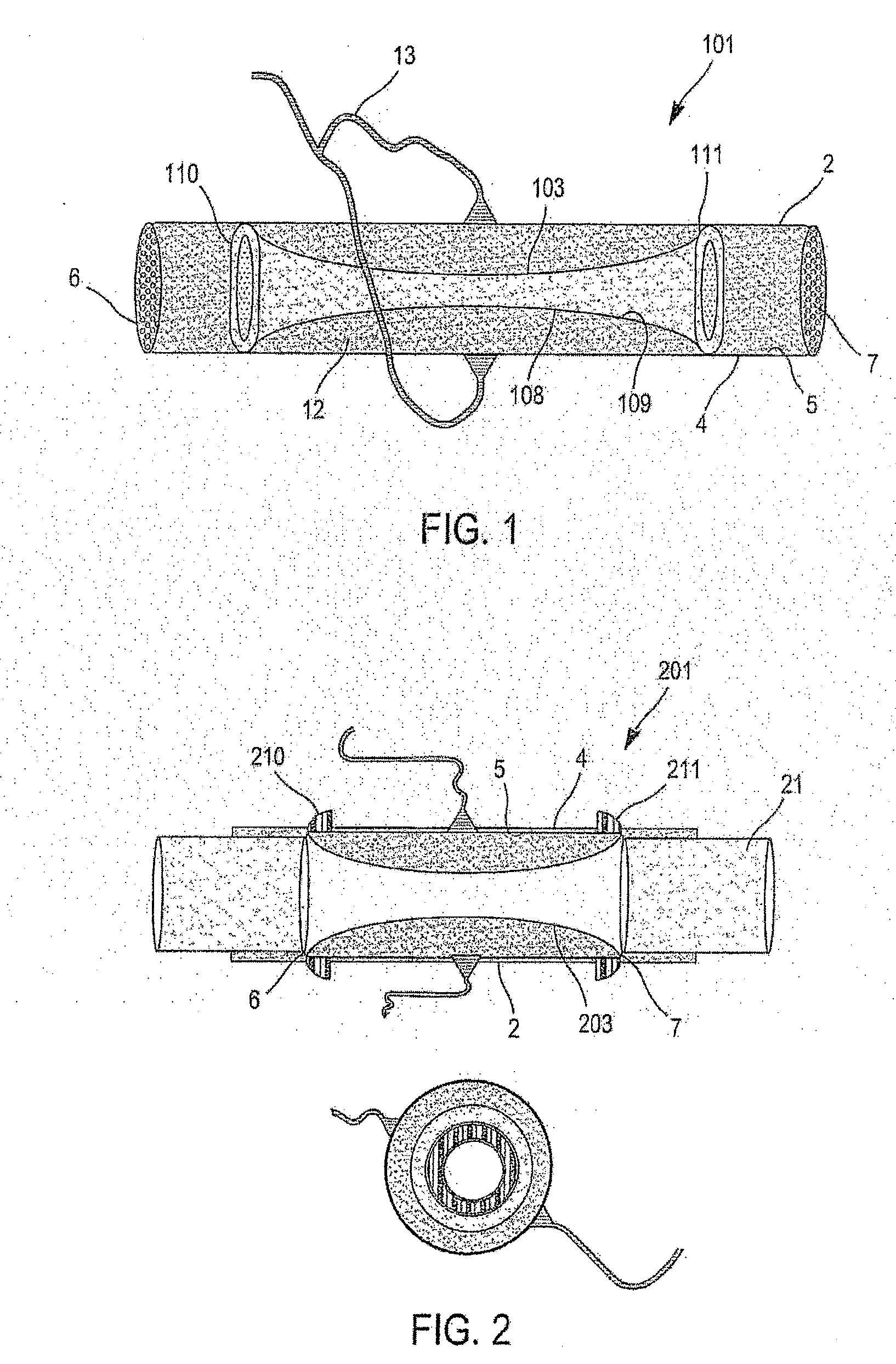
[0085]FIG. 1 shows a pulsatile medical device 101 of the invention. The device comprises an external pipe 2 in which an internal pipe 103 is inserted. The internal pipe 103 is a compressible pipe made of flexible biocompatible material, such as polyurethane, for example. The external pipe 2 is more rigid, e.g. being made of polyvinyl chloride (PVC) and serves as an outer shell.

[0086]The external pipe 2 has an outside wall 4, an inside wall 5, a first end 6 for connection to an ECC type machine, for example, and a second end 7 for connection to the body of a patient. The internal pipe 103 also has an outside wall 108 and an inside wall 109 as well as ends 110 and 111. The internal pipe 103 in FIG. 1 is shorter than the external pipe 2, and the zone where both pipes 2 and 103 are present is a zone referred to as a “two-lumen” zone. In the embodiment of FIG. 1, the ends 110 and 111 of the internal pipe 103 are fastened around their entire periphery to the inside wall 5 of the external ...

second embodiment

[0090]FIG. 2 shows the pulsatile medical device 201 that is identical with the embodiment of FIG. 1 except that the internal pipe 203 is as long as the external pipe 2, thus making its possible to use a different configuration for the ends 210 and 211 of the internal pipe 203. These ends are not fixed to the inside wall 5 of the external pipe 2, but rather to the outside wall 4 of the external pipe 2 by being folded over. This pulsatile medical device 201 is then fastened at each of its two ends 6 / 210 and 7 / 211 to standard ECC connector ports 21 via couplings so as to avoid intravascular accidents in the event of fluid leakage.

third embodiment

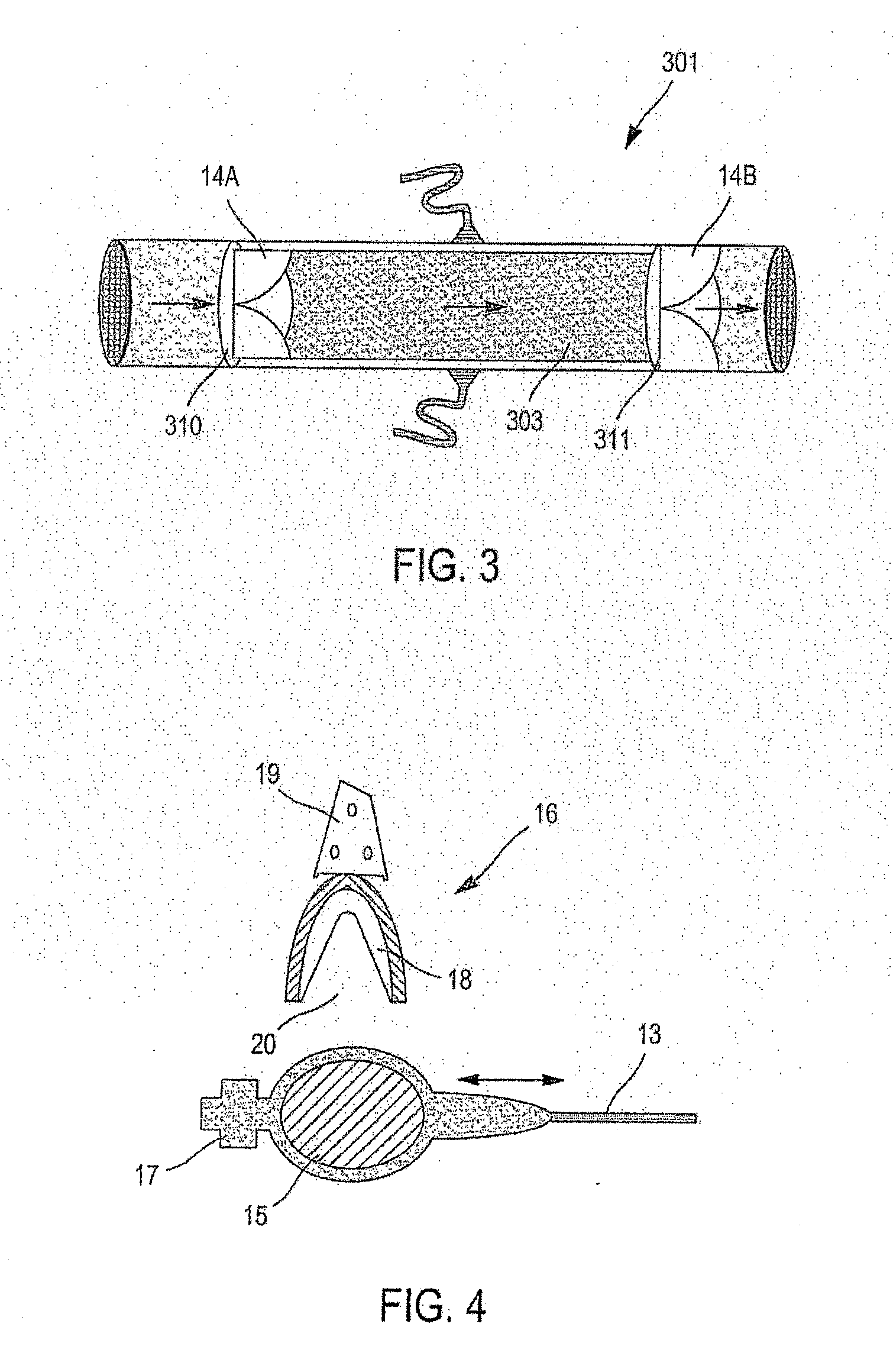
[0091]FIG. 3 shows the pulsatile medical device 301 in which two unidirectional type valves 14A and 14B are placed at the ends 310 and 311 of the internal pipe 303.

[0092]The presence of these valves 14A and 14B makes it possible to obtain a unidirectional blood flow (represented by arrows), thereby reducing vortex effects and head losses.

[0093]FIG. 4 shows an example of an appliance for creating pulsations through the pulsatile medical device of the invention. The appliance comprises:[0094]a first portion including a pouch 15 filled with fluid and connected at one end to the connector port 13 and its other end to an anti-reflux valve 17 enabling the pouch 15 to be filled with fluid or to be emptied; and[0095]a second portion constituted by means 16 for compressing said pouch 15 and comprising a pouch compressor compartment 18 and a control 19, e.g. electromechanical, for controlling said compressor compartment 18. The compressor compartment 18 has a recess 20.

[0096]In operation, the...


PUM


Abstract

The invention relates to a pulsatile medical device enabling a blood flow to be circulated. The invention consists in a device comprising:
    • an external pipe presenting an inside wall, an outside wall, and two ends, one end being for connection to an ECC type machine or to a cardiac assistance system or to the patient's body, and one end being for connection to the patient's body;
    • an internal pipe inserted in said external pipe, the internal pipe presenting an inside wall, an outside wall, and two ends that are fastened all around their peripheries to the external pipe all around its periphery, the blood flow passing through said internal pipe;
    • the outside wall of the internal pipe and the inside wall of the external pipe defining a space for filling with fluid, said space being connectable via a connector port to an appliance for creating one or more inflations/deflations of said space, thereby creating one or more pulses in the blood flow.

Description

FIELD OF THE INVENTION[0001]The present invention relates to a pulsatile medical device for use in extracorporeal surgery and in other cardiac assistance systems using a pneumatic or an electric pump as a source. By way of example, the device enables regular pulsatile perfusion to be obtained in the organism of a patient being subjected to open heart surgery, or suffering from heart failure.[0002]The cardiovascular system is a closed hydraulic circuit under pressure and that is internally lined with endothelial cells. The endothelium is continuously subjected to tangential shear stress forces that are essential for maintaining the endothelial function that includes vascular tonus by nitride oxide synthesis (NOS), blood coagulation, the inflammatory response, arteriosclerosis, angiogenesis, and apoptosis.[0003]An extracorporeal circulation (ECC) is a medical device that is used to take the place of the heart and the lungs in operating blocks, cardiac catheterization laboratories, or ...

Claims


Application Information

Patent Timeline
Patent Type & Authority Applications(United States)
IPC IPC(8): A61M1/12
CPCA61M1/106A61M1/3667A61M1/107A61M60/113A61M60/274A61M60/38A61M60/851A61M60/427A61M60/232A61M60/279A61M60/405A61M60/569
Inventor NOUR, SAYED
Owner CHASTANIER PIERRE
Why Eureka
Try Eureka