
Analyte detection devices and methods with hematocrit-volume correction and feedback control

a technology of hematocrit and volume correction, applied in the direction of instruments, fluorescence/phosphorescence, etc., can solve the problems of pre-programmed catalysts producing either insufficient or excessive blood, and the development of accurate glucose monitoring may be subject to errors, etc., to achieve accurate, efficient, and economical determination of the presence and/or concentration of analytes

Active Publication Date: 2011-08-18
INTUITY MEDICAL INC
View PDF103 Cites 48 Cited by
  • Summary
  • Abstract
  • Description
  • Claims
  • Application Information

AI Technical Summary

Benefits of technology

This approach enhances the accuracy of glucose monitoring by correcting for hematocrit and sample volume variations, ensuring precise glucose concentration measurements and minimizing user discomfort and test inefficiencies.

Problems solved by technology

Glucose monitoring technology that relies on the photometric method of quantifying the glucose concentration in whole blood may be subject to errors associated with variations in hematocrit level, or concentration of red blood cells within the blood sample.
This presents a challenge for the development of accurate glucose monitoring.
If the user has not supplied a sufficient amount, then the meter generally displays an error code and the user must test again.
Further, a user may supply more than the typical amount of blood to the test strip, which may lead to an inaccurate result if the calibration of the strip is volume sensitive.
For others, this pre-programmed catalyst may produce either an insufficient or excessive amount of blood, as well as other undesired outcomes, such as excessive bruising (for those with fragile capillary networks), an unnecessary delay in obtaining results (for fast bleeding individuals), as well as excessive residual blood left on the skin.
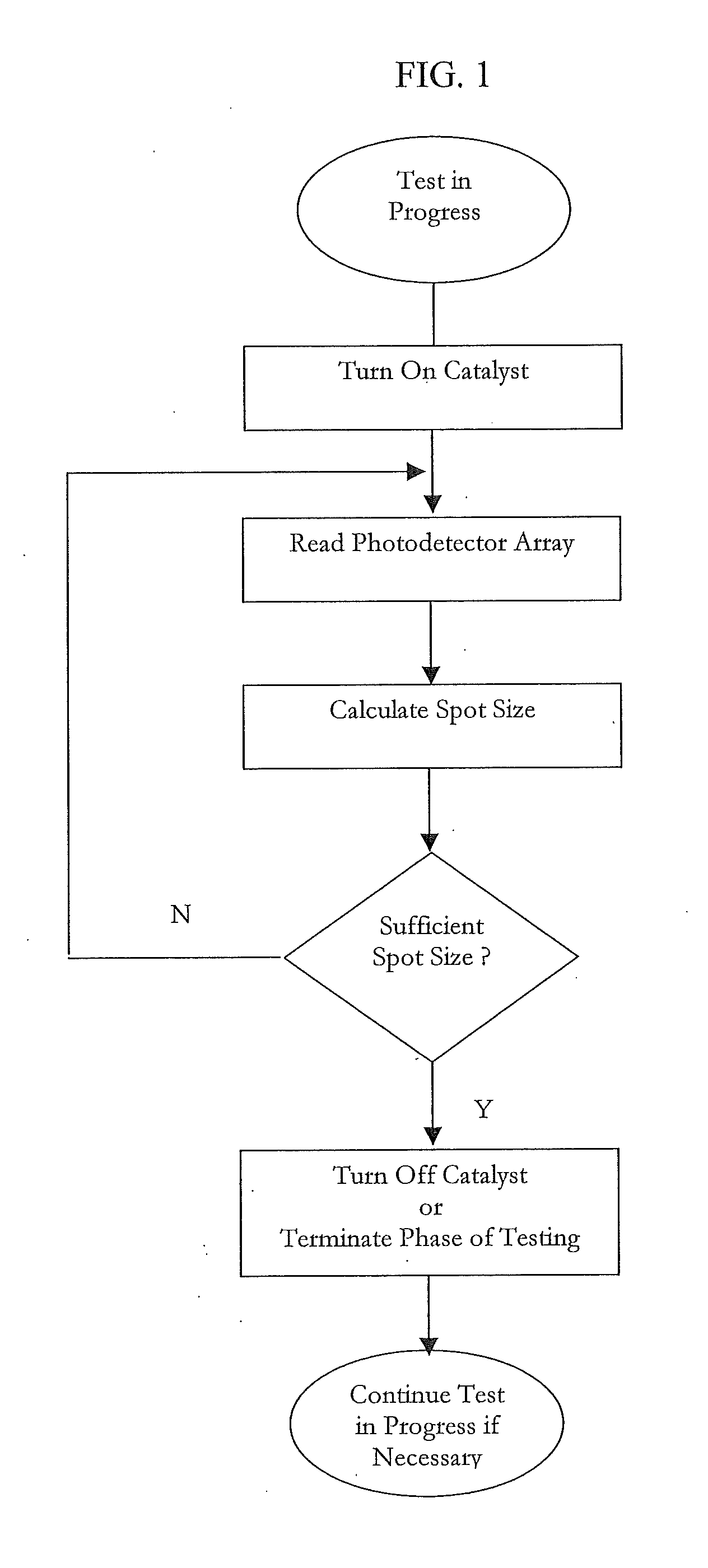
For a variety of reasons, the quality of a spot developed as a result of an analyte reacting with the reagent membrane may not be ideal as described above.
Most devices on the market cannot detect or correct for low quality spots.
Their sensors, typically one or more photodiodes, do not have the ability to discretely analyze the flaws within a reagent spot.
Thus, there exists a risk that these systems may not provide an accurate reading in circumstances of a non-ideal spot.

Method used


Image

  • Analyte detection devices and methods with hematocrit-volume correction and feedback control
  • Analyte detection devices and methods with hematocrit-volume correction and feedback control
  • Analyte detection devices and methods with hematocrit-volume correction and feedback control

Examples


Embodiment Construction

[0036]Exemplary arrangements and methods for the detection and measurement of the presence and / or concentration of a target analyte, such as glucose, bilirubin, alcohol, controlled substances, toxins, hormones, proteins, etc., will now be described.

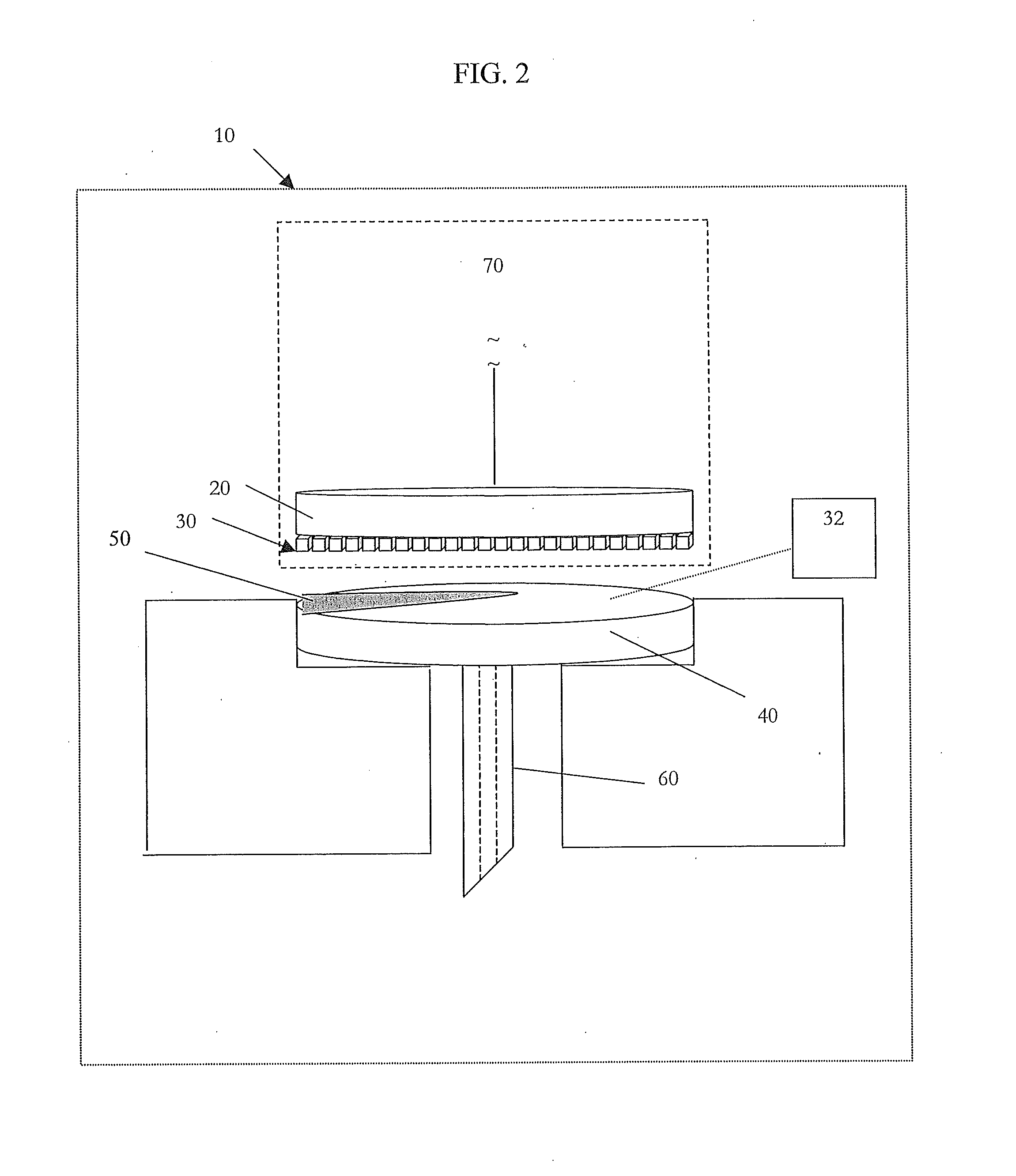
[0037]In broader aspects, the current invention provides the ability to correct for broad variations in sample hematocrit levels in the measurement of an analyte, such as glucose, during the course of the test. The invention takes advantage of an imaging array of detectors to perform this correction and does not require any additional hardware. In other words, no other distinct sensors or detectors, other than the imaging array, are required to calculate the correction. The very sensor that is used to quantify the analyte within the sample may also correct for hematocrit. Strategic algorithms that process the data from the imaging array provide real-time or near real-time information about the sample hematocrit level. Thus, more accurate ...


PUM

PropertyMeasurementUnit
volumeaaaaaaaaaa
volumeaaaaaaaaaa
lengthaaaaaaaaaa

Abstract

Disclosed are devices, arrangements and methods for quantifying the concentration of an analyte present in bodily fluid, including: an assay pad having at least one chemical reagent capable of producing a detectable signal in the form of a reaction spot upon reaction with the analyte; a light source; a detector array; a processor; and a memory in communication with the processor, the memory comprising: (a) at least one value indicative of one or more of: (i) the level of hematocrit contained in the sample; (ii) the volume of the sample applied to the assay pad; or (iii) imperfections present in the reaction spot; and (b) at least one algorithm for calculating the concentration of the analyte contained in the sample.

Description

[0001]The present application claims priority pursuant to 35 U.S.C. §119(e) to provisional application Ser. No. 60 / 689,546 filed Jun. 13, 2005, the entire content of which is incorporated herein by reference.FIELD OF THE INVENTION[0002]The present invention is directed to techniques and devices for detection of the presence and / or concentration of an analyte.BACKGROUND OF THE INVENTION[0003]In the following discussion certain articles and methods will be described for background and introductory purposes. Nothing contained herein is to be construed as an “admission” of prior art. Applicant expressly reserves the right to demonstrate, where appropriate, that the articles and methods referenced herein do not constitute prior art under the applicable statutory provisions.[0004]According to the American Diabetes Association, diabetes is the fifth-deadliest disease in the United States and kills more than 213,000 people a year, the total economic cost of diabetes in 2002 was estimated at...

Claims


Application Information

Patent Timeline
Patent Type & Authority Applications(United States)
IPC IPC(8): A61B5/157G01N21/78
CPCA61B5/14532A61B5/14535A61B5/14546Y10S435/895A61B2562/0295G01N33/54373G01N33/66A61B5/681A61B5/155A61B5/157A61B5/6802G01N21/78G01N2021/752A61B5/1455A61B5/1495A61B5/150358A61B5/6831G01N33/523
Inventor EMERY, JEFFREY L.STIVERS, CAROLE R.
Owner INTUITY MEDICAL INC
Learn More