
Optimized and defined method for isolation and preservation of precursor cells from human umbilical cord

Active Publication Date: 2010-08-26
LAB MEDINFAR PROD FARMS
View PDF2 Cites 9 Cited by
  • Summary
  • Abstract
  • Description
  • Claims
  • Application Information

AI Technical Summary

Benefits of technology

[0026]In addition to the absence of mechanical manipulation, the introduction of three independent phases for dissociated cell recovery and the optimization of several technical parameters along the process make the method simple, robust, 100% reliable in terms of efficacy, and highly efficient in terms of number of cells obtained related to time. As a result the method can be applied in cell therapy services, as well as cryopreservation of mesenchymal stem cells resulting from isolation for future autologous use.
[0035]4—Composition of the digestion solution: several enzymatic digestion solution compositions were tested, namely Alpha-Mem supplemented with 20% FBS and 1% Penicillin / streptomycin; saline buffer solution e.g. HBSS, supplemented with EDTA, more specifically with 186 mg / ml CaCl2.2H2O, 400 mg / ml KCl, 60 mg / ml KH2PO4, 200 mg / ml MgSO4.7H2O, 8000 mg / ml NaCl, 350 mg / ml NaHCO3, 90 mg / ml NaH2PO4.7H2O, 1000 mg / ml glucose, and 76 mg / ml (0.260 mM) EDTA; the previous HBSS solution supplemented with 5 mM CaCl2; 25mM HEPES buffer supplemented with 5 mM CaCl2. The saline buffer solution (HBSS), supplemented with 0.260 mM EDTA, produced the best yields.
[0038]7—Type of agitation (moderate or vigorous) during the incubation period of the digestion reaction: the incubation was performed in a water bath with orbital shaking with either moderate or vigorous oscillation rates, consisting of 100 oscillations per minute (opm) and 140 oscillations per minute (opm), respectively. The moderate agitation of 100 opm led to better results in terms of cell yields.
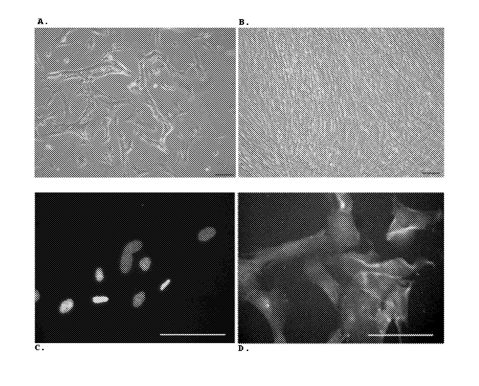
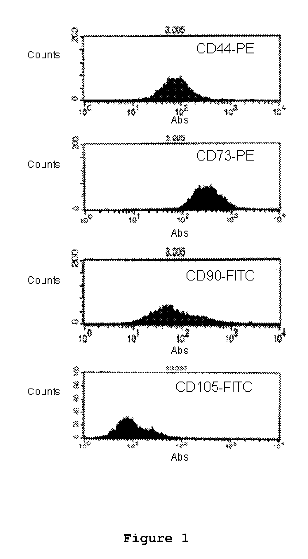
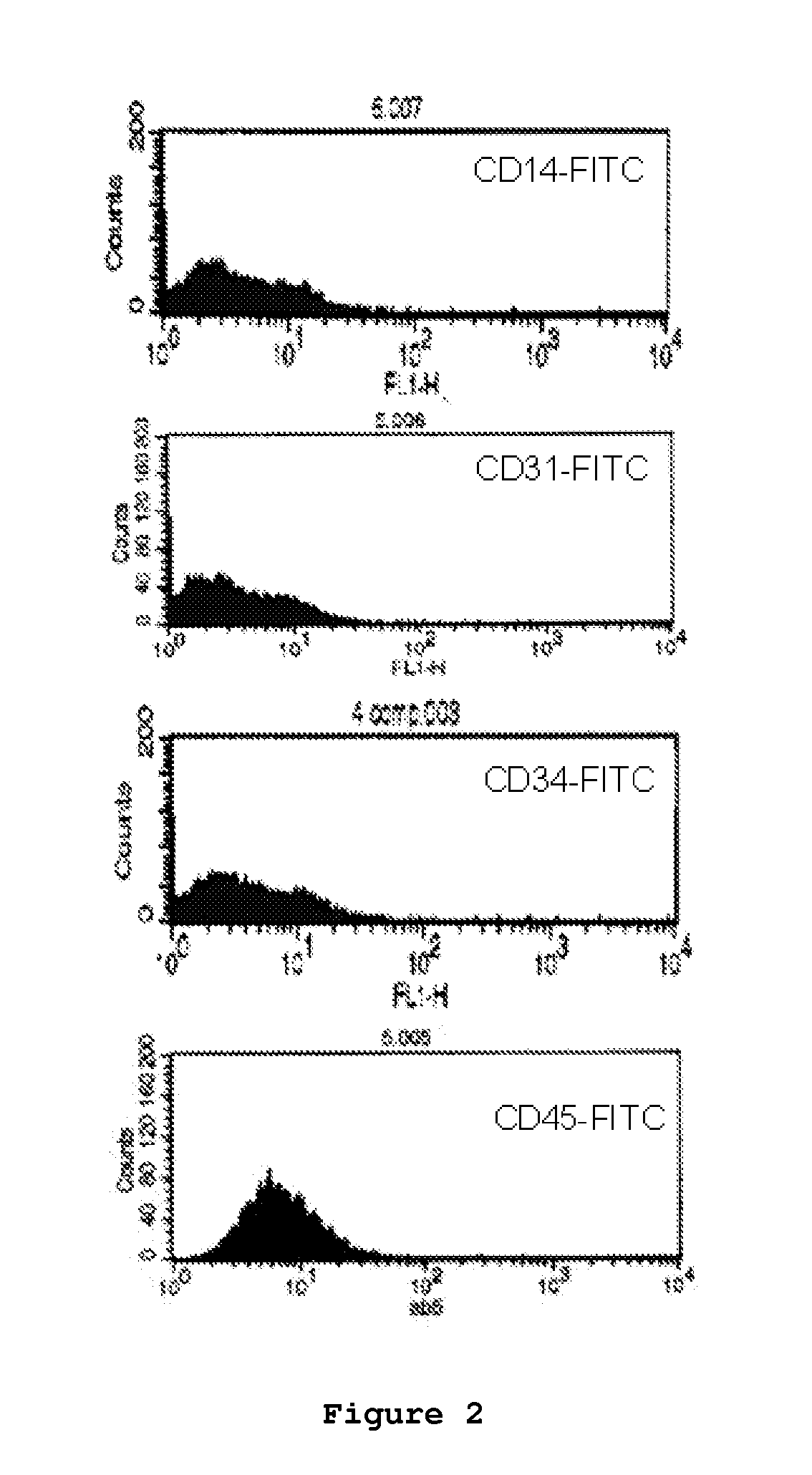
[0048]Sequential cell recovery and all the other optimized factors described previously were optimized so that the method presents 100% efficacy and maximum efficiency in terms of number of cells selected and isolated. In turn, the cell population obtained can be identified by the cells being capable of adhering and expanding / multiplying in a surface and defined growth medium, where the majority of cells in culture express the cell surface markers CD44, CD73, CD90 and CD105, and the majority of cells present only residual expression of the cell surface markers CD14, CD31, CD34 and CD45, where the majority of cells are able to undergo up to 18 expansion / multiplication phases, maintaining a constant duplication factor of approximately 1.7 / 24 h, a constant fibroblast-like morphology, and the capacity of partial or terminal differentiation into specialized cells, such as osteoblasts, chondrocytes, adipocytes, cardiomyocytes, and glial cells.

Problems solved by technology

Despite the enormous differentiation potential of embryonic precursor cells, their utilization for research or / and therapy is controversial and has raised serious ethical and safety issues.
The major limitations to the utilization of mesenchymal precursor cells arise during clinical practice, namely during cell harvesting.
Furthermore, because mesenchymal stem cells are rare, the final number of cells obtained is generally low.
Since mesenchymal stem cells are present in umbilical cord blood in limited numbers, attempts to isolate these cells from this tissue have resulted in some frustration, and even the most successful attempts, using very high amounts of blood, have not surpassed the 60% success rate, relative to the total number of tissue samples processed.
A detailed analysis reveals that the protocols described are, in one way or the other, restrictive in terms of the nature of the cells obtained, or rather vague in terms of success rate and efficiency in the number of cells isolated.
In fact, the restrictive nature of these protocols invariably resulted in loss of phenotypic diversity of the cell populations isolated, mainly due to unnecessary focus on specific tissues or geographic locations within the umbilical cord structure.
Furthermore, uncertainly invariably remained about the actual number of stem cells that could be obtained in the end.
Additionally, none of the methods described so far has demonstrated efficacy in terms of number of successful tissue samples processed in order to be reliable enough so as to be applied in cell therapy protocols.
Furthermore, the introduction of unnecessary steps of structural manipulation, such as extraction of umbilical vessels (Purchio et al., 1998; Mitchell et al., 2003; Davies et al., 2004; Wang et al., 2006), or mechanical maceration (Seyda et al., 2006), makes existing protocols hard to standardize and reproduce, never assuring enough cell numbers for cell therapy application.
Furthermore, excessive tissue manipulation induces cell differentiation which is undesirable if one wants to maintain precursor cell phenotype (Gardner et al., 2000; Claes et al., 2002; Cullinane et al., 2003).
Also showing limitations are the protocols based on the umbilical vessels themselves.
These reports are not consistent, also contributing to the lack of definition and criteria of the applied methodologies.
The discrepancy between these approaches is unjustified and the excess tissue manipulation of the existing tissues in either of these two protocols undermines desirable effects on precursor phenotype maintenance, and consequently endangers the utilization of the isolated precursor cells in isolation and cryopreservation services for the population in general.

Method used


Image

  • Optimized and defined method for isolation and preservation of precursor cells from human umbilical cord
  • Optimized and defined method for isolation and preservation of precursor cells from human umbilical cord
  • Optimized and defined method for isolation and preservation of precursor cells from human umbilical cord

Examples


example 1

Optimization of Type / Amount of Mechanical Manipulation and Fraction Dimension

[0070]The type of mechanical manipulation and size of initial tissue fragments were optimized maintaining a constant proportion of tissue mass (g), surface area of the bottom of the digestion flask (cm2), digestion volume (ml), and the total volume of the flask (ml), of approximately 1:2:2:37, considering that a fraction of 1 cm of human umbilical cord weighs about 1 g. After removing the amniotic membrane, several types of umbilical cord fractionation were tested: low (5 cm fractions); medium (2.5 cm fractions); high (0.3 cm fractions); and minced tissue. All fractionations were performed with the help of a scalpel and the samples processed using exactly the same conditions. It was concluded that the best yields, in terms of total cells at the end of P0 / umbilical cord mass / time, were obtained when using 2.5 cm fractions. Table 1 summarizes the results obtained qualitatively.

TABLE 1Fractionation optimizatio...

example 2

Optimization Relative to the Presence or Absence of Blood Clots within Umbilical Cord Vessels (1 Vein and 2 Arteries)

[0071]It is known that lysis of red-blood cells is toxic, reducing cell viability in vitro. Therefore, cell yields were compared when the digestion was performed in the presence or absence of blood clots. For the latter experiment, blood clots were removed with the help of a scalpel. It was concluded that blood clots had a negative effect upon yield, in terms of total cells at the end of P0 / umbilical cord mass / time. Table 2 summarizes the results obtained qualitatively.

TABLE 2Blood clots: effects upon cell yield.PresenceAbsence−++Key: +++ = Excellent,++ = very good,+ = good,− = reasonable,−− = bad,0 = unsuccessful

example 3

Optimization Relative to Enzyme Nature, Individual or Combined Enzyme Action, and Enzyme Concentration in the Digestion Solution

[0072]In order to maximize yields, in terms of the number of cells with the desired characteristics isolated from the initial tissue, two initial approaches were adopted related to enzyme digestion: direct cell adhesion to the culture flask, in the presence of culture medium, with no digestion, and therefore in the absence of enzymes; and tissue dissociation with a single enzyme: 0.075% (w / v) collagenase II or 2.0% (w / v) pronase.

[0073]Since the utilization of collagenase II alone was the most efficient approach, this enzyme was then combined with other enzymes, specifically with Trypsin 0.125% (w / v) (in the presence or absence of EDTA 0.260 mM), with hyaluronidase 0.5% (w / v) alone, and with hyaluronidase 0.5% (w / v), combined with pronase 2.0% (w / v).

[0074]For these tests the optimal fraction size of 2.5 cm was used and the proportion of tissue mass (g), surf...


PUM


Abstract

The present invention refers to an optimized and defined method for isolation and preservation of precursor cells from human umbilical cord. Besides being reproducible and 100% reliable, in terms of the number of samples processed, the method results in a high and defined number of precursor cells, being the majority obtained after a single adhesion and expansion / multiplication phase ex vivo (thus granting cell phenotype), in a shorter time frame than what was previously described in the state-of-the-art. With this method, it is possible to obtain, in 9 days, after direct freezing of a cell fraction, and after one expansion / multiplication phase ex vivo (end of P0) of the majority of the cells, about 8.6(±0.1)×105 cells / gram of processed umbilical cord. In turn, the characteristics of the cells allow, for example, after 35 days, obtaining an average of 7.7×10¹⁵ cells, with precursor phenotype, from 100% of processed umbilical cord samples. The method, because it is simple, robust and 100% reliable, can be performed under good manufacturing practices (GMP) in laboratories dedicated to cell therapy in humans. Furthermore, the method has applications in the pharmaceutical, cosmetic and biotechnology areas.

Description

TECHNICAL FIELD OF THE INVENTION[0001]This application is a U.S. national phase filing under 35 U.S.C. §371 of PCT Application No. PCT / IB2008 / 054067, filed Oct. 3, 2008, and claims priority thereto under 35 U.S.C. §119 to Portuguese patent application no. 103843, filed Oct. 4, 2007, the entireties of both of which are incorporated by reference herein.[0002]It is an aspect of the present invention an optimized and defined method for isolation and preservation of precursor cells from the human umbilical cord matrix.[0003]By “precursor cell” in the present invention, it is meant a type of cell capable of adhering and expanding / multiplying in a surface and defined growth medium, where the majority of cells in culture express the cell surface markers CD44, CD73, CD90 and CD105, and the majority of cells present only residual expression of the cell surface markers CD14, CD31, CD34 and CD45, where the majority of cells are able to undergo up to 18 expansion / multiplication phases, maintaini...

Claims


Application Information

Patent Timeline
IPC IPC(8): C12N5/073C12N5/0775
CPCC12N5/0605C12N5/0668C12N2509/00A61P43/00
Inventor GANCHAS SOARES, RITA ISABELBAPTISTA COELHO, MARIA CONSTANCASILVA SANTOS, JORGE MIGUELMARTINS, JOSE PAULOBASTO, VERA ALEXANDRAESTILITA MONTEIRO DA CRUZ, PEDROSOARES DA CRUZ, HELDER JOAQUIM
Owner LAB MEDINFAR PROD FARMS
Learn More