Cerebral protection during carotid endarterectomy and methods of use

Inactive Publication Date: 2002-09-12
EDWARDS LIFESCIENCES CORP
View PDF7 Cites 230 Cited by
  • Summary
  • Abstract
  • Description
  • Claims
  • Application Information

AI Technical Summary

Benefits of technology

[0006] A dramatic improvement in the neurologic outcome of patients undergoing carotid endarterectomy, and arteriotomy procedures generally, can be achieved by using a blood filter device to capture and remove dislodged embolic material and a shunt to maintain vascular perfusion during the surgical procedure in accordance with our invention. Thus, the invention provides novel methods and apparatus for protecting a patient from embolization during arteriotomy procedures. In one embodiment, the invention provides a bypass tubing or indwelling shunt, having a main lumen for blood bypass and a second, branching lumen adapted to receive an elongated blood filter and to allow passage of same into an artery distal to the endarterectomy region. The branching secondary lumen can either merge and communicate with the main lumen of the shunt, or may extend to a distal opening separate from the blood bypass lumen of the device. A hemostatic valve is included in the proximal end of the second lumen to prevent blood loss from the shunt.
[0008] In another embodiment, the shunt comprises two tubular members. The first tubular member has a lumen that communicates with a distal port and first and second proximal ports. The second member has a lumen, which communicates with a proximal port and a distal end, which is adapted for releasable attachment to the first proximal port of the first tubular member. A filter device comprising an elongate member is insertable through the hemostatic valve included in the second proximal port of the first tubular member. In certain embodiments, the first and / or second tubular member includes a valve for regulating blood flow through the shunt, thereby maintaining optimal vascular perfusion. A manometer may be included in the distal end of the first tubular member to monitor blood pressure downstream the atheromatous lesion. This apparatus is especially useful in performing endarterectomy in patients having tenuous cerebral perfusion pressure (i.e., having ICA pressure slightly above 50 mmHg) and initially not requiring a shunt. The second tubular member can easily be connected to the first tubular member intra-operatively when unexpected cerebral hypoperfusion occurs, e.g., as in systemic hypotension or cardiogenic shock, to maintain blood flow to the brain.
[0013] It will be understood that there are several advantages to using the apparatus and methods disclosed herein for performing open endarterectomy, especially the on the extracranial carotid arteries. For example, the apparatus (1) provides a filter device to capture embolic debris generated during the procedures, thereby minimizing the risk of perioperative stroke, (2) provides a shunt for maintaining cerebral perfusion, thereby further reducing the risk of perioperative morbidity, (3) provides balloon occluders as an alternative to using vascular clamps which commonly cause injuries to vessel, such as hematoma or dissection, (4) provides a filter device which can be used in conjunction with a standard single-lumen indwelling shunt, and (5) can be used in performing endarterectomy on various arteries, including the aorta, the iliac, the femoral, and the popliteal arteries.

Problems solved by technology

The atheromatous lesions in the extracranial carotid vessels, i.e., the common carotid arteries (CCA), the internal carotid arteries (ICA), and the external carotid arteries (ECA), progress through a stage in which brain blood flow is marginal to the stage of occlusion, which results in insufficient cerebral perfusion, with brain death and clinical stroke.
If the stump pressure is lower than 50 mm Hg, brain perfusion during carotid occlusion may be inadequate, thereby requiring a shunt to bypass the clamped region of the artery to maintain cerebral perfusion.
The above-described procedure, however, suffers from a deficiency which relates to the escape of embolic material which may lead to devastating neurologic complications, particularly when emboli pass through the internal carotid artery.

Method used


Image

  • Cerebral protection during carotid endarterectomy and methods of use
  • Cerebral protection during carotid endarterectomy and methods of use
  • Cerebral protection during carotid endarterectomy and methods of use

Examples


Embodiment Construction

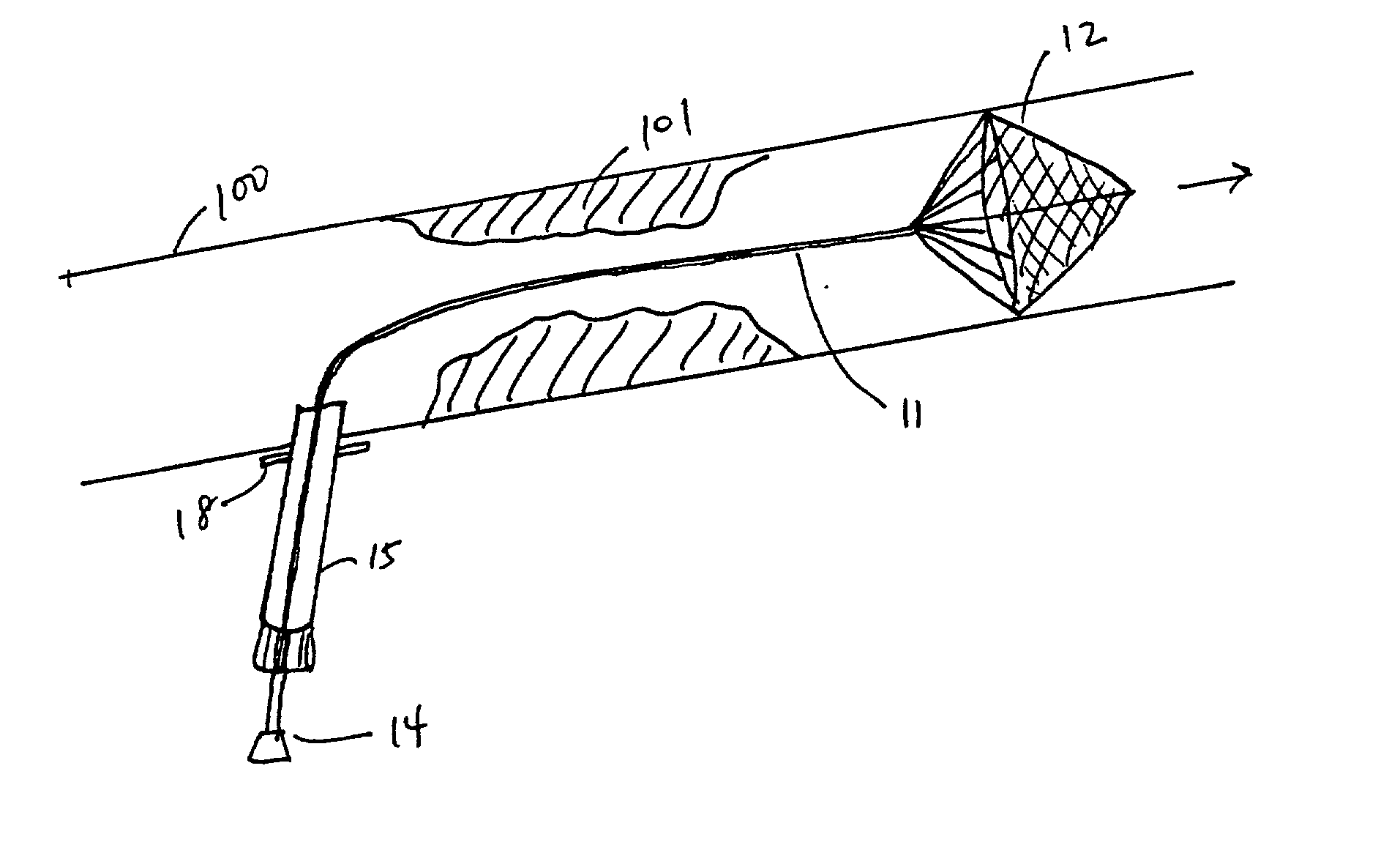
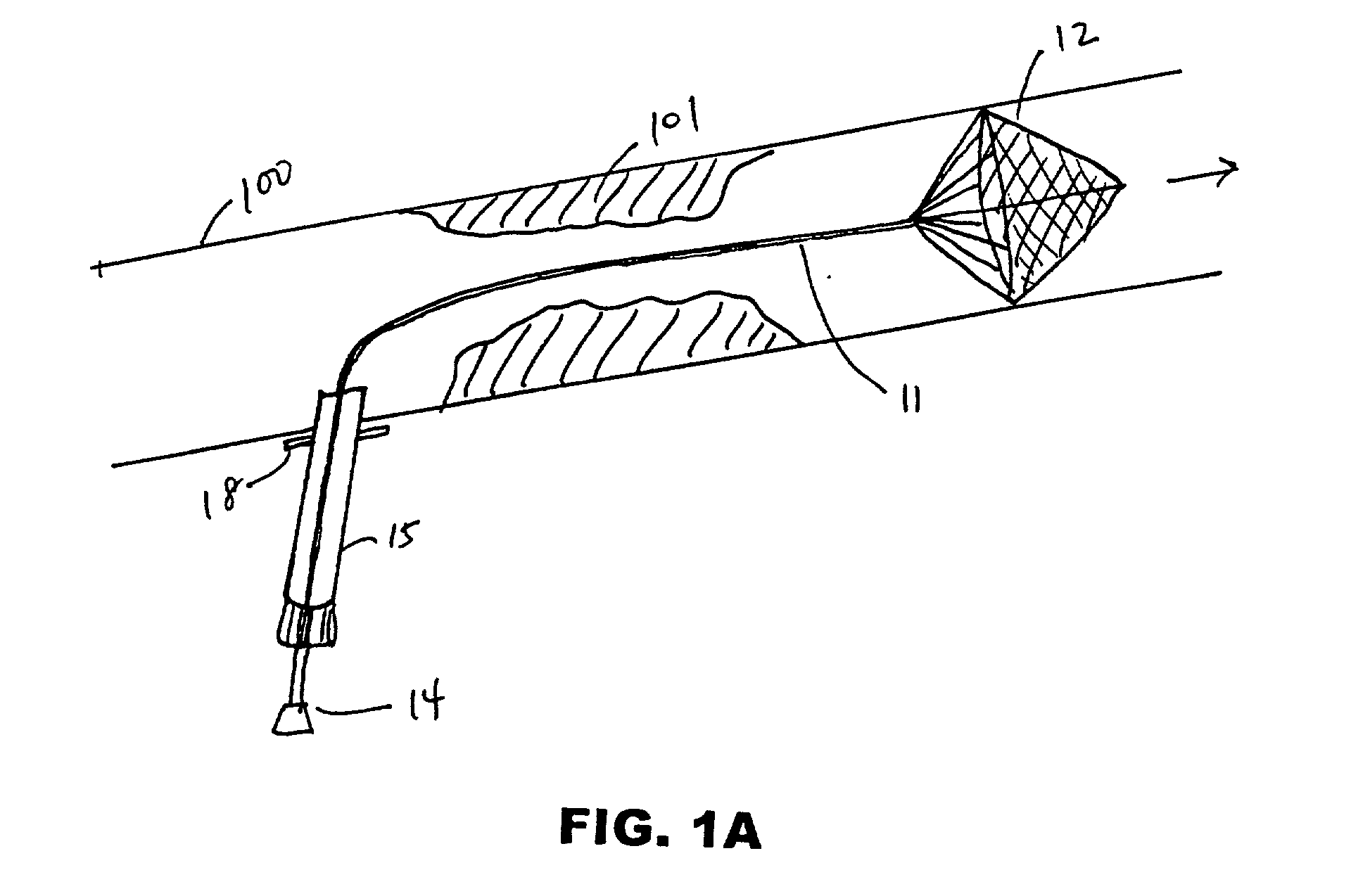
[0058] The devices and methods disclosed herein function to prevent embolic material from migrating downstream (into the brain, the kidneys, the lower extremities, etc.) during vascular surgery. The devices and methods herein are useful during any procedure where vessels are incised for the purpose of removing occlusions or performing other types of repair that may require the use of shunting to maintain distal blood flow.

[0059] FIG. 1A depicts an embodiment of a filter device deployed in carotid artery 100 proximal to atheromatous lesion 101. The device comprises filter assembly 12 mounted on elongate member 11 (guidewire, sheath, etc.). Elongate member 11 is insertable in the lumen of introducer 15 and is connected to filter delivery cartridge 14 at its proximal end. Suture flange 18 is mounted on a distal region of introducer 18.

[0060] The device of FIG. 1A is useful in performing endarterectomy in patients with adequate cerebral perfusion and not requiring a shunt. Introducer 15...


PUM


Abstract

A shunt and method of use for maintaining distal blood flow during an arteriotomy procedure is disclosed. The shunt includes first and second tubular members having proximal ports, distal ports, and lumens therebetween. The distal port of the second tubular member is adapted for releasable attachment to the proximal port of the first tubular member. A second lumen merges and communicates at its distal end with the lumen of the first tubular member and includes a hemostatic valve attached to its proximal end. In using the apparatus for performing open endarterectomy, a filter device is inserted into the vessel and deployed downstream the region of interest in the internal carotid artery. The distal end of the shunt is advanced over the filter device and secured onto the artery. The proximal end of the shunt is inserted upstream the region of interest, typically in the common carotid artery.

Description

FIELD OF THE INVENTION[0001] The present invention relates to open carotid endarterectomy. More particularly, it relates to methods and apparatus for improving endarterecomy procedures by using blood filtration to protect the patient from embolization and vascular shunting to maintain blood perfusion during these vascular surgeries.BACKGROUND OF THE INVENTION[0002] Arteriosclerosis, generally known for thickening and hardening of the arterial wall, is responsible for the majority of deaths in the United States and most westernized countries. Atherosclerosis, one type of arteriosclerosis, is the cause for disorder of the larger arteries that underlies most coronary artery disease, aortic aneurysm, arterial disease of the lower extremities, and cerebrovascular disease. Atherosclerosis is characterized by an accumulation of lipid-filled smooth muscle cells, macrophages, and fibrous tissues, commonly known as atheroma or plaque, in focal areas of cardiovascular tissues, especially the c...

Claims


Application Information

Patent Timeline
IPC IPC(8): A61B17/22A61F2/01
CPCA61B17/22A61F2/013A61F2002/018A61F2230/0006A61F2230/0065A61F2230/0067A61F2230/008A61F2230/0093
Inventor TUROVSKIY, ROMANBALCETA, JOBERTTSUGITA, ROSS S.THORNTON, PETERNUEL, BRIAN P.MARTINEZ, LORRAINE M.
Owner EDWARDS LIFESCIENCES CORP
Why Eureka
Try Eureka