Magnetic resonance imager using cylindrical offset region of excitation, and method

a magnetic resonance imager and offset region technology, applied in the field of magnetic resonance imagers using cylindrical offset regions of excitation, can solve the problems of inferiority of mri and ct tomographic approaches to the current catheter based examination

Inactive Publication Date: 2010-04-29
ALLEGHENY SINGER RES INST
View PDF52 Cites 31 Cited by
  • Summary
  • Abstract
  • Description
  • Claims
  • Application Information

AI Technical Summary

Benefits of technology

[0025]FIGS. 15a and 15b show the principle of reconstruction of tomosynthesis views. FIG. 15a shows that the three projective views of FIGS. 14a and 14b are shifted such that the rectangular features which corresponds to a certain depth in the body is aligned in

Problems solved by technology

Currently, CT offers tomographic resolution of approximately 0.5×0.5×0.5 mm3, and MRI offers tomographic resolution of approximately 1×1×1 mm3 Despite the ability to view the coronary arteries with isotropic resolution, both MRI and CT tomographic approaches have been deemed inferior compared to the current catheter based examination.

Method used


Image

  • Magnetic resonance imager using cylindrical offset region of excitation, and method
  • Magnetic resonance imager using cylindrical offset region of excitation, and method
  • Magnetic resonance imager using cylindrical offset region of excitation, and method

Examples


Embodiment Construction

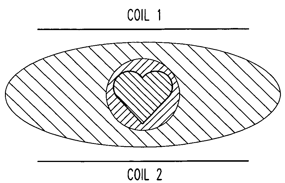
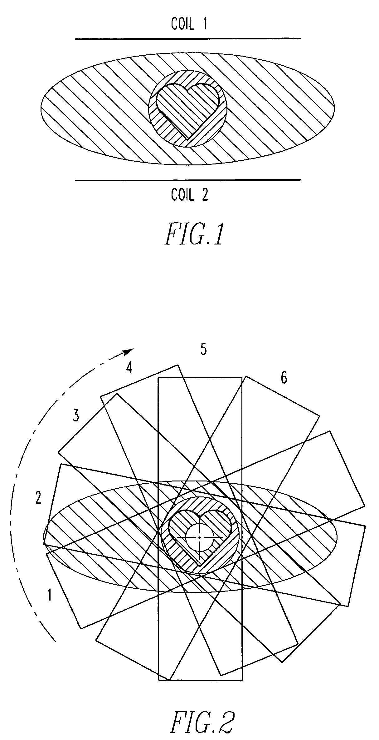
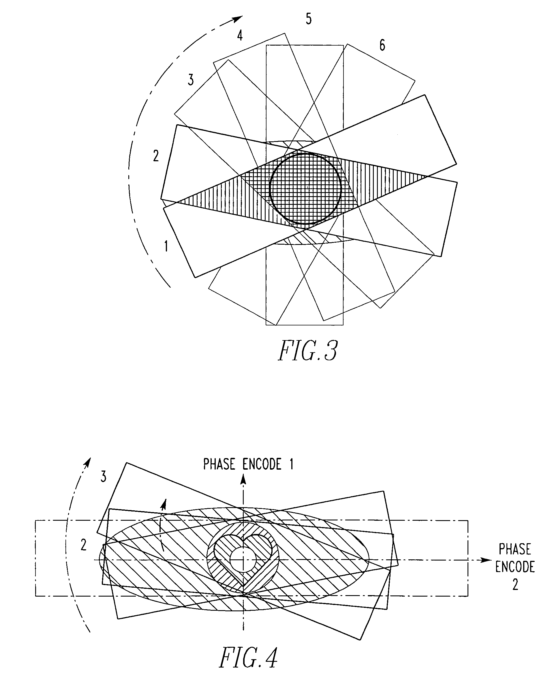
[0027]Referring now to the drawings wherein like reference numerals refer to similar or identical parts throughout the several views, and more specifically to FIG. 16 thereof, there is shown a magnetic resonance imager 10 performing images of a patient. The imager 10 comprises imaging coils 12. The imager 10 comprises receiver coils 14. The imager 10 comprises a computer 16 that causes the imaging coils 12 to produce a first steady-state free precession excitation slab with respect to a first position regarding a target of the patient during a first repetition time, and a second steady-state free precession excitation slab with respect to a second position different from the first position regarding the target during a second repetition time; and forming a first 3-D dataset of the target associated with the first excitation slab and a second 3-D image dataset of the target associated with the second excitation slab from information received from the receiver coils 14. The first 3-D ...


PUM


Abstract

A magnetic resonance imager performing images of a patient imager includes imaging coils. The imager includes receiver coils The imager includes a computer that causes the imaging coils to produce a first steady-state free precession excitation slab with respect to a first position regarding a target of the patient during a first repetition time, and a second steady-state free precession excitation slab with respect to a second position different from the first position regarding the target during a second repetition time; and forming a first 3-D dataset of the target associated with the first excitation slab and a second 3-D image dataset of the target associated with the second excitation slab from information received from the receiver coils. The first 3-D image dataset and the second 3-D image dataset, together defining a series of 3-D image datasets for each repetition time; and producing an image of the target from the series of 3-D image datasets. A method for analyzing a patient. A method to evaluate coronary arteries to assess their degree of stenosis. A computer program which, when executed by a computer of a Magnetic Resonance Imager, forms an image of a patient.

Description

FIELD OF THE INVENTION[0001]The present invention is related to obtaining data with a magnetic resonance imager of each of a plurality of steady-state free precession excitations slabs rotated about a target of a patient. (As used herein, references to the “present invention” or “invention” relate to exemplary embodiments and not necessarily to every embodiment encompassed by the appended claims.) More specifically, the present invention, Cylindrical Offset Region of Excitation (CORE), is related to obtaining data with a magnetic resonance imager of each of a plurality of steady-state free precession excitations slabs during respective repetition times rotated about the target of a patient to obtain a series of 3-D image datasets for each repetition time, and producing an image of the target from the series of 3-D image datasets.BACKGROUND OF THE INVENTION[0002]This section is intended to introduce the reader to various aspects of the art that may be related to various aspects of th...

Claims


Application Information

Patent Timeline
IPC IPC(8): G06K9/00G06T15/20
CPCG01R33/4826G01R33/4833G01R33/5635G01R33/5611G01R33/5614G01R33/4838A61B5/055G06T17/00
Inventor DOYLE, MARK
Owner ALLEGHENY SINGER RES INST
Why Eureka
Try Eureka