
Bispecific fusion protein having therapeutic and diagnostic potential

a fusion protein and bispecific technology, applied in the direction of fusions for specific cell targeting, antibody medical ingredients, drug compositions, etc., can solve the problems of reducing the integrity of the vessel wall, and reducing the blood supply to the tissue, so as to prevent the erosion of arteriosclerotic plaques and maintain the integrity of the endothelial body

Inactive Publication Date: 2010-03-18
UNIV TUBINGEN
View PDF2 Cites 4 Cited by
  • Summary
  • Abstract
  • Description
  • Claims
  • Application Information

AI Technical Summary

Benefits of technology

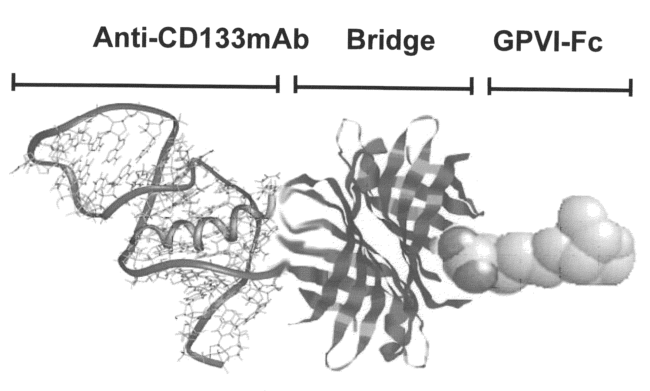
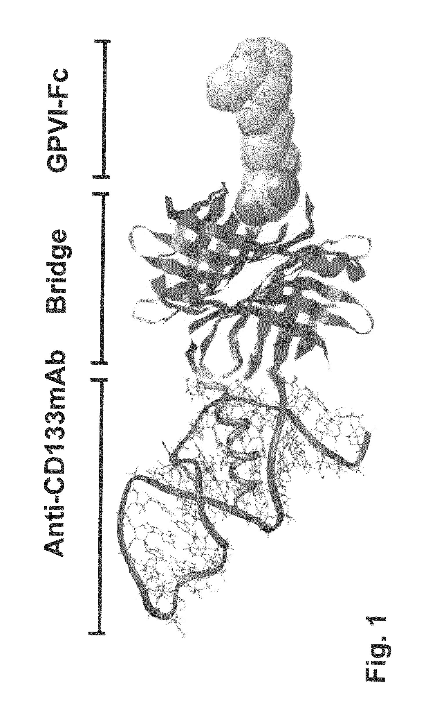
The present invention provides a novel agent for maintaining endothelial integrity to prevent arterosclerotic plaque erosion. This is achieved by a bispecific fusion protein which contains a first polypeptide that binds to collagen and a second polypeptide that binds to endothelial precursor cells. This fusion protein can be administered directly to damaged tissue or can be co-incubated with stem cells before administration. The invention also provides a method for recruiting stem cells to damaged vessels or tissue and maturing them into endothelial cells to prevent or treat vessel-damaging reactions caused by stent implant or chemical agents. The invention is effective in preventing arterosclerotic diseases and can be easily administered without the need for costly coating of the coronary stents.

Problems solved by technology

Damage to the vessels of the cardiovascular system occurs in particular as a consequence of stent or stent graft implants into the vessels, which in turn have to be inserted into the vessels affected because of other diseases or events in order to ensure supply of the surrounding tissue or to organs.
Needless to say, damage in the vessel wall leads to the integrity of the vessel wall being eliminated and to subsequent hemorrhaging into surrounding tissue.
The supply of blood to the tissue is no longer ensured due to the occurrence of such thrombi, so that ischemic states of the tissue lying distally to the thrombus may occur.
After implantation of the stent, free flow through the vessel is indeed ensured again, but the vascular endothelium which represents a barrier between the circulating blood cells and the subendothelial matrix under physiological conditions is still damaged.
Adhesion of blood platelets and subsequent formation of thrombi and the resulting acute myocardial infarction are therefore a major complication after a stent implant.
On reperfusion of the region previously ischemic due to blockage of a vessel, this is supplied with oxygenated blood again, as a result of which on the one hand cell damage is limited, but this process is associated with continuing damage to the myocardium.
Since stents coated with medicaments which are released gradually after implantation are currently also employed, the re-endothelialization of the treated vessel is also delayed by the medicaments released, so that stent thrombosis is an extremely critical complication of this method.

Method used


Image

  • Bispecific fusion protein having therapeutic and diagnostic potential
  • Bispecific fusion protein having therapeutic and diagnostic potential
  • Bispecific fusion protein having therapeutic and diagnostic potential

Examples


example 1

Preparation of a Bispecific Protein / Monoclonal Antibody Construct for Recruiting of Bone Marrow Stem Cells to Vessel Lesions

Material and Methods

Reagents

[0066]Biocoll separating solution was obtained commercially from Biochrom AG (Berlin, Germany), and EBM as “BulletKit” (EGM) from Cambrex Bio Science (East Rutherford, N.J.). Collagen I, collagen III, laminin, vitronectin, fibrinogen and fibronectin were obtained commercially from BD Sciences (Heidelberg, Germany), human VEGF from PeproTech Inc. (Rocky Hill, N.J.), and the primary mouse antibody anti-vWF and the phalloidin-AlexaFluor 488 from Chemicon (Temecula, Calif.). DAPI, the Cy3-labeled secondary antibody (goat anti-mouse) and the “Celltracker Vybrand DiD” was obtained commercially from Molecular Probes / Invitrogen GmbH (Karlsruhe, Germany).

[0067]Isolation and Culturing of CD34+ Cells and CD133+ Cells

[0068]Human CD34+ cells and CD133+ cells were isolated from human umbilical cord blood and cultured as described by Lang, et al. (...


PUM

PropertyMeasurementUnit
total weightaaaaaaaaaa
permeabilityaaaaaaaaaa
hemostasis adhesionaaaaaaaaaa

Abstract

The present invention relates to a bispecific fusion protein, comprising (a) a first polypeptide which binds to collagen, and (b) a second polypeptide which binds to endothelial precursor cells. Also, pharmaceutical compositions are disclosed, comprising the fusion protein of the invention, as well as methods for using the fusion protein, in particular for treating or preventing lesions of vessels and tissues.

Description

RELATED APPLICATION[0001]This application is a continuation of copending International Patent Application PCT / EP2008 / 001369 filed on Feb. 21, 2008 and designating the United States, which was not published in English, and claims priority of German Patent Application DE 10 2007 010 306.0, filed on Feb. 22, 2007, both of which are incorporated herein by reference in their entireties.BACKGROUND OF THE INVENTION[0002]The invention relates to a bispecific fusion protein having therapeutic and diagnostic potential for treatment / diagnosis of lesions of vessels or tissues; the invention furthermore relates to a nucleic acid molecule encoding this fusion protein, a pharmaceutical and diagnostic composition which comprises the fusion protein or nucleic acid molecule encoding therefore, and a method for using the bispecific fusion protein or nucleic acid molecule for treatment or prevention of lesions of vessels / tissues of a mammalian subject and a method for therapy of acute or chronic vascul...

Claims


Application Information

Patent Timeline
Patent Type & Authority Applications(United States)
IPC IPC(8): A61K49/00C07K14/00C07K16/00C07H21/04A61K39/395C07K1/00A61P9/10
CPCC07K2319/33A61K47/48415C07K16/2896C07K14/705A61K47/48561A61K47/6811A61K47/6849A61P19/08A61P9/00A61P9/10A61P9/14
Inventor LANGER, HARALDGAWAZ, MEINRADBUHRING, HANS-JORGSKUTELLA, THOMASJUNG, GUNDRAM
Owner UNIV TUBINGEN
Learn More