Adaptive Medical Image Acquisition System

a medical imaging and image acquisition technology, applied in the field of medical imaging systems, can solve the problems of r wave not being the best signal, unable to accurately synchronize image acquisition for diagnosis of specific cardiac functions, and unable to achieve the best signal,

Inactive Publication Date: 2009-12-17
SIEMENS HEALTHCARE GMBH
View PDF26 Cites 59 Cited by
  • Summary
  • Abstract
  • Description
  • Claims
  • Application Information

AI Technical Summary

Problems solved by technology

However, an electrophysiological signal, such as an R wave may not be the best signal for cardiac function diagnosis and may lack precision for synchronizing image acquisition for diagnosis of specific cardiac function or tissue pathologies.
For example, an electro-cardiac spike (such as a QRS complex) may not provide an accurate time gating signal for maximum volume image capture and calculation of cardiac chambers, such as a left ventricle for systolic and diastolic volume.
Additionally, known systems used in image diagnosis, characterization and evaluation are subjective and need extensive medical expertise and clinical experience for accurate interpretation and appropriate cardiac rhythm management.
However, in known clinical procedures and applications, there is a lack of an efficient, effective method for cardiac function based gating and synchronization for image acquisition and diagnosis.
Known systems lack accurate gating to measure maximum volume of a chamber and may also use retrospective image evaluation to extract an image of maximum volume, for a left ventricle chamber, for example.
However, an image database used for retrospective analysis may not contain an image of maximum volume and size.
Known medical imaging systems usually use gating for elimination and reduction of patient noise and artifacts (such as from respiration and heart beating) but lack comprehensive function gating and imaging synchronization capability.
Tuning image acquisition rate and the use of high speed scanning in known systems may cause inefficient usage of a scanning system which may reduce the life of the image system, such as an X-ray machine.
This may cause radiation over-dose and inefficient usage of an image acquisition system.

Method used


Image

  • Adaptive Medical Image Acquisition System
  • Adaptive Medical Image Acquisition System
  • Adaptive Medical Image Acquisition System

Examples


Embodiment Construction

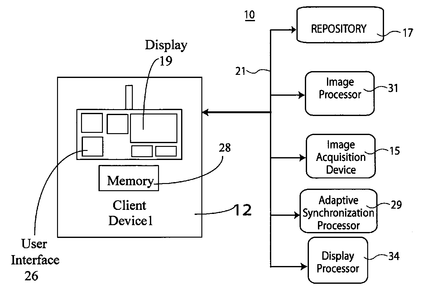
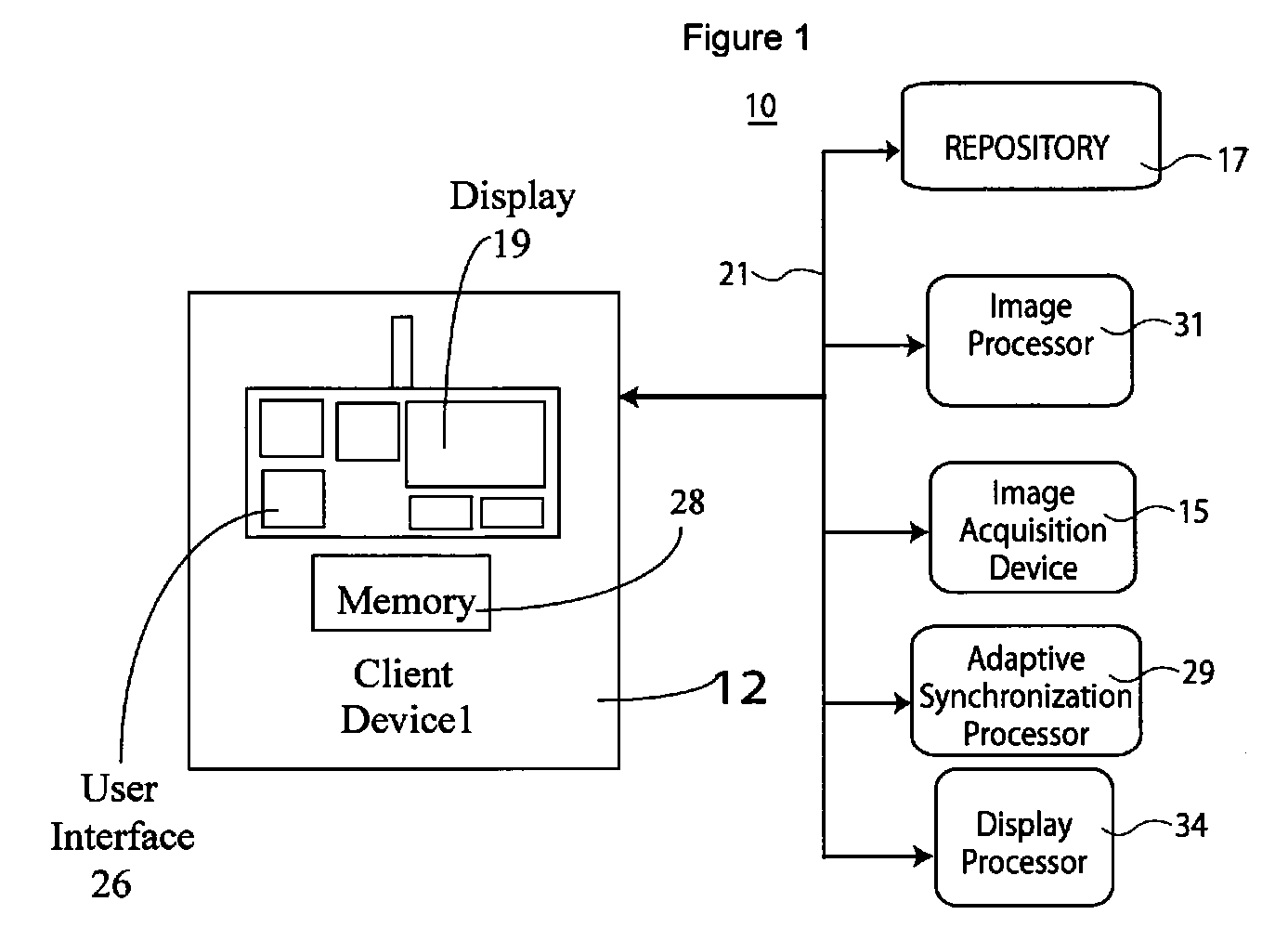
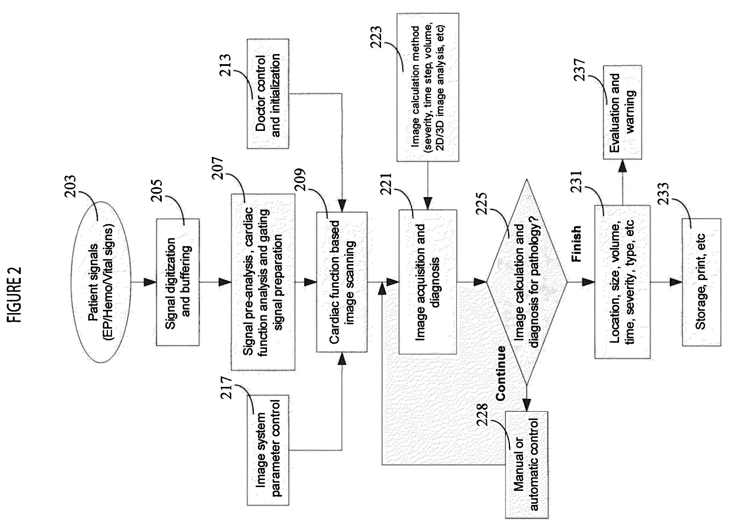
[0015]A system synchronizes and gates image acquisition with cardiac function signals (hemodynamic, electrophysiological, intra-cardiac blood pressure & vital signs signals) to improve precision, quality and reliability of image acquisition and diagnosis. Thereby the system advantageously excludes patient motion induced noise (heart beating and respiration) to determine an accurate image acquisition time as well as provide stable image capture, accurate image diagnosis, and cardiac tissue and function characterization (such as pathology type and severity). Cardiac function signals provide a precise and reliable time and phase of image acquisition synchronization for capturing and characterizing cardiac function and tissue status. The cardiac function signals synchronize imaging to help a physician provide a more objective and accurate diagnosis and medical treatment. Furthermore, the system provides safer use of an image system (such as an X-ray system) with less power usage and rad...


PUM


Abstract

A medical imaging system adaptively acquires anatomical images. The system includes a synchronization processor for providing a heart rate related synchronization signal derived from a patient cardiac function blood flow related parameter. The synchronization signal enables adaptive variation in timing of acquisition within successive heart cycles of each individual image frame of multiple sequential image frames. An image acquisition device initiates acquisition of anatomical images of a portion of patient anatomy in response to the synchronization signal. A display processor presents images, acquired by the acquisition device and synchronized with the synchronization signal, to a user on a reproduction device. The image acquisition device adaptively selects image pixel resolution of individual image frames of the multiple sequential image frames in response to data identifying a heart cycle segment so that successively acquired image frames have different image pixel resolution within a single heart cycle.

Description

[0001]This is a non-provisional application of provisional application Ser. No. 61 / 061,780 filed Jun. 16, 2008 and of provisional application Ser. No. 61 / 101,373 filed Sep. 30, 2008, by H. Zhang et al.FIELD OF THE INVENTION[0002]This invention concerns a medical imaging system for adaptively acquiring anatomical images in response to a synchronization signal derived from a patient cardiac function blood flow related parameter enabling adaptive variation in timing of acquisition.BACKGROUND OF THE INVENTION[0003]Angiography (or arteriography) is an imaging process used to visualize cardiac chamber size and segmental wall mobility and coronary size, morphology, flow, anatomy and arterial luminal size by displaying static and dynamic image silhouettes. This provides the ability to assess cardiac and coronary arterial function and calculate estimations of cardiac chamber volumes (Ventricular and Atrial), which facilitates diagnosis of cardiac disease. In known systems, electrophysiologic...

Claims


Application Information

Patent Timeline
Patent Type & Authority Applications(United States)
IPC IPC(8): A61B5/044A61B5/026A61B5/0205
CPCA61B5/0205A61B5/021A61B6/541A61B6/503A61B6/504A61B5/026
Inventor ZHANG, HONGXUANWADE, HAROLD JAMESCHAN, JINGHUA
Owner SIEMENS HEALTHCARE GMBH
Why Eureka
Try Eureka