Eureka AIR delivers breakthrough ideas for toughest innovation challenges, trusted by R&D personnel around the world.

Methods and apparatus for analysis of angiographic and other cyclical images

a technology of cyclical images and methods, applied in the field of diagnostic medical imaging, can solve problems such as edge detection

Inactive Publication Date: 2010-04-13
GE MEDICAL SYST GLOBAL TECH CO LLC
View PDF6 Cites 104 Cited by
  • Summary
  • Abstract
  • Description
  • Claims
  • Application Information

AI Technical Summary

Benefits of technology

[0011]It will be appreciated that configurations of the present invention directed to cardiac image analysis provide fully automatic left ventricle analysis using multiple images of systole and diastole. Using these multiple images, a single representative image of each phase proves more accurate and repeatable edge detection and dependent measures, such as ejection fraction than is known in current methods and apparatus. Corresponding advantages can be obtained in configurations of the present invention directed to other imaging applications, including non-medical applications in which various forms of cyclical data and images of an object are recorded.

Problems solved by technology

Automatic (i.e., computerized) edge detection is possible, but is often difficult due to lack of edge definition or obscuration by musculature inside the ventricle.

Method used


Image

  • Methods and apparatus for analysis of angiographic and other cyclical images
  • Methods and apparatus for analysis of angiographic and other cyclical images
  • Methods and apparatus for analysis of angiographic and other cyclical images

Examples


Embodiment Construction

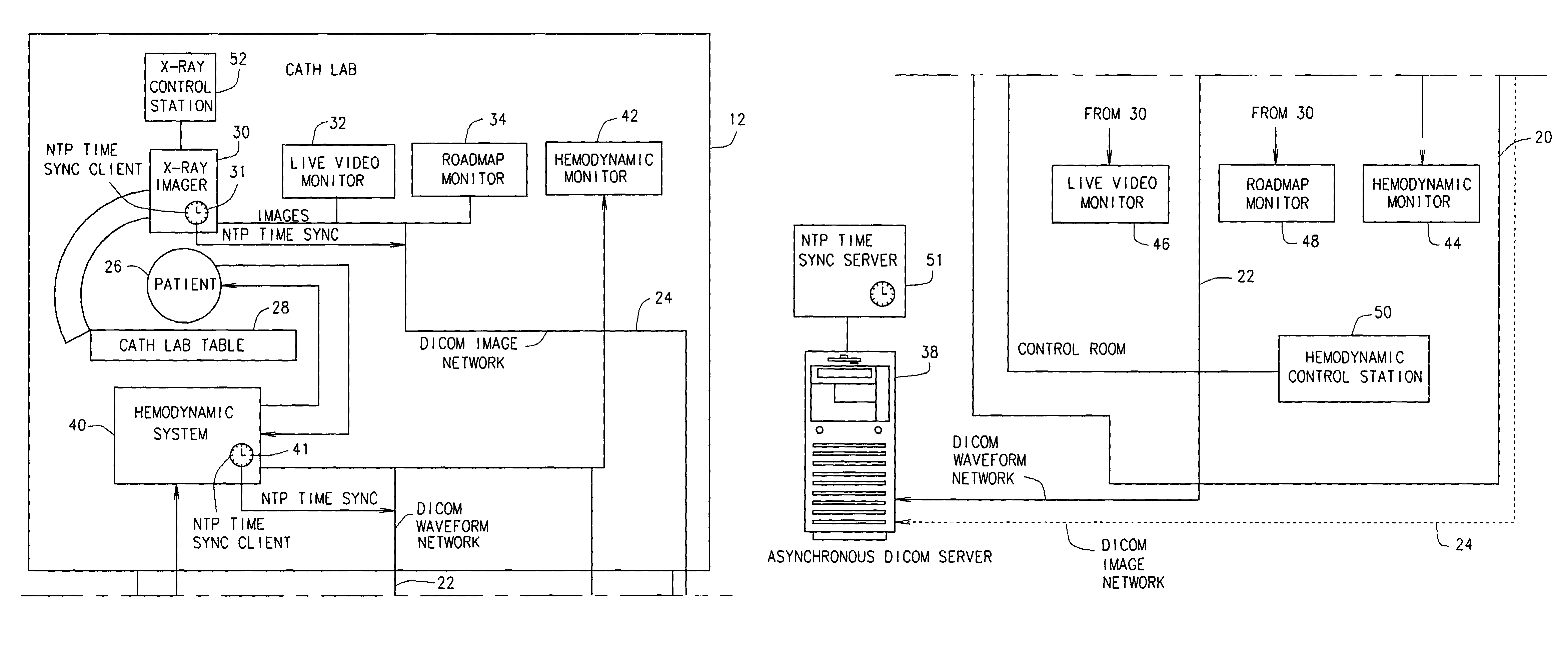
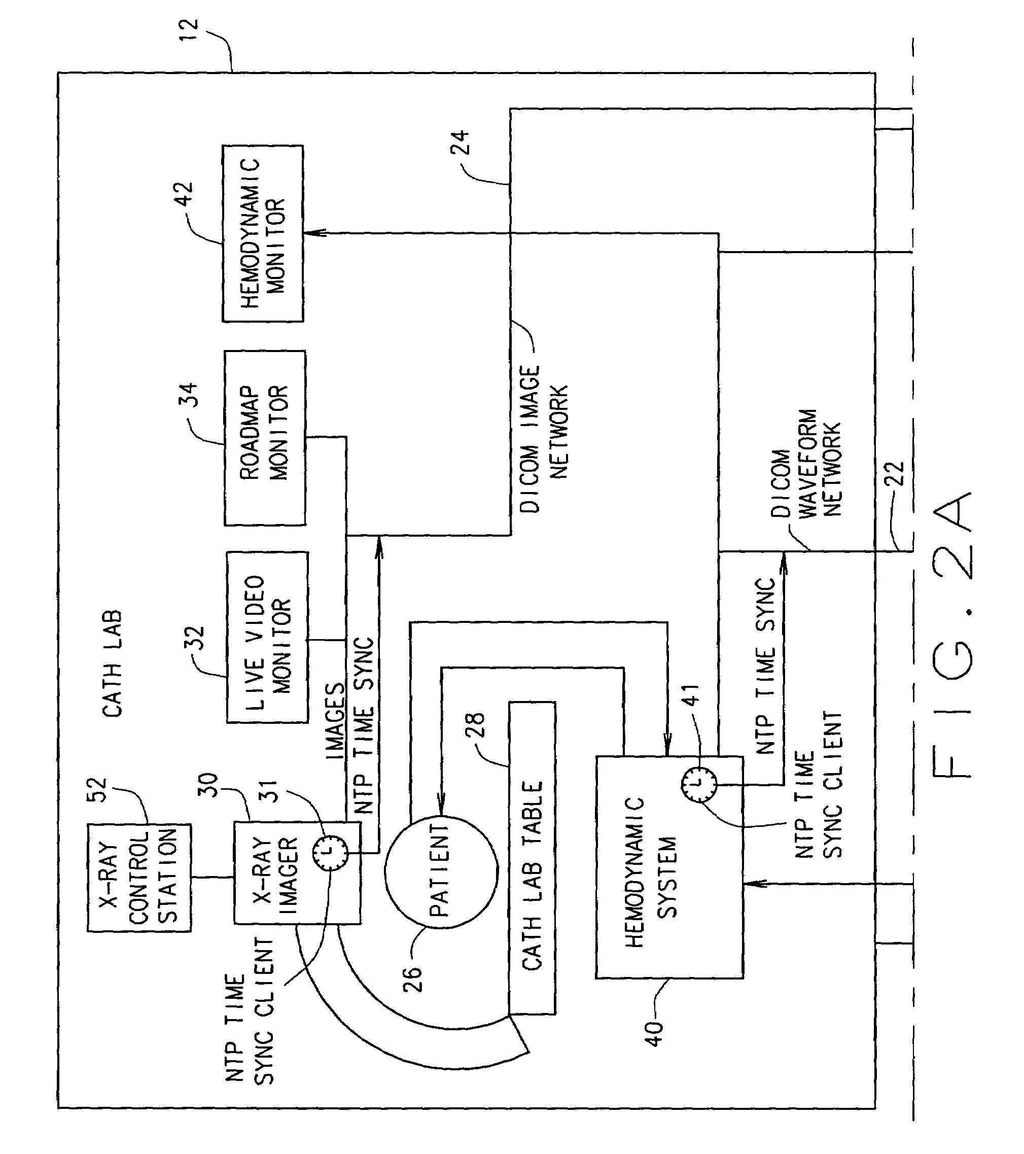
[0017]A technical effect of the systems and processes described herein include at least one of producing a single representative frame of image data from a plurality of stored images, providing left ventricle analysis using multiple images of systole and diastol, and facilitating measurement of ejection fraction.

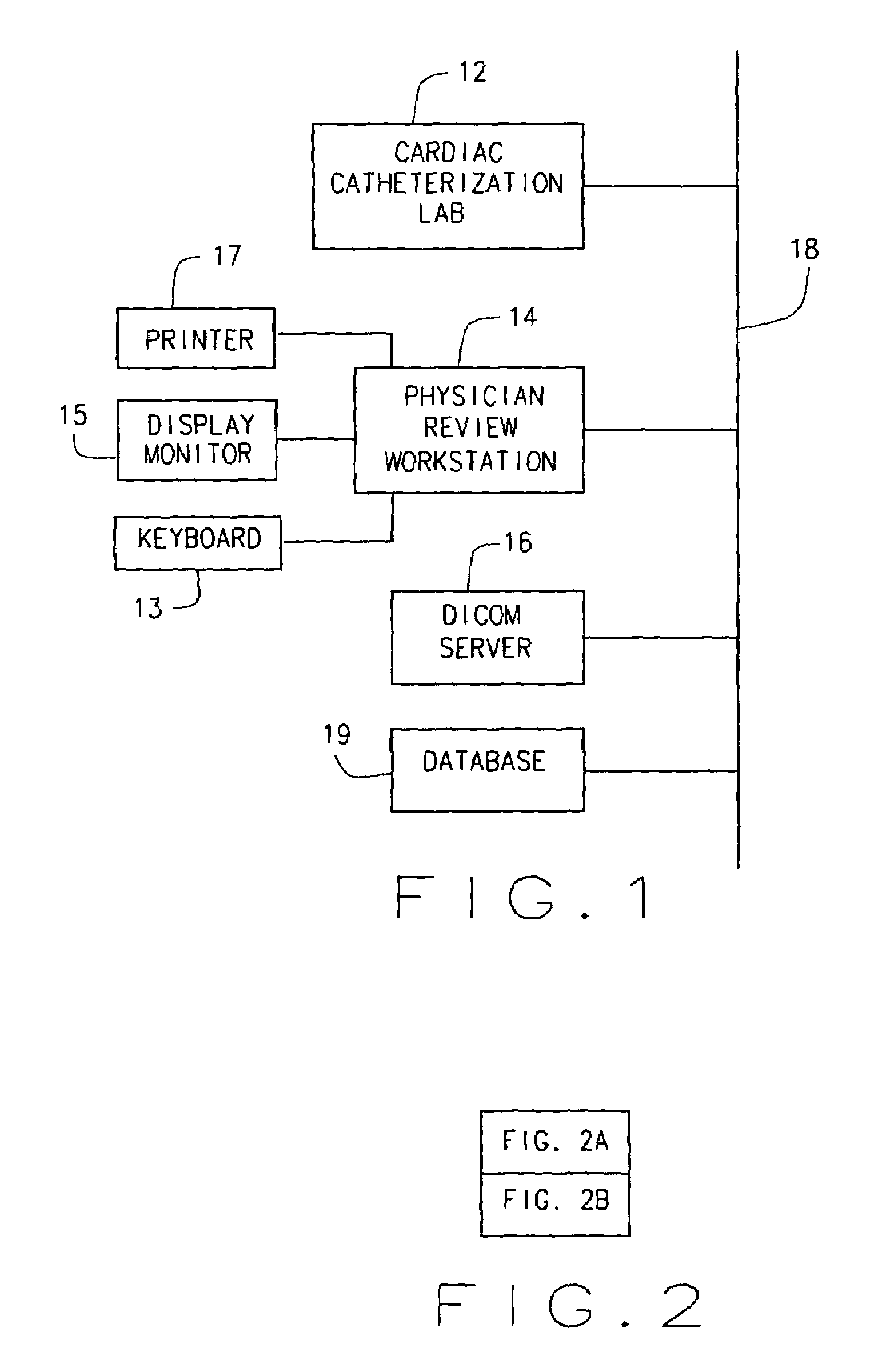
[0018]In some configurations of the present invention and referring to FIG. 1, a local area network (LAN) 18 facilitates communication between a systems housed in a cardiac catheterization laboratory 12, a physician review or overview workstation 14 and a DICOM server 16. For example, angiographic x-ray images acquired by imaging equipment in laboratory 12 and formatted as DICOM objects are transmitted by LAN 18 to a database 19 accessed by DICOM server 16. Workstation 14 can be used by a physician to display and perform quantitative analysis on retrieved images. Certain analysis, including left ventricular analysis, require the selection of images corresponding to particula...


PUM


Abstract

A method for analyzing stored frames of time-stamped imaging data and time-stamped physiologic data obtained during a study of a patient during a plurality of cardiac cycles. The imaging data includes frames representative of a succession of images of the patient's heart. These configurations use a computer to analyze the stored physiologic data to determine times of a selected phase in the plurality of cardiac cycles, select, in the plurality of cardiac cycles, stored images corresponding to the determined times of the selected phase, and align and average images corresponding to the selected phase across the plurality of cardiac cycles to produce a single representative frame of image data.

Description

BACKGROUND OF THE INVENTION[0001]This invention relates generally to methods and apparatus for diagnostic medical imaging and more particularly to methods and apparatus for automated angiographic analysis. However, the invention is also more generally useful for analysis of other cyclical images.[0002]A coronary angiogram (or synonymously, an arteriogram) is an x-ray of the coronary arteries. Such images help the physician observe blocked arteries for the diagnosis of coronary artery disease. An angiogram is often part of a cardiac catheterization procedure including other tests and measurements. A patient undergoing catheterization also undergoes physiological monitoring. Typically, an electrocardiogram (ECG or, synonymously, EKG) is produced from electrical leads attached to a patient and analyzed to detect irregularities indicative of heart damage or disease. Data from the ECG is stored in a computer database.[0003]To perform an angiogram, a physician injects a contrast medium in...

Claims


Application Information

Patent Timeline
Patent Type & Authority Patents(United States)
IPC IPC(8): A61B5/05A61B6/00G06T5/00G06T5/50G06T7/60
CPCA61B6/481A61B6/504G06T5/50G06T7/602G06T7/0012G06T2207/30048G06T2207/10121G06T7/62
Inventor JENKINS, JOHN H.PALMA, MARK D.
Owner GE MEDICAL SYST GLOBAL TECH CO LLC
Why Eureka
Eureka Blog
Learn More