
Method for Detection of Cancer Cells Using Virus

Inactive Publication Date: 2009-12-17
MEMORIAL SLOAN KETTERING CANCER CENT
View PDF24 Cites 39 Cited by
  • Summary
  • Abstract
  • Description
  • Claims
  • Application Information

AI Technical Summary

Benefits of technology

[0007]The invention relates to compositions and methods for cancer cell detection in bodily samples wherein a cancer cell can be detected within a mixed population of cancer cells and non-cancer cells. The invention also relates to compositions and methods that may be used in cancer cell detection, specifically viruses that are replication-competent conditional to a cancer cell, in particular an oncolytic herpes virus, such as NV1066 and a vaccinia virus, such as GLV-1 h68. Provided are methods and kits for using these viruses that preferentially replicate in cancer cells and may also preferentially infect cancer cells for specific identification of such cancer cells, even when a cancer cell is present, for example, at a ratio of one infected cancer cell in a background of ten thousand non-cancer cells, thus further providing a reproducible and sensitive screening method for cancer detection, monitoring and prognosis.

Problems solved by technology

When cancers develop, they are often not found until they have metastasized and are no longer curable.
However, examination of these specimens by current cytological techniques is labor intensive and is low in sensitivity.
Furthermore, conventional cytology relies on the morphological identification of malignant cells by a skilled pathologist and is subject to interpretive error and interpathologist variability (Khalid et al., (2005) Clin Lab Med; 25(1):101-16; Harewood et al., (2004) Am J Gastroenterol; 99(8):1464-9; all of which are herein incorporated by reference).

Method used


Image

  • Method for Detection of Cancer Cells Using Virus
  • Method for Detection of Cancer Cells Using Virus
  • Method for Detection of Cancer Cells Using Virus

Examples


example 1

Materials And Methods

I. Construction of Viruses for Use in the Present Inventions:

[0184]A. Construction of a Heroes Simplex Virus for Use in the Present Inventions for Example, NV1066, is Briefly Described Below.

[0185]NV1066 refers to an attenuated oncolytic herpes virus that expresses enhanced green fluorescent protein (eGFP). See, FIG. 1. Specifically, an engineered herpes virus was constructed when a transgene encoding eGFP, under a constitutive CMV promoter, was inserted into the internal repeat sequence of the parent virus, resulting in deletions in one copy each of the viral genes encoding ICP-4, ICP-0, and γ134.5. These deletions rendered the virus selective for infection and replication in tumor cells while attenuating its' potential neurovirulence.

[0186]More specifically, NV1066 was constructed by transfecting a BAC mid (BAC 17-28), a cosmid (cos12a), and a plasmid (pUL56-GFP-US1) containing overlapping and contiguous HSV-1 sequences into Vero cells with LipofectAMINE accor...

example 2

Herpes Simplex Viral Mutant, NV1066, Infects a Range of Cancer Cell Lines in Vitro

[0204]NV1066 infectivity was tested in numerous cancer cell lines as described in Table 8 below. One hundred and eleven cancer cell lines were infected in vitro at an MOI of 0.01, 0.1 and 1.0. The cell lines listed became infected and expressed eGFP that was detected upon examination under fluorescent microscopy and by flow cytometry.

[0205]The inventors observed that in vitro, NV1066 infected the cell lines listed in Table 8 at MOIs of 0.01, 0.1 and 1.0. Specifically, NV1066 infected cancer cells expressed eGFP within 1 to 2 hours of incubation. After 18 hours incubation, the majority of cancer cells were infected and expressed eGFP. Although the virus was able to infect and express eGFP at a lower MOI of 0.1, at a higher MOI of 1.0, the majority of cancer cells in the sample were infected at an earlier time point and expressed strong eGFP fluorescence. The expressed eGFP was intracellular such that th...

example 3

Mean Intensity of NV1066 Infected Cancer Cells is 11-344-Fold Higher Than Background Autofluorescence

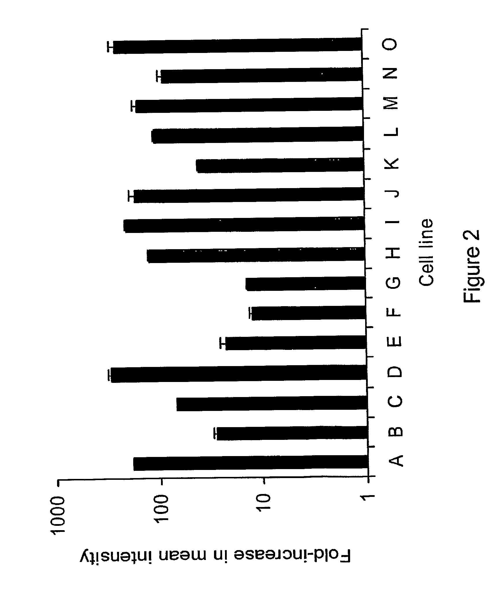
[0207]In order to determine the level of eGFP expression over background cellular autofluoreseence, fifteen representative human cancer cell lines (A-O, described below) were infected in vitro at an MOI of 1.0 (multiplicity of infection, ratio of viral particles per cancer cell), incubated for 18 hours, and analyzed by flow cytometry. Compared to background autofluorescence, infected eGFP expressing cancer cells showed higher mean intensity of green fluorescence (11-344-fold higher, represented in logarithmic scale).

[0208]Because of this strong expression of eGFP, cancer cells in body fluids can be easily identified, even in a background of millions of cells or in cell clumps. See, FIG. 2. (A-O cancer cells: lung—A549, H1299; bladder—UMUC-3, KU19-19; stomach—OCUM-2MD3; colorectal —HT29; hepatoma—HepG2; mesothelioma—MSTO-211H, JMN, H-Meso, H-28; breast MCF-7; head and neck —SCCVII, SC...


PUM

PropertyMeasurementUnit
Timeaaaaaaaaaa
Timeaaaaaaaaaa
Timeaaaaaaaaaa

Abstract

The invention relates to compositions and methods for cancer cell detection in bodily samples wherein a cancer cell can be detected within a mixed population of cancer cells and non-cancer cells. The invention also relates to compositions and methods that may be used in cancer cell detection, specifically viruses that are replication-competent conditional to a cancer cell, in particular an oncolytic herpes virus, such as NV 1066 and a vaccinia virus, such as GLV-1h68. Provided are methods and kits for using these viruses that preferentially replicate in cancer cells and may also preferentially infect cancer cells for specific identification of such cancer cells, even when a cancer cell is present, for example, at a ratio of one infected cancer cell in a background often thousand non-cancer cells, thus further providing a reproducible and sensitive screening method for cancer detection, monitoring and prognosis.

Description

FIELD OF THE INVENTION[0001]The invention relates to compositions and methods for cancer cell detection in bodily samples wherein a cancer cell can be detected within a mixed population of cancer cells and non-cancer cells. The invention also relates to compositions and methods that may be used in cancer cell detection, specifically viruses that are replication-competent conditional to a cancer cell, in particular an oncolytic herpes virus, such as NV1066 and a vaccinia virus, such as GLV-1 h68. Provided are methods and kits for using these viruses that preferentially replicate in cancer cells and may also preferentially infect cancer cells for specific identification of such cancer cells, even when a cancer cell is present, for example, at a ratio of one infected cancer cell in a background of ten thousand non-cancer cells, thus further providing a reproducible and sensitive screening method for cancer detection, monitoring and prognosis.BACKGROUND[0002]Screening methods for detect...

Claims


Application Information

Patent Timeline
IPC IPC(8): C12Q1/70
CPCA61K35/13C12N2710/16611G01N33/574C12Q1/6897C12N2710/24111
Inventor FONG, YUMANADUSUMILLI, PRASAD
Owner MEMORIAL SLOAN KETTERING CANCER CENT
Learn More