
Medical image processing apparatus and x-ray diagnosis apparatus

a technology of image processing and diagnosis apparatus, which is applied in the field of medical image processing apparatus and x-ray diagnosis apparatus, can solve the problems of large, complicated apparatus, and inability to arrange the configuration of the apparatus, and achieve the effect of reducing exposur

Inactive Publication Date: 2009-12-17
TOSHIBA MEDICAL SYST CORP
View PDF19 Cites 32 Cited by
  • Summary
  • Abstract
  • Description
  • Claims
  • Application Information

AI Technical Summary

Benefits of technology

[0049]The present invention will be described below with reference to embodiments.

Problems solved by technology

Either of the techniques disclosed in these references requires a large, complicated apparatus to grasp the position of a catheter.
Consequently, an apparatus to obtain images to be referred to by using such position detection is inevitably a large, complicated apparatus.
That is, it is too expensive to arrange an apparatus configured to simply display a medical image of a portion corresponding to an affected part so as to support a medical treatment.
The above technique, however, has the following problem.
Using only two-dimensional projection images makes it impossible to accurately check information of an object to be examined in the depth direction such as the thickness of a blood vessel wall and a state in the thickness direction in a blood vessel, which are required for a medical treatment and diagnosis.

Method used


Image

  • Medical image processing apparatus and x-ray diagnosis apparatus
  • Medical image processing apparatus and x-ray diagnosis apparatus
  • Medical image processing apparatus and x-ray diagnosis apparatus

Examples


embodiment 1-1

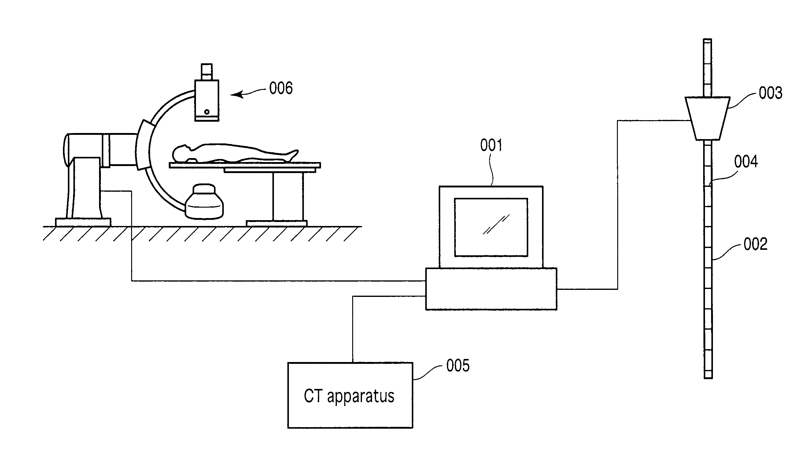
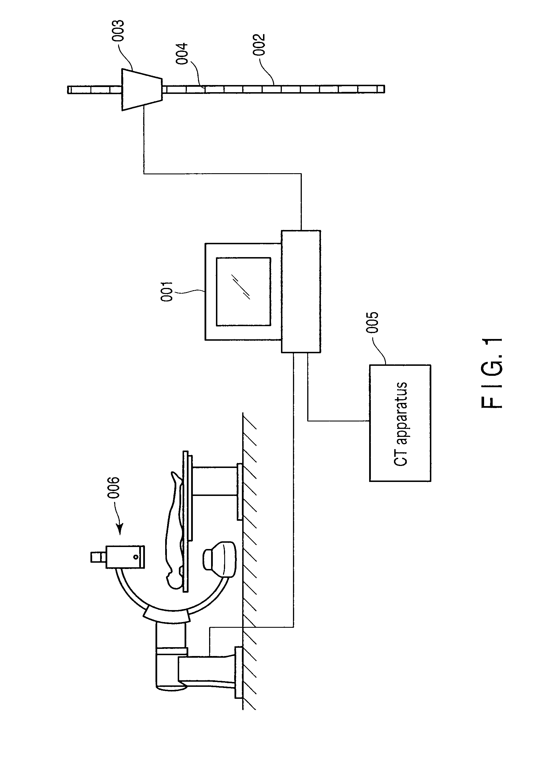
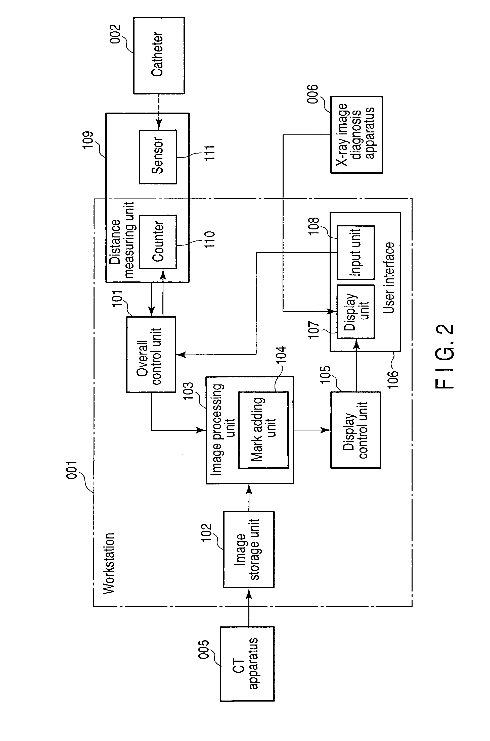
[0052]The overall configuration of an X-ray diagnosis apparatus according to this embodiment will be described with reference to FIG. 1. As shown in FIG. 1, an image diagnosis system according to this embodiment includes a medical image diagnosis apparatus such as a CT apparatus 005, a workstation 001 connected to the CT apparatus 005 via a network, an X-ray image diagnosis apparatus 006 connected to the workstation 001 via the network, a hub 003 incorporating a sensor 111 connected to the workstation 001, and a catheter 002. This embodiment uses the CT apparatus 005 as an apparatus to form a medical image for supporting a medical treatment. However, it suffices to use any other apparatus to form medical images which can be used for supporting a medical treatment. For example, an MRI apparatus or a 3D (three-dimensional) angiography apparatus can be used. This embodiment makes the workstation 001 display images formed by the X-ray image diagnosis apparatus 006. However, it suffices ...

embodiment 1-2

[0095]A medical image processing apparatus according to embodiment 1-2 of the present invention will be described below. The medical image processing apparatus according to this embodiment is configured to be different from the first embodiment in that upon receiving the designation of a reference position using one type of medical image by an operator, the apparatus can determine a reference position in another medical image. The following description is mainly directed to the designation of a reference position and the selection of a medical image associated with the position of the distal end after movement. The arrangement of the functional units of the medical image processing apparatus according to this embodiment is the same as that in the block diagram of FIG. 2.

[0096]This embodiment selects and creates one type of medical image of volume rendering, 2D projection, CPR, SPR, fly-through, MIP, and cross-cut images based on the three-dimensional image data (volume data) stored ...

embodiment 2-1

[0101]An X-ray diagnosis apparatus, medical image processing apparatus, and image processing method according to embodiment 2-1 of the present invention will be described below with reference to FIGS. 6 to 13. FIG. 6 is a block diagram showing the arrangement of an X-ray diagnosis apparatus 1. The apparatus 1 includes a medical image processing apparatus 10, a CT apparatus 20, and an X-ray diagnosis apparatus 30. The three-dimensional image data recorded by the CT apparatus are sent to the medical image processing apparatus 10 via a network or the like.

[0102]The medical image processing apparatus 10 includes a CT image data acquisition unit 11 (image acquisition means), an system control unit 12, an operation unit 13, an image storage unit 14, a computation unit 15, an image creating unit 16 (creating means / extraction means), a storage unit 17, a display control unit 19, a two-dimensional X-ray image acquisition unit 21 (image acquisition means), and a position information input uni...


PUM


Abstract

A medical image processing apparatus includes an image storage unit which stores the data of a three-dimensional image associated with an object receiving an operation using a lumen insertion device, a lumen detection unit which detects a lumen centerline associated with a specific lumen from the three-dimensional image, a current position specifying unit which specifies the current position of the lumen insertion device based on the insertion distance from a reference position associated with the lumen insertion device and the detected lumen centerline, a cross-sectional image generating unit which creates, from the data of the three-dimensional image, the data of a cross-sectional image associated with a cross-section passing through at least one of the current position and an expected insertion position located ahead of the current position, and a display unit which displays the generated data of the cross-sectional image.

Description

CROSS-REFERENCE TO RELATED APPLICATIONS[0001]This is a Continuation Application of PCT Application No. PCT / JP2009 / 056007, filed Mar. 25, 2009, which was published under PCT Article 21(2) in Japanese.[0002]This application is based upon and claims the benefit of priority from prior Japanese Patent Applications No. 2008-077405, filed Mar. 25, 2008; and No. 2008-196178, filed Jul. 30, 2008, the entire contents of both of which are incorporated herein by reference.BACKGROUND OF THE INVENTION[0003]1. Field of the Invention[0004]The present invention relates to a medical image processing apparatus and X-ray diagnosis apparatus suitable for supporting catheterization.[0005]2. Description of the Related Art[0006]Recently, as a medical treatment for a coronary artery disease, a PCI (Percutaneous Coronary Intervention) treatment has been often performed, in which when a catheter is inserted into an aorta and reaches an impairment, a medical treatment is performed. In this PCI treatment, an op...

Claims


Application Information

Patent Timeline
Patent Type & Authority Applications(United States)
IPC IPC(8): G06K9/00
CPCA61B6/504A61B6/12
Inventor MATSUZAKI, TAKEOHAYASHI, YOSHIYASUSAKAGUCHI, TAKUYANISHIKI, MASAYUKISATO, TOSHIYUKI
Owner TOSHIBA MEDICAL SYST CORP
Learn More