
Methods of stabilizing the sacroiliac joint

a sacroiliac joint and joint stabilization technology, applied in the field of sacroiliac joint stabilization, can solve the problems of degenerative arthritis pain, wear and tear of the joint, and the lower back is susceptible to injury, and achieves the effect of allowing fusion of the sacroiliac joint and not easily displaced

Active Publication Date: 2009-04-16
SPINAL INNOVATIONS +1
View PDF8 Cites 255 Cited by
  • Summary
  • Abstract
  • Description
  • Claims
  • Application Information

AI Technical Summary

Benefits of technology

[0002]The sacroiliac (SI) joint is a diarthrodial joint that joins the sacrum to the pelvis. In this joint, hyaline cartilage on the sacral side moves against fibrocartilage on the iliac side. The joint is generally C shaped with 2 lever arms that interlock at the second sacral level. The joint contains numerous ridges and depressions, indicating its function for stability more than motion. In fact, the SI joint's main function appears to be providing shock absorption for the spine through stretching in various directions. Stability in the SI joint is provided by the large ridges present in the joint and by the presence of generously sized ligaments, which offer resistance to shear and loading. The deep anterior, posterior, and interosseous ligaments resist the load of the sacrum relative to the ilium and the more superficial ligaments, such as the sacrotuberous ligament react to dynamic motions.
[0007]Therefore, there is a need in the art for a minimally invasive device that provides for long term joint stability.

Problems solved by technology

The stress placed on this area in the upright position makes the lower back susceptible to injury.
In fact, one of the most common causes of problems at the SI joint is an injury.
The force from such an injury can strain the ligaments around the joint potentially causing tearing which can lead to hypermobility in the joint.
Such hypermobility can eventually lead to wear and tear of the joint and pain from degenerative arthritis.
Injuries can also cause direct injury of the articular cartilage lining the joint, which too, over time, can lead to degenerative arthritis in the joint.
In these cases, two or more of the vertebra that should fuse together remain separated, which can give rise to problems with the SI joint.
Furthermore, women may be at risk for developing SI joint problems later in life due to childbirth since the long dorsal sacroiliac ligament can become stretched in periods of such reduced lumbar lordosis (i.e. pregnancy).
However, this stretching can cause changes to the SI joints, making them hypermobile.
Over a period of years, these changes can eventually lead to wear-and-tear arthritis.
As would be expected, the more pregnancies a woman has, the higher her chances of SI joint problems.
This historically has been a large surgical procedure without great success.

Method used


Image

  • Methods of stabilizing the sacroiliac joint
  • Methods of stabilizing the sacroiliac joint
  • Methods of stabilizing the sacroiliac joint

Examples


Embodiment Construction

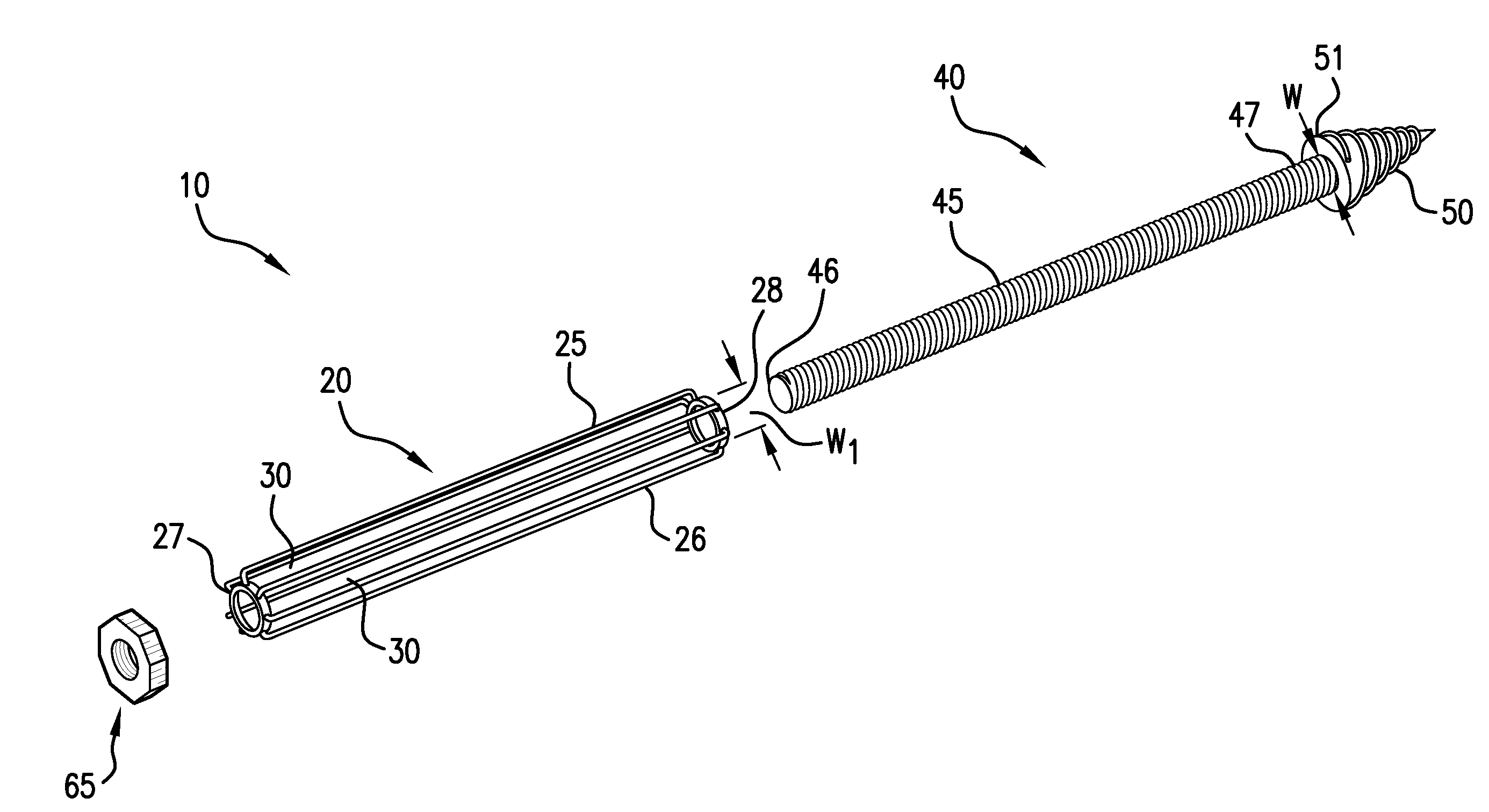
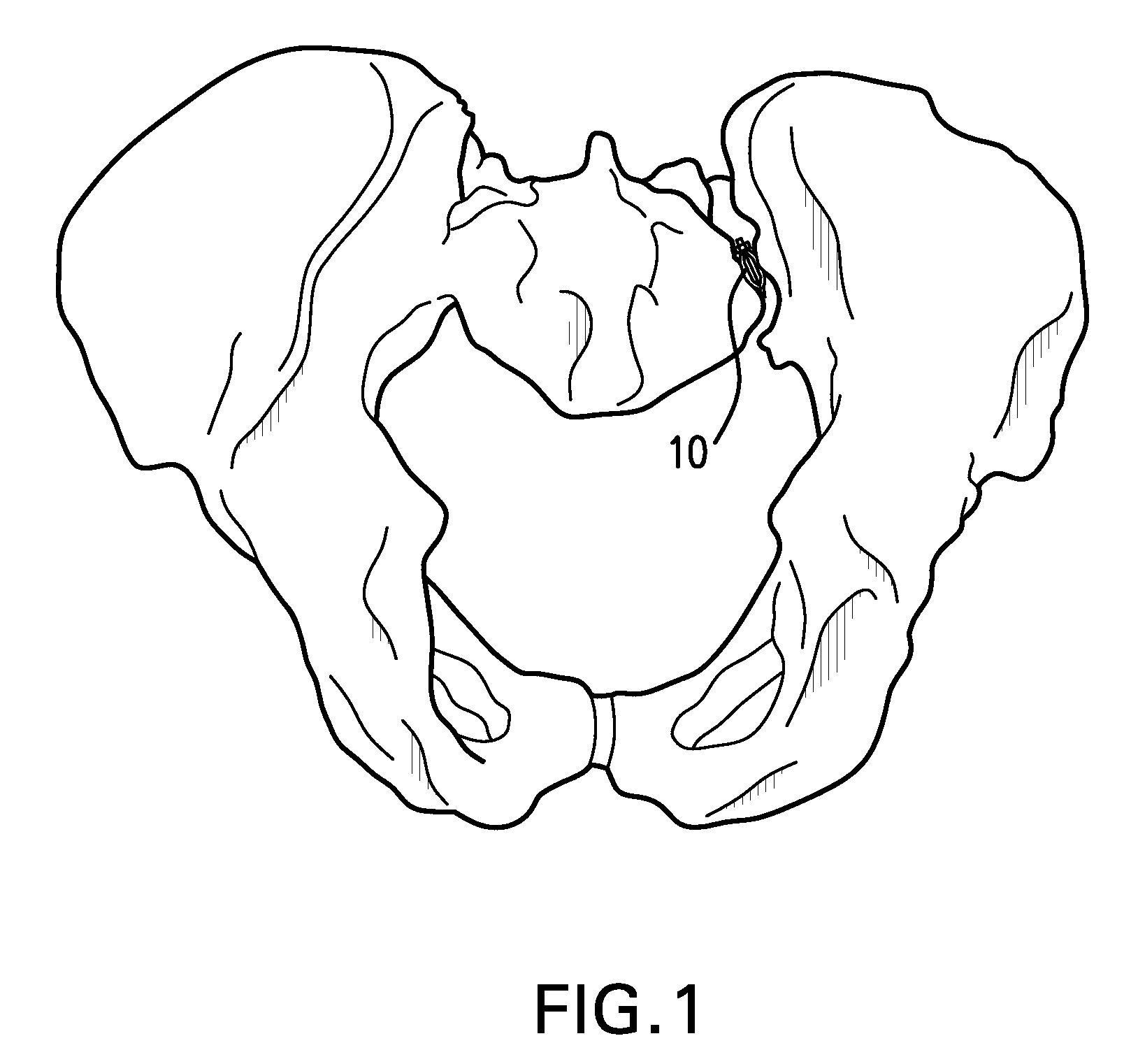
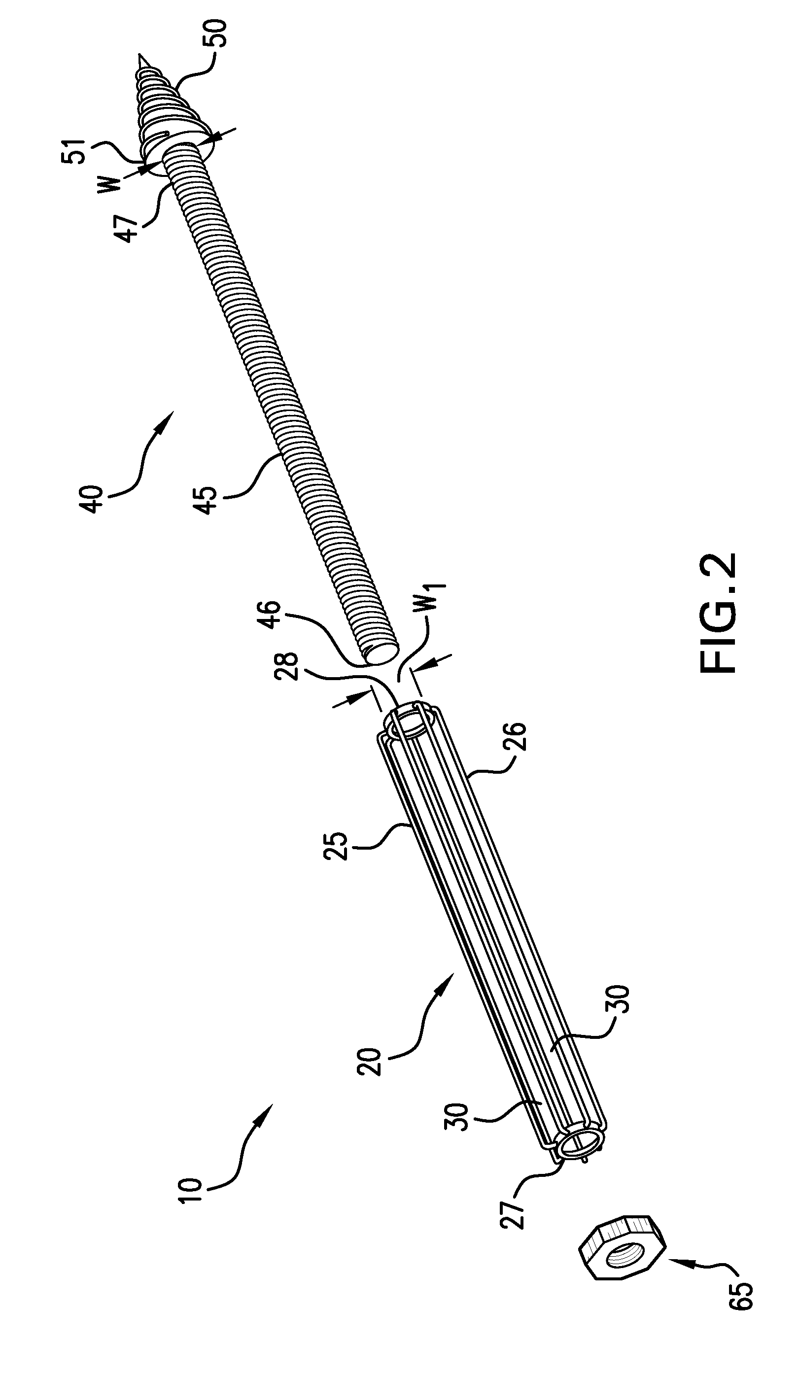
[0036]In general, the present invention provides methods of stabilizing the sacroiliac joint in a patient in need thereof using a device having an expandable portion that engages the sacral and iliac surfaces of the sacroiliac joint. Specifically, referring to FIG. 1, the methods involve generating laterally opposing forces against the iliac and sacral surfaces of the SI joint to securely seat a device 10 in a plane generally parallel to the SI joint. Prior methods, in contrast, involved disposing a bone screw perpendicular to the joint to direct compressive forces against the iliac and sacral surfaces to draw the surfaces closer together. The expandable portion of devices used in the methods of the present invention is either coated with or otherwise contains a bone material, such as bone graft or a substrate containing a bone morphogenic protein, to facilitate fusion of the joint. By stabilizing the sacroiliac joint, the methods of the present invention eliminate or reduce motion ...


PUM


Abstract

Methods of stabilizing the sacroiliac joint by placing an expandable device in the joint to generate laterally opposing forces against the iliac and sacral surfaces of the SI joint to securely seat the device in a plane generally parallel to the SI joint. The expandable device is coated with or otherwise contains a bone material to promote fusion of the joint. The expandable device used in methods of the present invention can be, for example, an expandable cage, a balloon, a balloon-expandable stent or a self-expanding stent.

Description

TECHNICAL FIELD[0001]The present application is directed to various methods for stabilizing the sacroiliac joint including via fusing the joint.SUMMARY OF THE INVENTION[0002]The sacroiliac (SI) joint is a diarthrodial joint that joins the sacrum to the pelvis. In this joint, hyaline cartilage on the sacral side moves against fibrocartilage on the iliac side. The joint is generally C shaped with 2 lever arms that interlock at the second sacral level. The joint contains numerous ridges and depressions, indicating its function for stability more than motion. In fact, the SI joint's main function appears to be providing shock absorption for the spine through stretching in various directions. Stability in the SI joint is provided by the large ridges present in the joint and by the presence of generously sized ligaments, which offer resistance to shear and loading. The deep anterior, posterior, and interosseous ligaments resist the load of the sacrum relative to the ilium and the more sup...

Claims


Application Information

Patent Timeline
IPC IPC(8): A61F5/00A61B17/00A61M29/00
CPCA61B17/844A61B17/7055
Inventor JOHNSON, DONALDBUTLER, DANIEL
Owner SPINAL INNOVATIONS
Learn More