Methods for diagnosing and treating neuroendocrine cancer
a neuroendocrine cancer and cancer technology, applied in the field of neuroendocrine cancer diagnosis and treatment, can solve the problems of increased cell death, decreased cell division, and toxic effects of glutamate and nmda agonists on these neuroblastoma cells, so as to inhibit or decrease the activity or expression, and reduce the proliferation of neuroendocrine tumor cells
- Summary
- Abstract
- Description
- Claims
- Application Information
AI Technical Summary
Benefits of technology
Problems solved by technology
Method used
Image


Examples
example 1
Cell Culture
[0079]Breast cancer (i.e., MCF-7 and SKBR3) and SCLC (i.e., classical human SCLC cell lines NCI-H345, DMS-53, NCI-H146, and the variant SCLC cell line, NCI-H82,) cells were maintained at 37° C. in an atmosphere of 5% CO2 using Dulbecco's Minimal Essential Medium (DMEM; 0.8 mM Mg2+, 0.4 mM glycine; Sigma, St. Louis, Mo.) or RPMI 1640 (Mediatech, Inc., Herndon, Va.) supplemented with 10% fetal bovine serum and 50 μg / mL gentamicin. Every 3-4 days, cells received fresh DMEM or were subcultured using 0.06% trypsin with 0.02% EDTA. Cell densities were at 105 to 5×10⁵ / ml.
[0080]MCF-7 and SKBR3 are art-established cell cultures for analyzing the pathophysiology of breast cancer and preclinical analysis of drug efficacy (Hanauske (2004) Oncology (Huntingt). 18(13 Suppl 8):66-9). In particular, studies using MCF-7 cultures have correlated well with in vivo therapeutic studies. For example, Johnson, et al. ((1997) Semin Oncol. 24(1 Suppl 3):S22-5) teach that paclitaxel and 5-fluorou...
example 2
RNA Isolation, RT-PCR and Northern Blot Analysis
[0081]RNA Isolation. Poly(A)+ RNA was isolated from cells using oligo(dT) cellulose chromatography in accordance with well-established methods (Badley, et al. (1988) Biotechniques 5:114-116).
[0082]RT-PCR. Poly(A)+ RNA (2 μg) from breast cancer or SCLC cells was denatured at 70° C. for 10 minutes and chilled on ice. First strand cDNA synthesis was carried with a SUPERSCRIPT™ preamplification system (GIBCO-BRL®, Gaithersburg, Md.), using an oligo(dT) primer and 1 μL (200 U) of SUPERSCRIPT™ II reverse transcriptase (PROMEGA®, Madison, Wis.). The reverse transcriptase product was directly used as a template for the PCR reaction. PCR was carried out using GENEAMP® PCR reagents (PERKIN ELMER™, Foster City, Calif.) in a thermocycler (EASY CYCLER™ Series, ERICOMP, San Diego, Calif.). The templates were initially denaturated at 97° C. for 8 minutes and amplified for 30 cycles under the following conditions: denaturation at 95° C. for 30 seconds...
example 3
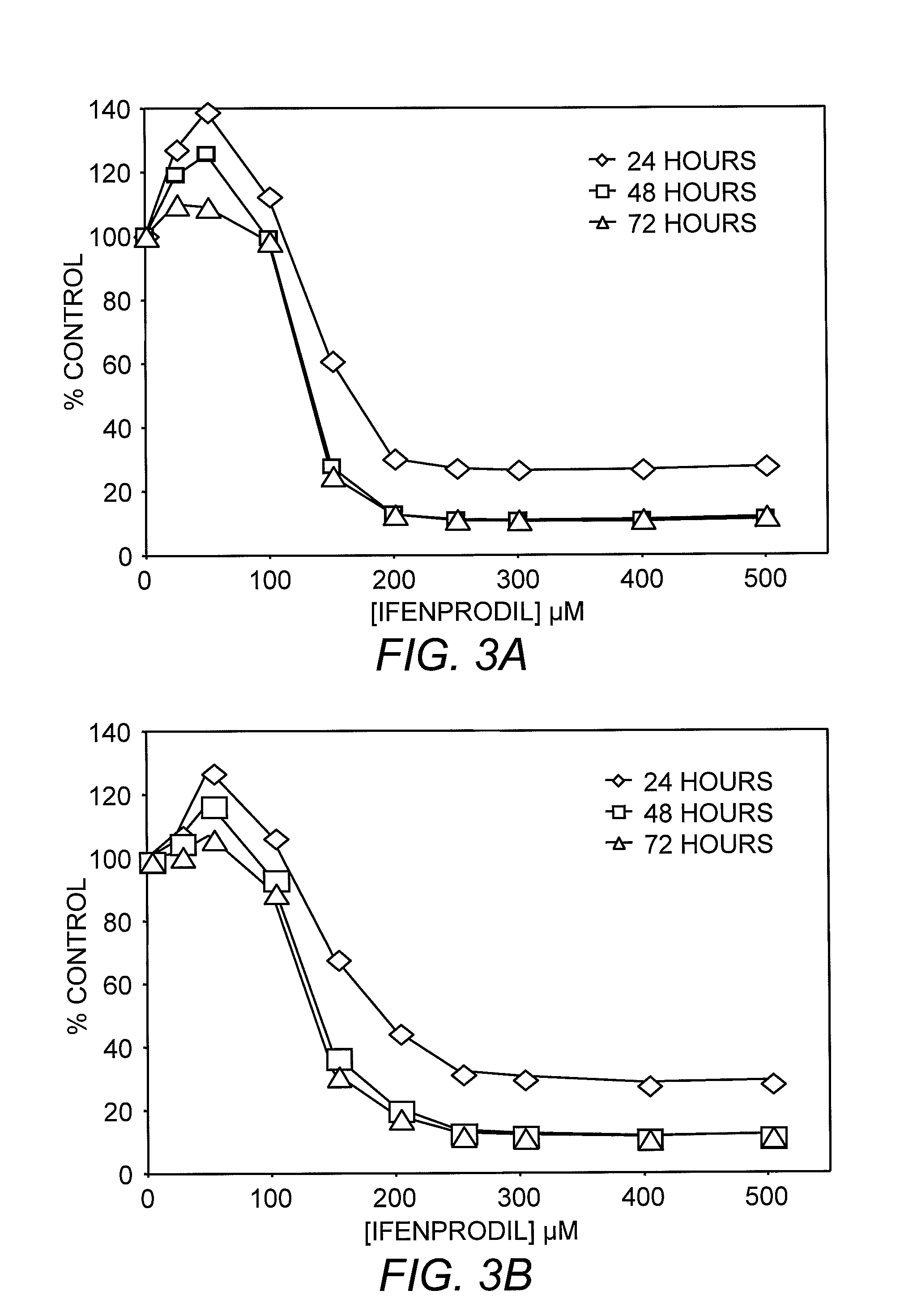
[0086]Breast cancer and SCLC cells were subcultured into 24-well plates (CORNING®, Corning, N.Y.). After 24 hours, the growth medium was aspirated and replaced with growth medium containing L-glutamic acid (Sigma, St. Louis, Mo.) at concentrations of 0, 1, or 10 mM, or N-phthalamoyl-L-glutamic acid (NPG; Research Biochemicals Inc., Natick, Mass.) at concentrations of 0, 0.1 or 1 mM. Following 48 hours of incubation at 37° C., the experimental medium was removed and replaced with DMEM containing 40 μg / mL neutral red (Sigma, St. Louis, Mo.). After a two hour incubation, the neutral red was aspirated and the cell monolayers carefully washed with phosphate-buffered saline (PBS). Incorporated dye was extracted from cells with 50% ethanol / 1% acetic acid. The absorbance of recovered dye was determined at 540 nm or fluorescence measured at excitation of 530 nm and emission 650 nm.
PUM
| Property | Measurement | Unit |
|---|---|---|
| body weight | aaaaa | aaaaa |
| temperature | aaaaa | aaaaa |
| concentrations | aaaaa | aaaaa |
Abstract
Description
Claims
Application Information
 - R&D
- Intellectual Property
- Life Sciences
- Materials
- Tech Scout
- Unparalleled Data Quality
- Higher Quality Content
- 60% Fewer Hallucinations
Browse by: Latest US Patents, China's latest patents, Technical Efficacy Thesaurus, Application Domain, Technology Topic, Popular Technical Reports.
© 2025 PatSnap. All rights reserved.Legal|Privacy policy|Modern Slavery Act Transparency Statement|Sitemap|About US| Contact US: help@patsnap.com