
Device to open and close a bodily wall

Active Publication Date: 2009-02-26
COOK MEDICAL TECH LLC
View PDF99 Cites 3 Cited by
  • Summary
  • Abstract
  • Description
  • Claims
  • Application Information

AI Technical Summary

Benefits of technology

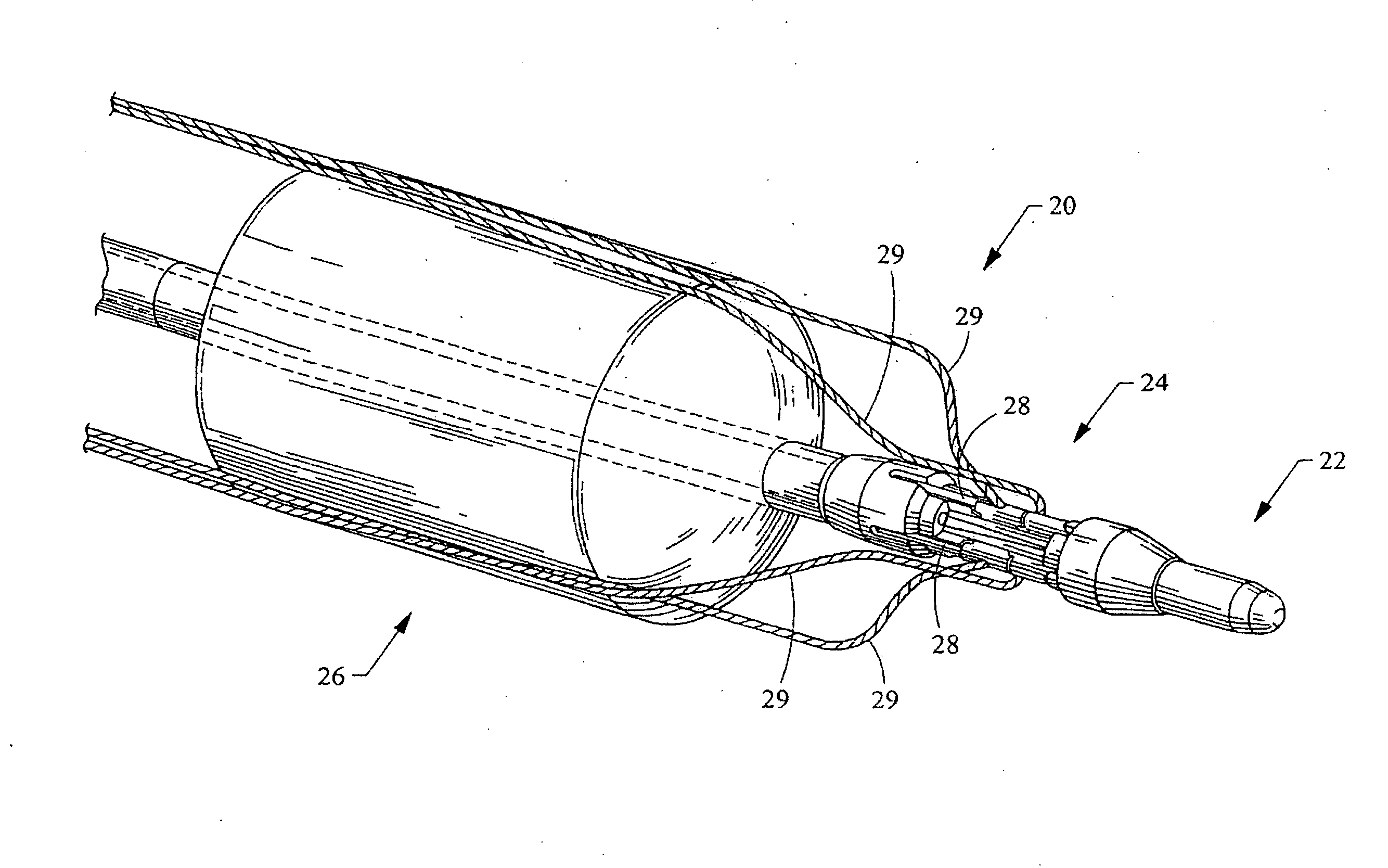
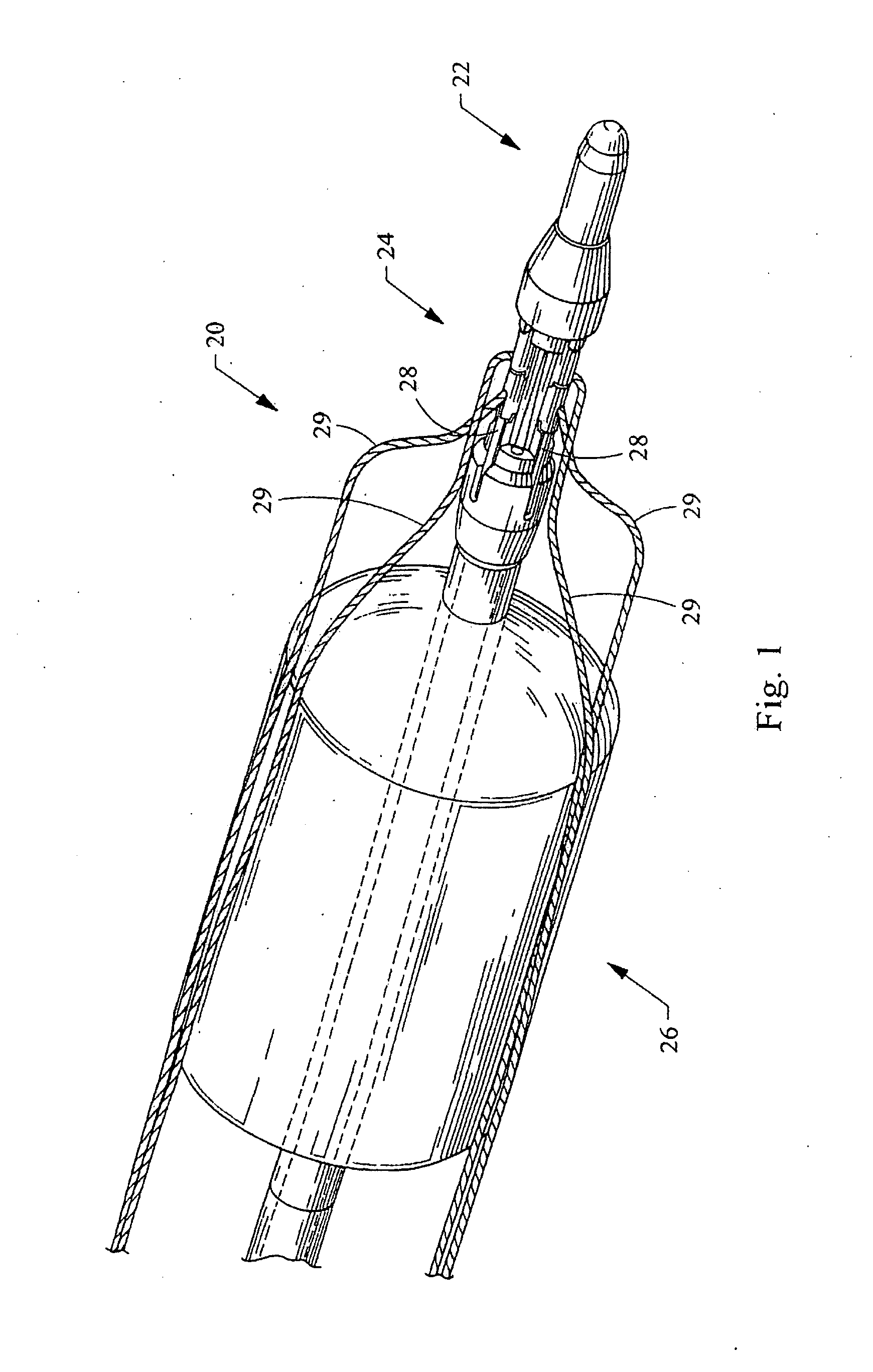
[0005]The present invention provides medical devices and methods for performing transluminal procedures that reduce procedure time as well as the number of instruments used. At about the same time, the medical device and method safely form a perforation and prepare the perforation for reliable closure. One embodiment of a method for opening and closing a visceral wall employs an elongate medical device having both a cutting tool and a suturing tool disposed at a distal end of the elongate medical device. The suturing tool includes a plurality of needles connected to one or more sutures. The elongate medical device is advanced through a bodily lumen to a position proximate the visceral wall. A perforation is formed in the visceral wall using the cutting tool. The elongate medical device and its suturing tool are advanced through the perforation. The plurality of needles are passed through the visceral wall around the periphery of the perforation by retracting the elongate medical device and its suturing tool. The plurality of needles are withdrawn through the bodily lumen and ultimately the perforation is closed using the suture.
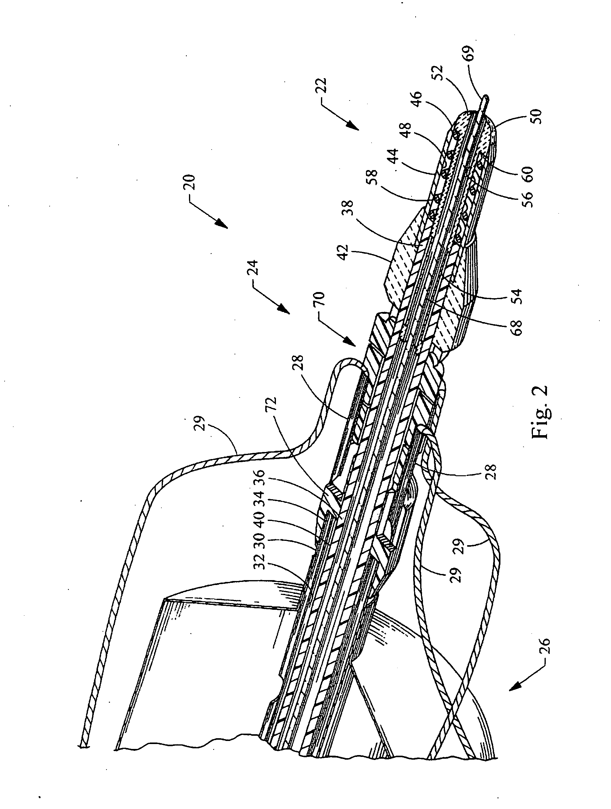
[0008]According to more detailed aspects of this embodiment, a protective tip is slideably disposed within the third lumen of the cutting tool. The protective tip is spring biased to project from a distal end of the cutting tool. The protective tip defines an access channel therethrough, the access channel being in communication with the second lumen. Preferably, the cutting tool is an electro-surgical cutting tool. Most preferably, the protective tip has a first contact attached thereto and the inner cannula has a second contact attached thereto. The first and second contacts form a switch which opens and closes to deenergize and energize the electrosurgical cutting tool.

Problems solved by technology

Generally, transluminal procedures require the use of several different medical instruments, and therefore can be time consuming.
There is also the risk of perforating structures that lie just beyond the bodily wall being cut.
For example, when incising the gastric wall, the potential of hitting blood vessels without knowing could lead to bleeding complications.
Accidentally puncturing the small intestines could lead to the spillage of bacteria into the peritoneal cavity.
Depending on the structure being cut, it has also proven difficult to adequately close the perforation and prevent leakage of bodily fluids to reduce the risk of infection.

Method used


Image

  • Device to open and close a bodily wall
  • Device to open and close a bodily wall
  • Device to open and close a bodily wall

Examples


Embodiment Construction

[0019]Turning now to the figures, an elongate medical device 20 for non-invasively opening and closing a visceral wall has been depicted in FIGS. 1-5, and is constructed in accordance with the teachings of the present invention. The medical device 20 generally includes a cutting tool 22, a suturing tool 24, and a dilation tool 26. The cutting tool 22 is used to form a perforation 12 in a visceral wall 10 (FIG. 3). The suturing tool 24 is used to place a plurality of needles 28 through the visceral wall 10, the needles 28 being connected to one or more sutures 29 for closing the perforation 12. The dilation tool 26 is used to dilate the perforation 12.

[0020]As shown in FIG. 2, the elongate medical device 20 is generally defined by a double-walled outer catheter 30 defining a first lumen 32, and an inner cannula 36 defining a second lumen 40. The inner cannula 36 projects beyond a distal end 34 of the outer catheter 30. The distal end 38 of the inner cannula 36 is connected to the cut...


PUM


Abstract

A medical device and method for performing transluminal procedures that reduces procedure time as well as the number of instruments used is disclosed. An elongate medical device has both a cutting tool and a suturing tool disposed at a distal end of the elongate medical device. The suturing tool includes a plurality of needles connected to one or more sutures. The elongate medical device is advanced through a bodily lumen to a position proximate the visceral wall. A perforation is formed in the visceral wall using the cutting tool. The elongate medical device and its suturing tool are advanced through the perforation and a plurality of needles are passed through the visceral wall around the periphery of the perforation. The plurality of needles are withdrawn through the bodily lumen and the perforation is closed using the suture.

Description

CROSS-REFERENCE TO RELATED APPLICATIONS[0001]This application claims the benefit of U.S. Provisional Application Ser. No. 60 / 956,569, filed on Aug. 17, 2007, entitled “DEVICE TO OPEN AND CLOSE A BODILY WALL”FIELD OF THE INVENTION[0002]The present invention relates generally to medical devices for cutting open and suturing closed the visceral walls of bodily lumens.BACKGROUND OF THE INVENTION[0003]Perforations in visceral walls may be formed to gain access to adjacent structures of the body, the methods commonly referred to as transluminal procedures. For example, culdoscopy was developed over 70 years ago, and involves transvaginally accessing the peritoneal cavity by forming a perforation in the Cul de sac. This access to the peritoneal cavity allows medical professionals to visually inspect numerous anatomical structures, as well as perform various procedures such as biopsies or other operations. Many transluminal procedures for gaining access to different body cavities using othe...

Claims


Application Information

Patent Timeline
IPC IPC(8): A61B17/32A61B17/04A61B18/12
CPCA61B17/0057A61B17/0469A61B17/0487A61B17/320016A61B2017/0472A61B18/14A61B2017/00659A61B2017/00663A61B2017/047A61B17/32053
Inventor DILLON, TRAVIS E.
Owner COOK MEDICAL TECH LLC
Learn More