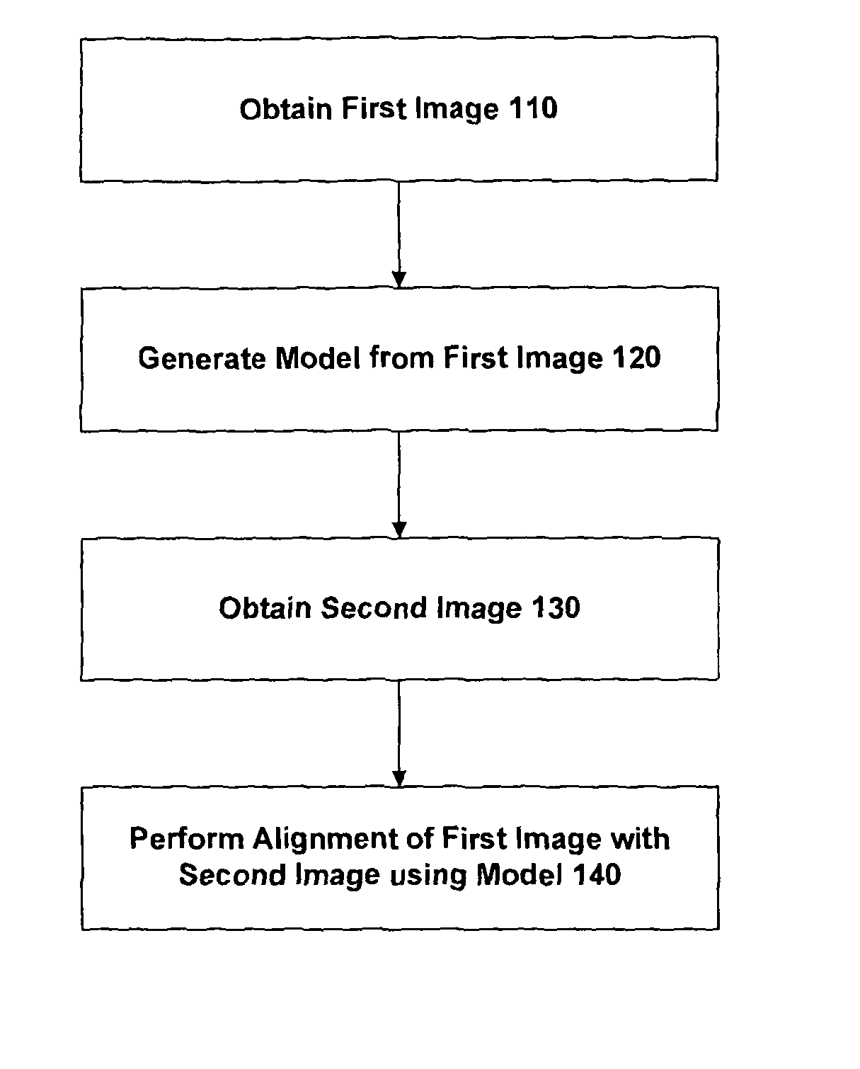

Apparatus and method for registering two medical images

a technology for medical images and apparatus, applied in the field of medical image registration, can solve the problems of difficult image registration, physical deformation and motion of neighbouring structures, and complicating the problem of image registration, so as to minimise the cost function and maximise the joint probability of noise

Active Publication Date: 2013-12-31
UCL BUSINESS PLC
View PDF6 Cites 250 Cited by
  • Summary
  • Abstract
  • Description
  • Claims
  • Application Information

AI Technical Summary

Benefits of technology

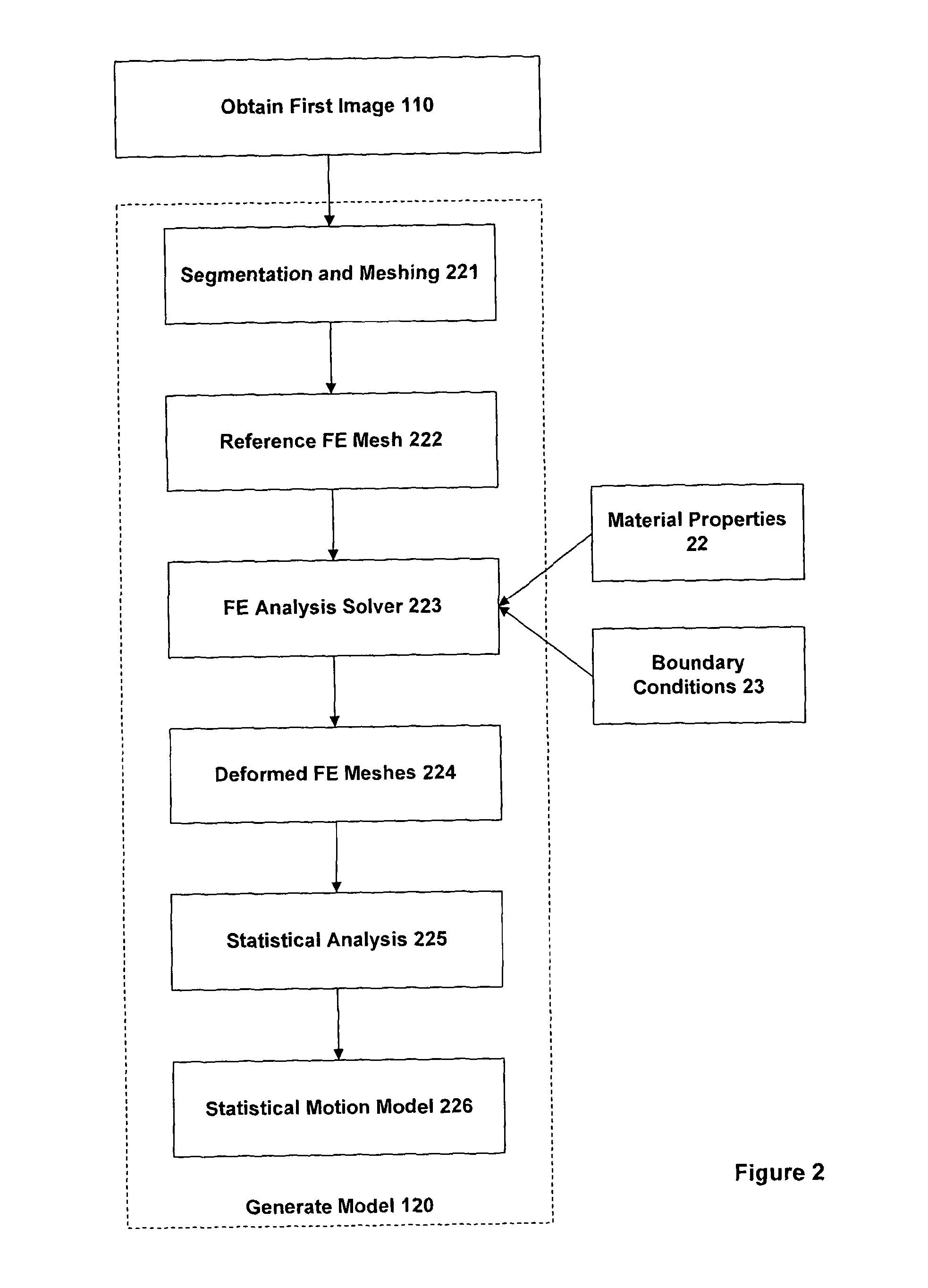
[0037]In one embodiment, a set of simulated deformations of the finite element model of the organ determined from the first image are performed using computational finite element analysis. Constructing a finite element model may include the use of solid modelling tools to convert the geometric surface model into a volumetric, finite element mesh representation of the organ(s) of interest, and assigning physical material properties, such as Young's Modulus and Poisson's Ratio, that are within the known physiological range of such properties, to elements of the model. Each simulation calculates the physical deformation of the organ model for particular material properties and boundary conditions. The boundary conditions specify, for example, which parts of the model are fixed and how other parts move in accordance with externally applied forces. A statistical motion model can then be generated by performing principal component analysis of the displacements of the finite element mesh nodes, calculated by the simulations. The statistical motion model provides a 3D representation of the motion and deformation of the finite element model—and hence the motion and deformation of the organ—as predicted by finite element analysis. The use of the principal component analysis enables a simpler, low-dimensional representation of the predicted displacement of the node points of the underlying finite element model, which therefore reduces the processing requirements (and hence time required) when determining the alignment.
[0038]In one embodiment, determining the alignment includes initially identifying one or more points representing anatomical landmarks in the second medical image and matching them to corresponding points in the geometric model in order to approximately orientate the geometric model with respect to the second medical image. For example, in the case of the prostate gland, the anatomical landmarks may comprise the points of entry and exit of the urethra at the base and apex of the gland. Since the number of points to be identified is generally rather small (often a handful at most), this can be done within the time constraints of a surgical procedure. The use of this matching procedure helps to limit the search space when determining the alignment, thus reducing the time required for finding the alignment and also reducing the chances of not finding the appropriate alignment.
[0040]In one embodiment, the second vector field, derived by filtering the second medical image, is considered to be a noise-corrupted version of the first vector field derived from the geometric model. The alignment is then determined on the basis of maximising the joint probability of the noise. Other approaches for determining an alignment may be to minimise a cost function or to use some other form of numerical optimisation technique—e.g gradient-descent, genetic algorithms, etc.
[0041]In one embodiment, the alignment is determined using a vector similarity measure that quantifies the orientational alignment between the first and second vector fields. The vector similarity measure can account for direction-dependent artefacts in the second medical image when this second medical image is an ultrasound image. Note that US imaging is particularly susceptible to such artefacts and the similarity measure therefore provides a robust approach for determining the alignment in their presence.

Problems solved by technology

This is often highly challenging due to the large differences in the intensity characteristics between images obtained using different imaging techniques.
In addition, fundamental differences between the underlying physics and image formation processes peculiar to each imaging method may also give rise to modality-specific artefacts.
A further problem is that for a deformable structure, which includes most of the soft tissue organs of the body, physical deformations and motion with respect to neighbouring structures may occur between imaging sessions.
These effects further complicate the problem of image registration.
However, this assumption is often not reliable in a situation where different imaging methods that exploit different physical properties are used to obtain an image of the same anatomical region.
Therefore, the feature-based approach to registration is often impractical if available computer-based automatic segmentation methods are unavailable or fail, or if manual segmentation of at least one of the images is prohibitively time-consuming and labour-intensive.
The reliance on feature-based image registration is a particular problem in time-critical applications, such as image-guided surgery, since images obtained during such a procedure are typically of much poorer quality than those obtained outside the surgical setting.
These image are therefore very often difficult to segment automatically or within a clinically acceptable timescale (i.e. seconds to a few minutes).
However, if the diagnostic image information is not accurately aligned with intra-procedural images, errors may be introduced that limit the accuracy of the biopsy as a diagnostic test or that can severely limit clinical efficacy of the intervention.
In practice, such errors include: inaccurate placement of biopsy needles, failure to remove an adequate margin of tissue surrounding a tumour such that malignant cancer cells are not completely eradicated from the organ, and unnecessary damage to healthy tissue with an elevated risk of side-effects related to the procedure in question.
Unfortunately, standard intensity-based multimodal registration algorithms are known to perform poorly with ultrasound images, largely due to high levels of noise, relatively poor soft-tissue contrast and artefacts typically present in clinical ultrasound images.
Furthermore, image segmentation is challenging for the same reasons and therefore the use of many feature-based registration approaches is precluded for most clinical applications.
However, both methods are known not to work well when the unseen image is corrupted in some way such that object boundaries are occluded or the intensity characteristics of the unseen image differ substantially from the images used to train the model.
This situation is very common in medical image applications, particularly during image-guided interventions where (unseen) images obtained during an intervention are typically noisy, contain artefacts, and include medical instruments introduced into the patient.
There are also many situations where, due to noise, artefacts and variability between patients, the variation in image intensity around points on the boundary of an object in a reasonably-sized set of training images is too wide for meaningful parametric statistical measures to be determined.
In this case, the assumptions of the active appearance model method may break down.
However, to date these have been demonstrated only for a few organs and for specialised applications, and rely on automatically converting at least one of the images into a form that is more amenable to performing a registration using established intensity-based methods.
However, this conversion step is not trivial in many circumstances, and these alternative approaches have yet to be demonstrated for many medically significant applications, such as image-guided needle biopsy of the prostate gland and image-guided surgical interventions for the treatment of prostate cancer.
However, this approach assumes that the intensity variation at corresponding locations across different training images adopts a Gaussian distribution, which may not be the case, particularly for interventional images.
The geometric model is created by segmenting both of the images to be registered, which is potentially problematic for surgical applications.
Prostate cancer is a major international health problem, particularly affecting men in the Western World.
Alternative minimally-invasive interventions for prostate cancer, such as brachytherapy, cryotherapy, high-intensity focused US, radiofrequency ablation, and photodynamic therapy are also now available, but the clinical efficacy of most of these treatment approaches has yet to be fully established through randomised controlled trials.
However, conventional (so-called ‘B-mode’) TRUS imaging is two-dimensional and typically provides very limited information on the spatial location of tumours due to the poor contrast of tumours with respect to normal prostatic tissue.
Although there is some evidence that the use of microbubble contrast agents can improve the specificity and sensitivity of tumour detection, this method is not widely used and performing accurate, targeted biopsy and therapy using TRUS guidance alone is difficult in practice, particularly for the inexperienced practitioner.
However, the ability to accurately fuse anatomical and pathological information on tumour location, derived from MR images or a previous biopsy procedure, with TRUS images obtained during a procedure remains a significant technical challenge, mainly due to the differences in intensity between MR and TRUS images, which frustrate standard registration methods, as well as the significant deformation that occurs between the imaging sessions.
However, this approach was found not to produce such accurate image registration, especially if the prostate gland has deformed significantly between the MR and US images.
(2008) state: “Currently, there is no fully automatic algorithm that is sufficiently robust for MRI / TRUS [Transrectal Ultrasound] image registration of the prostate”.

Method used


Image

  • Apparatus and method for registering two medical images
  • Apparatus and method for registering two medical images
  • Apparatus and method for registering two medical images

Examples


Embodiment Construction

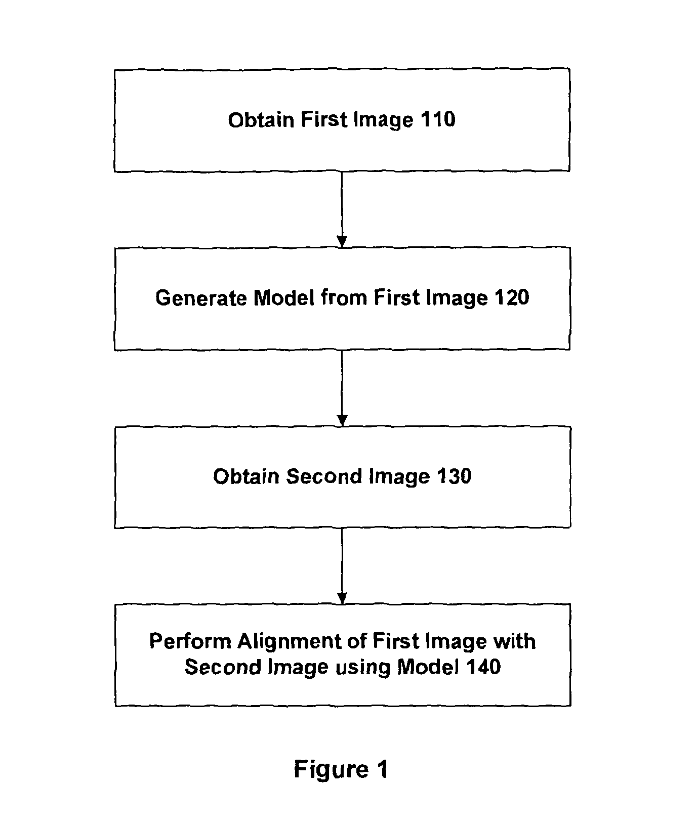
[0053]The approach described herein provides a computerised method for automatically registering, i.e. spatially aligning, two images of the same object by registering a geometric model of the object, derived from one image, to the other image. The method, referred to herein as a Model-to-Image Vector Alignment (MIVA) method, has been devised and tested in one embodiment for registering magnetic resonance (MR) images to transrectal ultrasound (TRUS) images in order to accurately guide surgical procedures for the diagnosis and treatment of prostate cancer. In this case, the geometric model is derived from an MR image and comprises surface meshes that represent the prostate gland and surrounding organs, including the rectum, pelvis and bladder. The model describes the shape and size of an individual's prostate, the prostate location relative to nearby anatomical structures, and the location of regions with a high probability of containing tumours (identified by an expert clinical obse...


PUM


Abstract

An embodiment of the invention provides a method and apparatus for registering two medical images with one another. A first medical image including a representation of a biological organ of a subject or for a population is obtained and the surface of the organ is identified in the first medical image. The identified surface is then used to construct a 3D geometric surface model of the organ. The geometric model is used to derive a motion model that incorporates information on the physical material properties of the organ and external forces that cause the organ to move and deform. A second medical image including a representation of the organ of the subject (or another subject, in the case that the first medical image is an atlas image) is obtained and an alignment is determined between a first surface normal vector field for the organ surface, derived from the geometric model, and a second surface normal vector field for the organ surface, derived by filtering the second medical image. The alignment accommodates deformation of the geometric model in accordance with the motion predicted by the motion model. The first and second medical images can then be registered with one another based on said determined model-to-image vector alignment (MIVA).

Description

FIELD OF THE INVENTION[0001]The present invention relates to registering two medical images with one another, especially where the two images are obtained by different imaging techniques.BACKGROUND OF THE INVENTION[0002]A variety of medical imaging techniques are known, including magnetic resonance (MR) imaging, X-ray computed tomography (CT), radionuclide imaging, optical imaging, and ultrasound (US). Other imaging techniques may be developed in the future. These imaging techniques may produce a two-dimensional (2D) array of pixels (a conventional image) or a three-dimensional (3D) array of voxels, which conceptually represent slices through a physical object. Each pixel or voxel is assigned a value or “intensity” related to one or more physical properties of tissue at a particular point, peculiar to the particular imaging method used. The term “image” as used herein encompasses both 2D and 3D data sets unless the context indicates otherwise.[0003]In some situations, it is desirabl...

Claims


Application Information

Patent Timeline
Patent Type & Authority Patents(United States)
IPC IPC(8): A61B5/05G06K9/00A61B6/00
CPCG06T2207/10136G06T2207/30081G06T7/0034G06T2207/10088G06T2207/30004G06T2207/10081G06T7/0032G06T7/344G06T7/35
Inventor BARRATT, DEANHU, YIPENG
Owner UCL BUSINESS PLC
Learn More