
Video microscopy system and multi-view virtual slide viewer capable of simultaneously acquiring and displaying various digital views of an area of interest located on a microscopic slide

a video microscopy system and virtual slide viewer technology, applied in the field of acquisition and analysis of digital images of objects and areas of interest located on microscopic slides, can solve the problems of user confusion as to what areas, close to impossible, and the drawbacks of conventional microscopic analysis, etc., to achieve cost efficiency, high resolution, cost efficient

Inactive Publication Date: 2009-01-01
TRIPATH IMAGING INC
View PDF20 Cites 14 Cited by
  • Summary
  • Abstract
  • Description
  • Claims
  • Application Information

AI Technical Summary

Benefits of technology

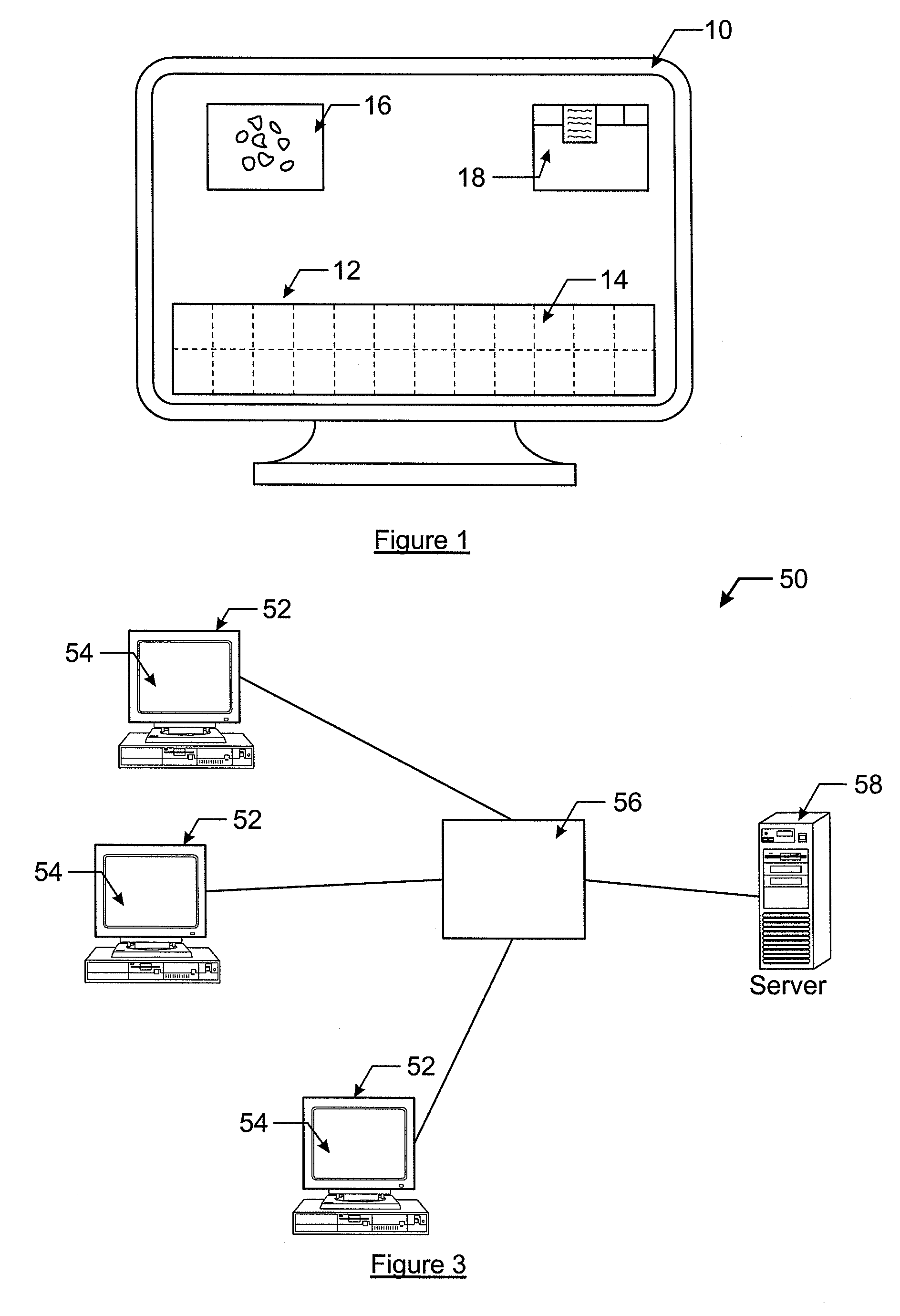
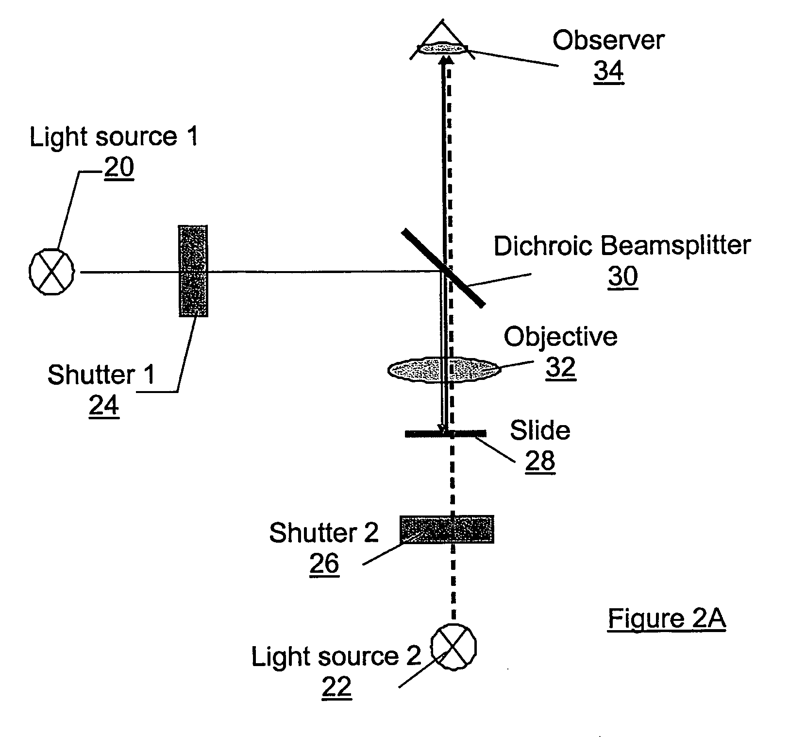
[0016]In view of the deficiencies with many conventional virtual slide viewing systems, the present invention provides a multi-view virtual slide viewing system that provides a display capable of illustrating multiple viewing windows containing different scans of an area of interest. The views may be either different magnifications of a selected area or different scans of the slide taken under different conditions. For example, the slide viewing system of the present invention may display a scan of the slide taken in with bright field illumination in one window and a scan of the same view with dark field illumination in another window of the display. This, in turn, not only allows the user to compare scans of varying magnification, but also to compare scans of the same slide taken under different illumination and optical conditions and with different markers, dyes, and other preparations and even scans taken from the same slide on different scan platforms. For example the slide viewer of the present invention is able to display a unitary low-resolution scan of the slide taken with a cost efficient flat bed scanner, as opposed to a display formed of tiles, and combine it in a correlated way with the image presentation of a tiled high resolution scan of the same slide taken with another scan platform. As such, the low-resolution display does not require processing to blend tiles together nor does it experience problems with resolution at tile boundaries. An additional advantage consists in the ability to add the high resolution scan at a later time and only on demand. In that respect a flat bed scanner can be used as a cost efficient pre scan device.
[0030]As will be described later, prior to the slide being scanned for the first time, the slide is provided with a zero point, i.e., (0, 0), for its coordinate system. This zero point is placed on the slide as a fiducial, typically in the form of an ink dot. All subsequent scans of the tissue sample are referenced from this zero point. Further, if the slide is removed for further preparations, the coordinate system for the new position of the slide is calibrated to the original zero point so that subsequent scans can be positionally correlated with the previous scans. Additionally, if the slide is moved to another microscope for acquiring specific scans, the microscope is first checked for calibration differences. The slide is then placed on the microscope and aligned with the microscope using the previously marked zero point on the slide. As the slide is maintained at the proper alignment for each scan, different scans for the same position on the slide can be positionally correlated with one another, such that the user may evaluate the various scans taken at a selected position of interest on the slide.
[0038]Generally, a data set for a tissue sample will include a large amount of data representing different scans using different lighting and contrast settings, magnification, and sample preparations. However, during analysis of the data, the user may determine that only a subset of the different scans is needed to report on their analysis of the sample. For this reason, the computing system of the present invention allows the user to save individual views of the sample in a snap shot gallery. Specifically, in one embodiment of the present invention, the user may indicate that they wish to save a particular scan. In this instance, the computing system of the present invention saves the scan in a separate file or creates a link to the file in the main header. The computing system may also associate a thumbnail of the scan on the viewing screen so that the user can more easily recall the scan.

Problems solved by technology

Although a desired research tool, conventional microscopic analysis does have some drawbacks.
This procedure may cause the user to become confused as to what areas have and have not been inspected in the sample.
This requires the user to constantly switch between different optical microscope settings and to compare and correlate the different types of information gleaned from that “multi-view” approach in his mind which, especially where details are concerned, is close to impossible.
While conventional virtual slide viewers, such as the one illustrated in FIG. 1, provide a user with both a low-resolution scan and higher-resolution scans simultaneously on a display, these virtual slide viewers are restricted to only the bright field view.
This is where conventional virtual slide viewers fall short.
They do not provide these additional digital views of a slide and deny the pathologist crucial diagnostic information.
As mentioned, conventional virtual slide scanner and viewer systems only provide different magnifications of the bright field view of a sample, they do not provide different scans of a sample in terms of use of different dye markers, or dark field and / or fluorescent scans.
A major reason for this failing of the prior art is due to the difficulty in matching coordinate systems for various scans.
This, in turn, makes it difficult, if not impossible, to positionally correlate the various scans of the same area of interest.

Method used


Image

  • Video microscopy system and multi-view virtual slide viewer capable of simultaneously acquiring and displaying various digital views of an area of interest located on a microscopic slide
  • Video microscopy system and multi-view virtual slide viewer capable of simultaneously acquiring and displaying various digital views of an area of interest located on a microscopic slide
  • Video microscopy system and multi-view virtual slide viewer capable of simultaneously acquiring and displaying various digital views of an area of interest located on a microscopic slide

Examples


Embodiment Construction

[0052]The present invention now will be described more fully hereinafter with reference to the accompanying drawings, in which preferred embodiments of the invention are shown. This invention may, however, be embodied in many different forms and should not be construed as limited to the embodiments set forth herein; rather, these embodiments are provided so that this disclosure will be thorough and complete, and will fully convey the scope of the invention to those skilled in the art. Like numbers refer to like elements throughout.

[0053]As discussed above, an important limitation of most conventional virtual slide viewers is that they only display different bright field magnifications of a sample. They do not display scans of the sample made with different contrast and illumination settings and methods or scans of the slide taken with different preparations or scan platforms. As such, the user of a conventional virtual slide viewer receives only limited information from these system...


PUM


Abstract

A slide viewer capable of simultaneous display of more than one representation of an area of interest of a slide is provided. The slide viewer includes a database containing at least two data files representing different representations for a same area of interest on one or multiple correlated slides or at least two different digital presentations of the same representation. The representations are views of different illumination and / or of different contrast. Associated with the database are a processor and a display. The processor retrieves data files representing different representations of the same of area of interest and displays them on the display. A user is allowed to simultaneously view representations of the same area of interest, where the representations are of views different from each other by either illumination and / or contrast or by the digital information content presented, and / or by the information acquired from multiple correlated slides.

Description

CROSS-REFERENCE TO RELATED APPLICATIONS[0001]This application is a continuation of U.S. application Ser. No. 10 / 143,735, filed May 10, 2002, which is hereby incorporated herein in its entirety by reference.FIELD OF THE INVENTION[0002]The present invention relates generally to the acquisition and analysis of digital images of objects and areas of interest located on a microscopic slide, and more particularly to a system and method capable of providing various digital views of the same areas of interest to a user, where each view provides digital images with different information for use in quantitative and qualitative analysis of the objects on the microscopic slide.BACKGROUND OF THE INVENTION[0003]Microscopic analysis is a widely used research tool in the field of cellular biology and pathology. Specifically, tissue samples and cell preparations are visually inspected by pathologists under several different conditions and test procedures with use of microscopes. Based on these visua...

Claims


Application Information

Patent Timeline
IPC IPC(8): G09G5/00G06F3/00G01B11/00G02B21/00G02B21/36G06F3/048G06F3/0487G06F17/30H04N1/04H04N1/21
CPCG02B21/367G06F17/30274H04N1/0402H04N2201/3277H04N1/32112H04N2201/3247H04N2201/3253H04N1/0408G06F16/54
Inventor GAHM, THOMASDORRER, RAINERSCHMID, JOACHIMWILLIAMS, RYAN
Owner TRIPATH IMAGING INC
Learn More