
Macrophage-Enhanced MRI (MEMRI)

a mri and macrophage technology, applied in the field of whole body mri scanning and cancer staging, can solve the problems of breast cancer remaining a leading cause of death in middle-aged women, breast cancer remains an incurable disease, and mortality rates have decreased

Inactive Publication Date: 2009-01-01
AMAG PHARMA
View PDF3 Cites 17 Cited by
  • Summary
  • Abstract
  • Description
  • Claims
  • Application Information

AI Technical Summary

Benefits of technology

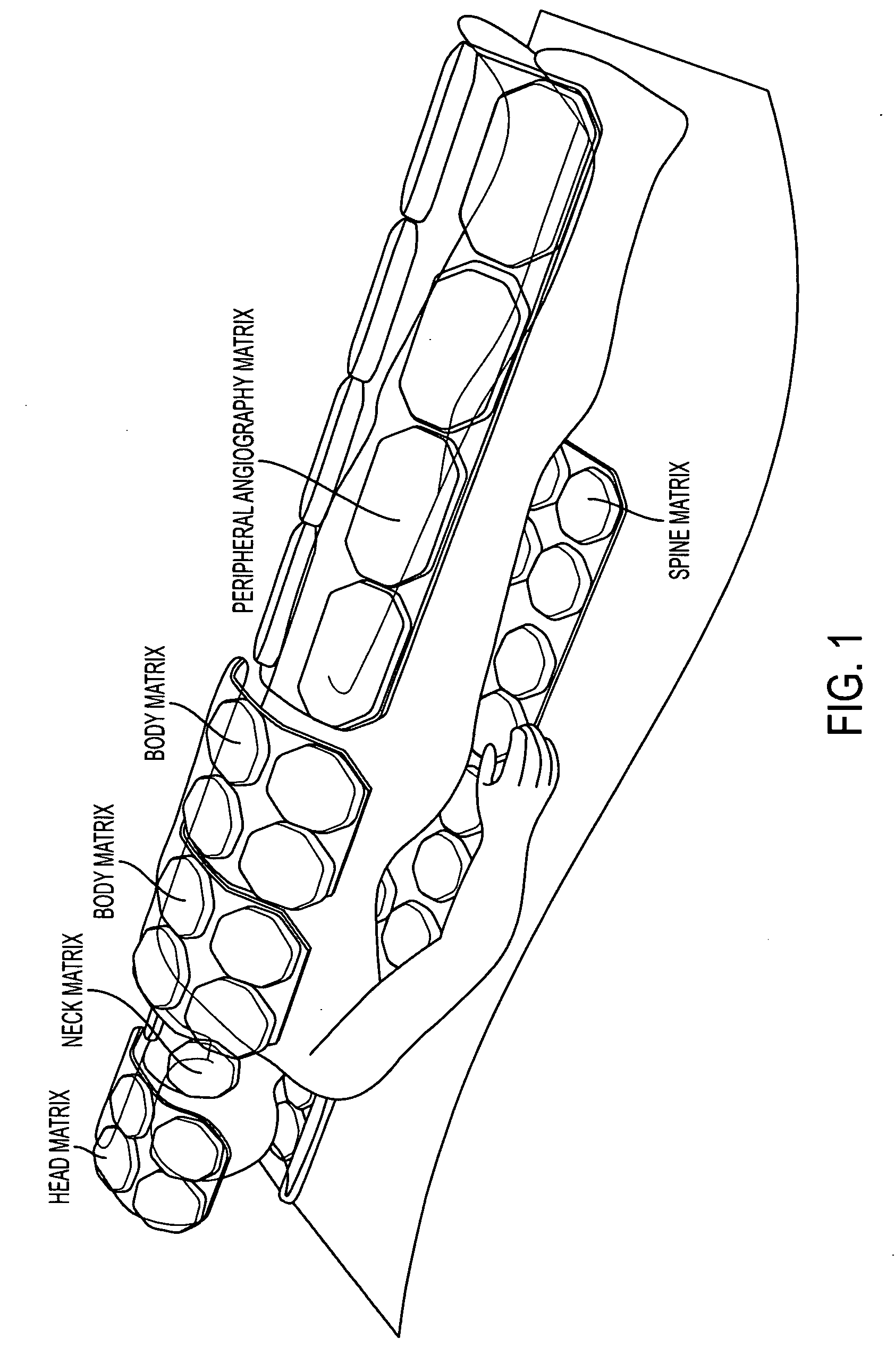
[0022]Another embodiment provides a method as described, wherein using the image includes observing macrophage activity associated with a primary tumor or with any metastatic tumor in bone, lymph node, spleen, liver, central nervous system, lung, or other organ. In particular embodiments, the regions collectively include the entire body. In other particular embodiments, the macrophage imaging agent is an ultrasmall superparamagnetic iron oxide particle and in still more particular embodiments, the macrophage imaging agent has a blood half-life sufficient to permit microphage trapping throughout the regions at cancer risk. In related particular embodiments the macrophage imaging agent is a complex of ultrasmall superparamagnetic iron oxide and a polysaccharide. In still other related embodiments, the polysaccharide is selected from the group consisting of dextran, reduced dextran and a derivative thereof.
[0023]Another particular embodiment provides a method of assessing efficacy of an anticancer treatment in a subject comprising administering a macrophage imaging agent to the subject before the anticancer treatment, making a magnetic resonance image of regions of the subject's body to be targeted by the anti-cancer treatment to establish a pre-treatment image, administering the anticancer treatment to the subject, administering the macrophage imaging agent to the subject after th

Problems solved by technology

Although mortality rates have decreased due to earlier detection and improved treatments, breast cancer remains a leading cause of death in middle-aged women.
Furthermore, metastatic breast cancer is still an incurable disease.
The morphologic appearance of the tumor can also be assessed but because tumors with similar histopathologic appearance can exhibit significant clinical variability, this approach has serious limitations.
Yet these analyses, though useful, are only partially predictive of the clinical behavior of breast tumors, and there is much phenotypic diversity present in breast cancers that current diagnostic tools fail to detect and current therapies fail to treat.
However, therapeutic options are very limited for advanced cases.
Indeed, mutations within a long-lived stem cell population may initiate the formation of cancer stem cells that underlie the growth and maintenance of tumors and whose presence contributes to the failure of current therapeutic approaches.
However, these agents have no cell targeting capabilities and their distribution and accumulation are not specific for cancer nor for the tissue at risk for cancer metastasis.
Bolus administration and dynamic MRI may provide some additional information about the degree of vascular leakiness, but such information can only be obtained for one body region of interest.
As is described in example 1 (vide infra), the use of such contrast-enhanced MRI may be insufficient to characterize the cancer stage even with respect to the primary tumor.

Method used


Image

  • Macrophage-Enhanced MRI (MEMRI)
  • Macrophage-Enhanced MRI (MEMRI)
  • Macrophage-Enhanced MRI (MEMRI)

Examples


example 1

MEMRI Evaluation of Patient with Suspected Cancer in Single Breast

[0069]This situation involves a patient presenting with known or suspected cancer of one breast but a normal mammogram of the opposite breast. Recently, it has been suggested that such patients should undergo contrast-enhanced breast MRI to rule out other cancer foci [See Lehman, et al (2007) N Eng. J Med 356, 1295-303. In such an evaluation, the contrast agent is usually a gadolinium chelate and abnormal breast tissue is expected to show a focal accumulation of gadolinium in the expanded extracellular space associated with the cancer that was not clinically or mammographically evident. Though sensitive, this procedure is fraught with false positives—the abnormal regions must be biopsied and four of five such regions will not be cancerous—and does not provide information on the possible metastasis to local lymph nodes. In the present invention, the patient at risk is administered the macrophage biomarker and the breas...

example 2

Patient with Breast Cancer and Bone Pain

[0074]When metastatic breast cancer is suspected, it is important to rule out the most common sites of metastasis, as well as recurrence or new cancer in the breast. If whole body MRI is performed during macrophage enhancement, in other words, if a whole body MEMRI is performed, identification of soft tissues where there is excessive macrophage density will identify where metastasis may be present. In addition, bone MRI is the best examination for bone metastasis, and the identification of macrophage dense areas by MEMRI would increase diagnostic accuracy in bone. Finally, displacement of normal macrophages in liver, spleen, or lymph nodes would suggest the presence of metastasis in these sites. One additional use of the macrophage imaging technique MEMRI is to identify regions where a tissue biopsy may be obtained for pathological information.

example 3

Patient with Bladder Cancer and / or Bone Pain

[0075]This example is similar to the above examples with breast cancer, with the additional advantage that regional nodes can be reexamined along with liver and lung, or other sites of potential metastasis.

[0076]FIGS. 5A and 5B show a patient with bladder cancer with macrophages around the primary tumors. The bladder is indicated generally with an arrow in FIGS. 5A and 5B as the large central light area in the center of the pelvis region. FIG. 5A is the MRI without contrast agent and FIG. 5B is the image with contrast agent of the invention. In FIG. 5A, the tumor's presence is only hinted at by the “crease” in the bladder (shown with an arrow) that seems to be an indication of pressure on or displacement of the bladder along this juncture. FIG. 5B, with contrast agent, clearly shows the line of demarcation for the tumor along that “crease”, with the massive tumor showing as a dark mass directly to the left of this line, continuing down to ...


PUM

PropertyMeasurementUnit
Timeaaaaaaaaaa
Timeaaaaaaaaaa
Densityaaaaaaaaaa

Abstract

Methods for assessing stage of cancer in a subject are provided, comprising administering a macrophage imaging agent to the subject, making a magnetic resonance image of regions of the subject's body at cancer risk, and using the image to assess macrophage density and displacement associated with any primary cancer or metastatic cancer in the subject, such density and displacement being indicative of neoplasia. The macrophage imaging agent may be an ultrasmall superparamagnetic iron oxide particle and in particular embodiments, the macrophage imaging agent has a blood half-life sufficient to permit microphage trapping throughout the regions at cancer risk. Additional embodiments provide methods for assessing efficacy of an anticancer treatment in a subject, methods for determining frequency of follow-up MEMRI evaluation in a subject, methods for determining metastatic potential of cancer foci in a subject, and methods for determining prognosis of cancer in a subject. Methods for directing site of biopsy in a subject by performing a whole body MEMRI evaluation of the subject to identify macrophage density at a tumor site of interest and assessing the macrophage density to identify the site of biopsy in the subject, macrophage density being an indicator of tumor growth are also provided, in addition to methods for providing individualized cancer treatment to a subject in need thereof using whole body MEMRI evaluation.

Description

CROSS REFERENCE TO RELATED APPLICATIONS[0001]The present application claims priority from provisional patent application U.S. Application Ser. No. 60 / 947,259, filed Jun. 29, 2007, which is hereby incorporated by reference herein.TECHNICAL FIELD[0002]The present invention relates to whole body MRI scanning and cancer staging using macrophage-seeking MRI agents to perform Macrophage-Enhanced MRI or “MEMRI”.BACKGROUND ART[0003]Cancer is one of the leading causes of death in the developed world, resulting in over 500,000 deaths per year in the United States alone. Over one million people are diagnosed with cancer in the U.S. each year, and overall it is estimated that more than 1 in 3 people will develop some form of cancer during their lifetime. Though there are more than 200 different types of cancer, four of them—breast, lung, colorectal, and prostate—account for over half of all new cases (Jemal et al., CA Cancer J. Clin. 53:5-26 (2003)). Cancer metastasis is considered to be due to...

Claims


Application Information

Patent Timeline
IPC IPC(8): A61K49/06A61K49/00A61B5/055
CPCA61B5/0515A61B5/055A61B5/415A61B5/416G01R33/5601A61B5/4504A61K49/1863B82Y5/00G01R33/3415A61B5/418A61B5/004
Inventor WOLF, GERALD L.
Owner AMAG PHARMA
Learn More