Eureka AIR delivers breakthrough ideas for toughest innovation challenges, trusted by R&D personnel around the world.

Internally administered therapeutic agents for cranial nerve diseases comprising mesenchymal cells as an active ingredient

a technology of mesenchymal cells and therapeutic agents, which is applied in the direction of drug compositions, extracellular fluid disorders, instruments, etc., can solve the problems of difficult preparation of neural stem cells from patients or other persons, difficult collection of tissues containing neural stem cells from the cerebrum, and complicated technique, etc., to prevent endothelial cells from ischemic damage, stimulate angiogenesis, and facilitate and safe obtained

Inactive Publication Date: 2008-11-20
NC MEDICAL RES
View PDF5 Cites 5 Cited by
  • Summary
  • Abstract
  • Description
  • Claims
  • Application Information

AI Technical Summary

Benefits of technology

[0107]The present inventors made further studies on the therapeutic effects of bone marrow stem cells (mesenchymal stem cells) on cranial nerve diseases by intravenously administering them to the above mentioned model animal in the same manner. They found that intravenous administration of bone marrow stem cells is very effective for treating cranial nerve diseases.
[0199]The present invention can be used to treat neurological damage caused by injury and neurological diseases including Krabbe's disease, Hurler's syndrome, metachromatic leukodystrophy, and stroke. Improved neurological function in experimental autoimmune encephalomyelitis (EAE) has been reported following intravenous infusion of human MSCs and neurosphere-derived multipotent precursors. Suggested mechanisms include reduction of inflammatory infiltration, remyelination, and elevation of trophic factors that may be neuroprotective or stimulate oligodendrogliosis. The present invention may have the advantage of exerting multiple therapeutic effects at various sites and times within the lesion as the cells respond to a particular pathological microenvironment.

Problems solved by technology

It is not impossible to prepare such cells from patients or other persons for use in cell therapy; however, it is problematic since tissue material must be collected from either the brain or nerves.
However, although it doesn't cause symptoms of neurological deficiency, collecting tissues that contain neural stem cells from the cerebrum is not easy.
This technique, however, is very complicated and dangerous.
While intravenous injection of BMSCs reduces infarction size and improves functional outcome in a rat stroke model, the therapeutic benefit of MSC-like multipotent precursor cells derived from peripheral blood (PMSCs) transplantation in cerebral ischemia is still uncertain.

Method used


Image

  • Internally administered therapeutic agents for cranial nerve diseases comprising mesenchymal cells as an active ingredient
  • Internally administered therapeutic agents for cranial nerve diseases comprising mesenchymal cells as an active ingredient
  • Internally administered therapeutic agents for cranial nerve diseases comprising mesenchymal cells as an active ingredient

Examples


example 1

Transient Middle Cerebral Artery Occlusion Model

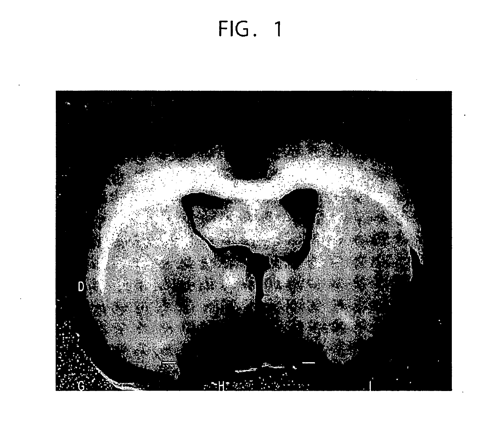
[0272]A rat middle cerebral artery occlusion model was used as a stroke model. Transient middle cerebral artery occlusion (MCAO) was induced for 45 minutes using the intravascular occlusion method (E. Z. Longa, P. R. Weinstein, S. Carlson, R. Cummins, Reversible middle cerebral artery occlusion without craniectomy in rats, Stroke 20 (1989) 84-91).

[0273]Adult male Sprague-Dawley rats (n=113) weighing 250 to 300 g were anaesthetized with 5% isoflurane, and the anesthesia was mechanically maintained with 1.5% isoflurane in a gaseous mixture of 70% N2O and 30% O2 under artificial ventilation. The rectal temperature was maintained at 37° C. using an infrared heat lamp. A cannula was inserted into the left femoral artery during surgery, for measuring blood pH, pO2, and pCO2. The tip of a 20.0 to 22.0 mm long 3-0 surgical suture (Dermalon: Sherwood Davis & Geck, UK) was rounded by heating near a flame, and was advanced from the external carot...

example 2

Preparation of Bone Marrow Cells

[0275]Autologous bone marrow was collected from the femur of MCAO rats, one and a half hours prior to bone marrow cell transplant.

[0276]The rats were anaesthetized with ketamine (75 mg / kg) and xylazine (10 mg / kg; i.p.). A 1 cm incision was made in the skin, a small hole (2×3 mm) was punctured in the femur using an air drill, and 1 ml of bone marrow was aspirated using a 22-gauge needle. The collected samples were diluted and suspended in a medium containing 2 ml of L-15 medium and 3 ml of Ficoll (Amersham Biosciences). After centrifugation at 2,000 rpm for 15 minutes, mononuclear cell fractions were collected and resuspended in 2 ml serum-free medium (NPMM: Neural Progenitor Cell Maintenance Medium; Clonetics, San Diego, Calif., USA). Following a second centrifugation (2,000 rpm, 15 minutes), cells were suspended in 1 ml of NPMM.

example 3

Experimental Groups

[0277]The experiment was conducted using 11 groups (n=88). Nothing was administered to the Group 1 (control) rats after MCAO (n=8). The rats in Groups 2 to 6 were intravenously administered with just the medium (without donor cell administration), 3, 6, 12, 24, and 72 hours after MCAO (n=8 for each group). The rats in Groups 7 to 11 were intravenously administered with the autologous bone marrow cells (1.0×10⁷ cells), 3, 6, 12, 24, and 72 hours after MCAO (each group n=8). Six rats in each group were used to calculate the infarct volume, and the others were used for other histological analyses.


PUM


Abstract

Intravenous administration of bone marrow cells collected from rat bone marrow or peripheral blood to a rat cerebral infarction model was found to be effective in treating cerebral infarction. Human and murine bone marrow stem cells showed similar effects. Mesenchymal cells such as bone marrow cells, cord blood cells, or peripheral blood cells can be used as agents for in vivo administration against cranial nerve diseases.

Description

[0001]This application is a Continuation of application Ser. No. 11 / 377,610, filed Mar. 17, 2006, which is a Continuation-in-Part of application Ser. No. 10 / 562,202, which is a national phase application of PCT / JP04 / 009386 filed on Jun. 25, 2004. The entire of contents of application Ser. No. 10 / 562,202, include the specification, drawing, claims, sequence listing and abstract are hereby incorporated by reference.TECHNICAL FIELD[0002]The present invention relates to cranial nerve disease therapeutic agents for in vivo administration, which comprise mesenchymal cells, particularly bone marrow cells, cord blood cells, or peripheral blood cells, or cells derived from these cells as active ingredients.BACKGROUND ART[0003]In recent years regenerative medical techniques have been in the limelight. In regenerative medical techniques, disorders that the natural, inherent regenerative-healing ability of the human body cannot cure can be cured by regenerating organs and such using artificial ...

Claims


Application Information

Patent Timeline
Patent Type & Authority Applications(United States)
IPC IPC(8): A61K35/12A61P7/00A61K35/28
CPCA61K35/28A61K2035/124G01N2800/285G01N2800/2871A61P7/00
Inventor HONMOU, OSAMUHAMADA, HIROFUMI
Owner NC MEDICAL RES
Why Eureka
Eureka Blog
Learn More