Medical device rapid drug releasing coatings comprising a therapeutic agent and a contrast agent

a technology of contrast agent and medical device, which is applied in the direction of catheters, blood vessels, therapy, etc., can solve the problems of affecting the effect of treatment effect, so as to improve the preservation of coating integrity and uniformity of drug distribution, and improve the resistance to mechanical disruption. , the effect of reducing the tendency to coat particles

Active Publication Date: 2008-10-16
LUTONIX INC
View PDF80 Cites 237 Cited by
  • Summary
  • Abstract
  • Description
  • Claims
  • Application Information

AI Technical Summary

Benefits of technology

[0025]Surprisingly, the present inventor has found that the at least one additive according to embodiments of the present invention, which comprises a hydrophilic part and a drug affinity part, in combination with a lipophilic or water-insoluble therapeutic agent and a contrast agent, forms an effective drug delivery coating on a medical device that avoids the disadvantages of conventional liposomal or encapsulation based coating formulations such as slow release, lipolysis dependence, and a very low limit to drug:lipid concentration ratio. Moreover, the coatings according to embodiments of the present invention avoid the disadvantages of prior formulation technologies for coatings of contrast agent and lipophilic or water-insoluble drug, such as the inability to preserve coating integrity and uniformity of drug distribution, and the unavoidable flaking off of particles from the coating during handling.
[0097]In a further embodiment, the present invention relates to a method for preparing a drug coated balloon catheter. In one aspect of this embodiment, the method comprises, (a) preparing a coating solution comprising an organic solvent, a therapeutic agent, a contrast agent, and an additive, (b) applying the coating solution to an inflated balloon catheter, and (c) deflating and folding the balloon catheter and drying the coating solution to increase uniformity of drug coating.

Problems solved by technology

While these medical devices initially appear successful, the benefits are often compromised by the occurrence of complications, such as late thrombosis, or recurrence of disease, such as stenosis (restenosis), after such treatment.
Over time, de-endotheliazation and injury to smooth muscle cells results in thrombus deposition, leukocyte and macrophage infiltration, smooth muscle cell proliferation / migration, fibrosis and extracellular matrix deposition.
The widespread use of coronary stents has altered the vascular response to injury by causing a more intense and prolonged inflammatory state, due to chronic irritation from the implanted foreign body, and in the case of drug eluting stents (DES), from insufficient biocompatibility of the polymer coating.
Moreover, local drug delivery may allow for the use of certain effective therapeutic agents, which have previously been considered too toxic or non-specific to use systemically.
Thus, a considerable disadvantage of a DES, or any other implanted medical device designed for sustained release of a drug, is that the drug is incapable of being rapidly released into the vessel.
Additionally, while drug-eluting stents were initially shown to be an effective technique for reducing and preventing restenosis, recently their efficacy and safety have been questioned.
A life-threatening complication of the technology, late thrombosis, has emerged as a major concern.
Drug eluting stents cause substantial impairment of arterial healing, characterized by a lack of complete re-endothelialization and a persistence of fibrin when compared to bare metal stents (BMS), which is understood to be the underlying cause of late DES thrombosis.
Concerns have also been raised that the polymeric matrix on the stent in which the anti-proliferative drug is embedded might exacerbate inflammation and thrombosis, since the polymers used are not sufficiently biocompatible.
Thus, combining a therapeutic agent with a polymer in a medical device coating may have significant disadvantages.
Another important limitation of the DES is that the water insoluble drugs are not evenly distributed in the polymeric matrix of the coating.
The non-uniform distribution of drug causes non-uniform drug release to the tissue of the vessel walls.
This may cause tissue damage and thrombosis in areas exposed to excess drug and hyperplasia and restenosis areas that are undertreated.
Yet another important limitation of the DES is that only a limited amount of an active agent can be loaded into the relatively small surface area of the stent.
Any component of the coating that inhibits rapid release, such as a crosslinked polymer, liposome, or encapsulating particle, is necessarily disadvantageous to the intended use of the balloon catheter, which is inflated for a very brief period of time and then removed from the body.
However, a polymeric hydrogel coating can not effectively deliver water insoluble drugs (such as paclitaxel and rapamycin), because they can not mix with the hydrogel coating.
However, for purposes of a medical device coating, all of these drug delivery formulations have significant disadvantages: drug loading is poor, and drug release from these preparations is slow.
Oils and lipids mix well with water-insoluble drugs such as paclitaxel or rapamycin, but when micelles or liposomes are then formed by interaction with aqueous media, the particles and particle sizes are relatively unstable, ranging in a broad particle size distribution from several hundred nanometers to several microns in diameter.
Another disadvantage of oil-based liposome formulations is the dependence of drug absorption on the rate and extent of lipolysis.
Lipolysis of oil-based triglycerides is difficult and dependent upon many factors, and triglycerides must be digested and drug released in order to be absorbed by diseased tissue.
The amount of hydrophobic drug delivered to tissues by these agents will be low, because liposomes and micelles cannot efficiently release hydrophobic drug, which they carry away before it can be absorbed by tissues.
Micelles, liposomes, or particles of oils and lipids are therefore not effective at rapidly and efficiently facilitating tissue uptake of drug during a very brief device deployment time, and no report has shown these types of coatings to be effective.
Loading capacity of conventional micelles and liposomes is low.
In addition, the inventor has found that drug release occurs far too slowly from a coating consisting essentially of just oil / lipid and lipophilic drug, because they bind to each other and to the external surface of the medical device so tightly that the drug cannot rapidly elute off the device during several minutes or less of deployment at the target site.
Drug that is encapsulated in polymeric particles may take even longer to diffuse from the coating (the reported range is months to years) and will have further difficulty permeating target tissues rapidly.
Thus, these polymeric microspheres are useful for sustained release of drug over a long period of time but cannot rapidly release drug and facilitate tissue uptake.
Notwithstanding this result, prior approaches to coating formulations with iodine contrast agent and a lipophilic or water-insoluble therapeutic agent have serious drawbacks.
The physical characteristics of these coatings are very poor.
There is a several-fold variability in drug levels in tissue after treatment, which results in inconsistent therapeutic outcomes.
Experimentally, the present inventor has found that the compatability or miscibility of lipophilic or water-insoluble therapeutic agents and iodine contrast agents is not good, and the integrity and uniformity of the coating is very poor.
Particles from the coating easily flake off and are lost during handling.
Furthermore, there is marked variability in adhesion of drug to the surface of the device, and clumps of contrast agent and drug of variable size are formed that easily flake off the device during minimal handling in the interventional cardiology suite.
These deficiencies adversely affect the consistency and uniformity of drug delivery to target tissues, which results in highly variable and inconsistent therapeutic effects and decreased therapeutic utility of the coated device.
Combining drugs and medical devices is a complicated area of technology.
It involves the usual formulation challenges, such as those of oral or injectable pharmaceuticals, together with the added challenge of maintaining drug adherence to the medical device until it reaches the target site and subsequently delivering the drug to the target tissues with the desired release and absorption kinetics.
The coating thickness must also be kept to a minimum, since a thick coating would increase the medical device's profile and lead to poor trackability and deliverability.
Thus, formulations that are effective with pills or injectables might not work at all with coatings of medical device.
If the drug releases from the device too easily, it may be lost during device delivery before it can be deployed at the target site, or it may burst off the device during the initial phase of inflation and wash away before being pressed into contact with target tissue of a body lumen wall.

Method used


Image

  • Medical device rapid drug releasing coatings comprising a therapeutic agent and a contrast agent
  • Medical device rapid drug releasing coatings comprising a therapeutic agent and a contrast agent
  • Medical device rapid drug releasing coatings comprising a therapeutic agent and a contrast agent

Examples


example 1

Preparation of Coating Solutions

[0259]Formulation 1—50-100 mg (0.06-0.12 mmole) paclitaxel, 1-1.6 ml acetone, 1-1.6 ml ethanol, 50-100 mg iopromide, and 50-100 mg Tween 20 are mixed.

[0260]Formulation 2—50-100 mg (0.06-0.12 mmole) paclitaxel, 1-1.6 ml acetone, 1-1.6 ml ethanol, 50-100 mg iopromide, and 50-100 mg Tween 40 are mixed.

[0261]Formulation 3—50-100 mg (0.05-0.11 mmole) rapamycin, 1-1.6 ml acetone, 1-1.6 ml ethanol, 50-100 mg iopromide, and 50-100 mg Tween 20 are mixed.

[0262]Formulation 4—50-100 mg (0.05-0.11 mmole) rapamycin, 0.5-1.0 ml acetone, 0.5-1.0 ml ethanol, 5-35 mg iopromide, and 35-70 mg Tween 40 are mixed.

[0263]Formulation 5—50-100 mg (0.06-0.12 mmole) paclitaxel, 1-1.6 ml acetone, 1-1.6 ml ethanol, 50-100 mg loversol, and 50-100 mg Tween 20 are mixed.

[0264]Formulation 6—50-100 mg (0.06-0.12 mmole) paclitaxel, 1-1.6 ml acetone, 1-1.6 ml ethanol, 50-100 mg loversol, and 50-100 mg Tween 40 are mixed.

[0265]Formulation 7—50-100 mg (0.05-0.11 mmole) rapamycin, 1-1.6 ml ...

example 2

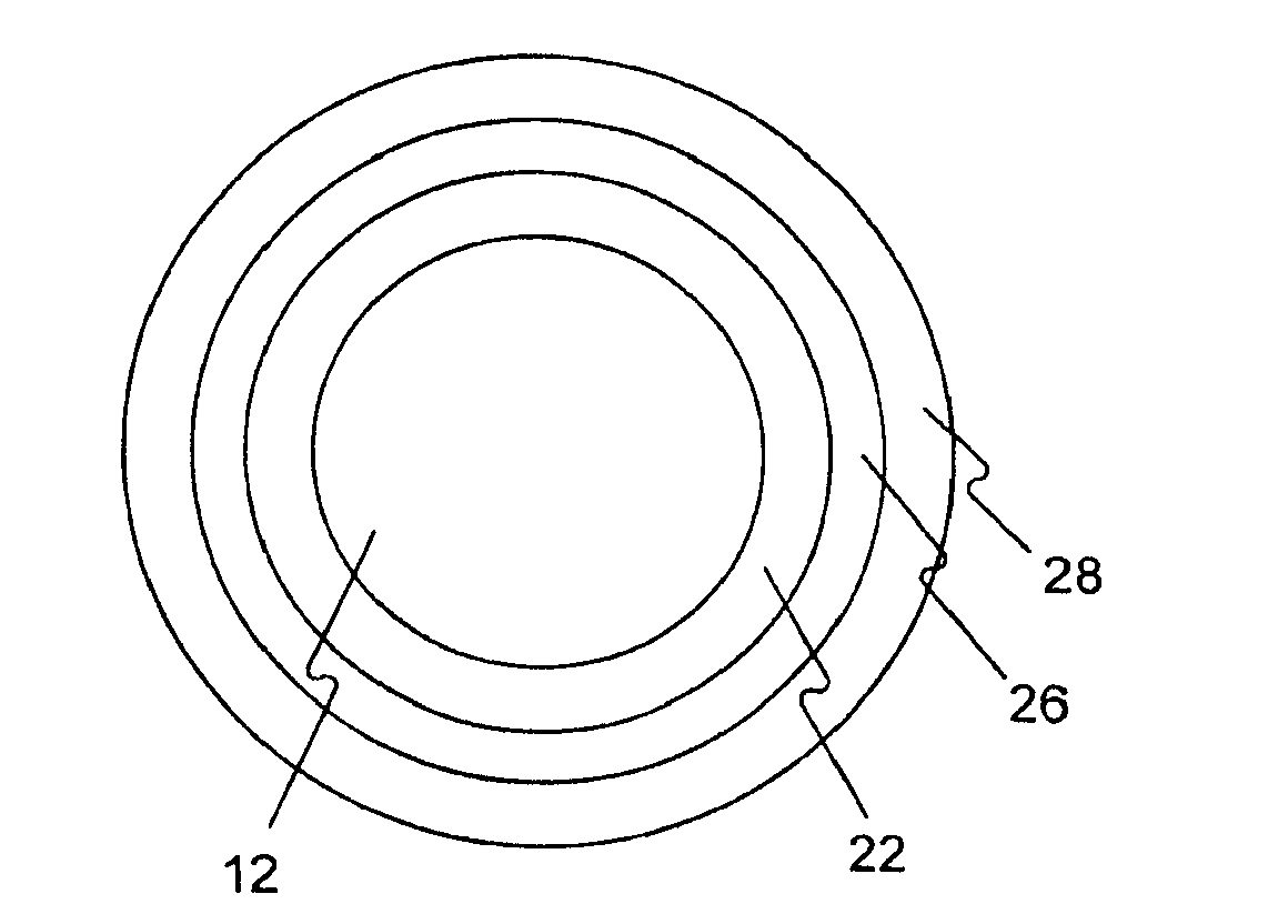
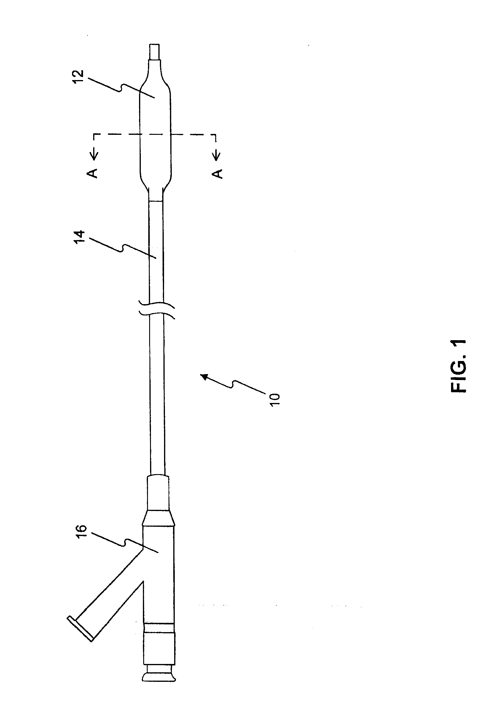
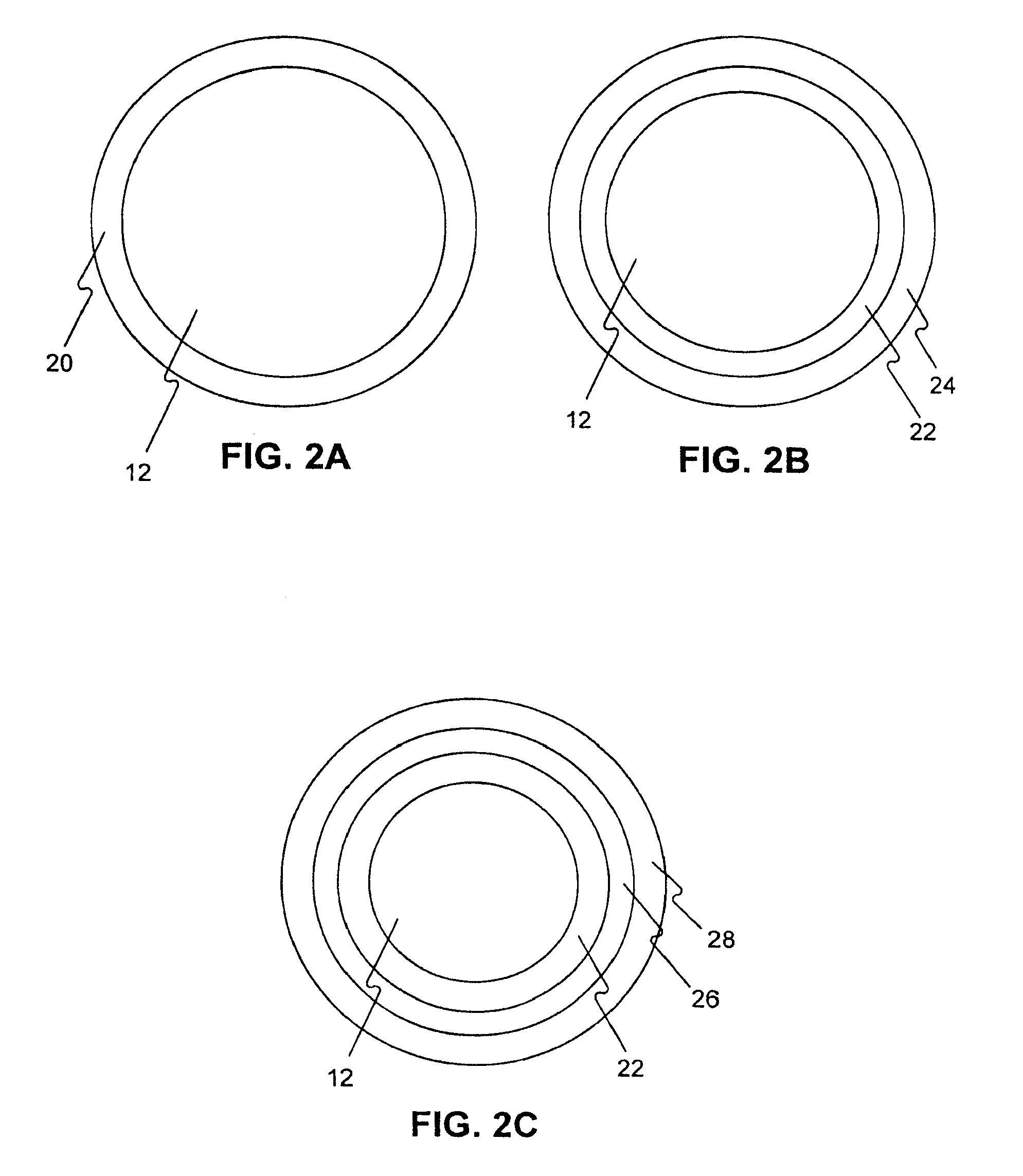
[0267]5 PTCA balloon catheters (3 mm in diameter and 20 mm in length) are folded with three wings under vacuum. The folded balloon under vacuum is sprayed or dipped in a formulation (1-8) in example 1. The folded balloon is then dried, sprayed or dipped again, dried again, and sprayed or dipped again until sufficient amount of drug on the balloon (3 microgram per square mm) is obtained. The coated folded balloon is then rewrapped and sterilized for animal testing.

example 3

[0268]Drug coated balloon catheters and uncoated balloon catheters (as control) are inserted into coronary arteries in pigs. The balloon is over dilated (1:1.2), and the inflated balloon is held in the vessel for 60 seconds to release drug and additive, then deflated and withdrawn from the pig. The animals are angiographed after 3 days, 31 days, 3 months, 6 months, 9 months and 12 months. The amount of drug in the artery tissues of the sacrificed animal is measured after 60 minutes, 3 days, 31 days, 3 months, 6 months, 9 months and 12 months.


PUM

PropertyMeasurementUnit
Solubility (mass)aaaaaaaaaa
Concentrationaaaaaaaaaa
Solubility (mass)aaaaaaaaaa

Abstract

The invention relates to a coated medical device for rapid delivery of a therapeutic agent to a tissue in seconds to minutes. The medical device has a layer overlying the exterior surface of the medical device. The layer contains a therapeutic agent, a contrast agent, and an additive.

Description

CROSS REFERENCE TO RELATED APPLICATIONS[0001]This application is a continuation-in-part of application Ser. No. 11 / 942,452, filed Nov. 19, 2007, which claims the benefit of priority of U.S. Provisional Application No. 60 / 860,084, filed on Nov. 20, 2006, U.S. Provisional Application No. 60 / 880,742, filed Jan. 17, 2007, U.S. Provisional Application No. 60 / 897,427, filed on Jan. 25, 2007, U.S. Provisional Application No. 60 / 903,529 filed on Feb. 26, 2007, U.S. Provisional Application No. 60 / 904,473 filed Mar. 2, 2007, U.S. Provisional Application No. 60 / 926,850 filed Apr. 30, 2007, U.S. Provisional Application No. 60 / 981,380 filed Oct. 19, 2007, and U.S. Provisional Application No. 60 / 981,384 filed Oct. 19, 2007, the disclosures of all of which are incorporated by reference herein.FIELD OF THE INVENTION[0002]Embodiments of the present invention relate to coated medical devices, and particularly to coated balloon catheters, and their use for rapidly delivering a therapeutic agent to par...

Claims


Application Information

Patent Timeline
IPC IPC(8): A61M25/10A61F2/82A61K31/337A61K31/4433A61K51/02A61K101/02
CPCA61K31/337A61K31/436A61L29/16A61L29/18A61L31/16A61L31/18A61M5/007A61L2300/608A61L2300/416A61K2300/00A61B17/320725A61B18/24A61B18/245A61B2018/00327A61B2018/00345A61B2018/00446A61B2018/00488A61B2018/00505A61F2/07A61F2/82A61L29/08A61L29/085A61L31/08A61L31/10A61L2300/606A61L2420/06A61L2420/08A61L2430/20A61M25/09A61M25/10A61M25/1002A61M31/005A61M37/00A61M2025/105
Inventor WANG, LIXIAO
Owner LUTONIX INC
Why Eureka
Try Eureka