
Analysis of methylation using selective adaptor ligation

a technology of adaptor ligation and methylation, which is applied in the field of arrays and methods for detecting methylation of nucleic acids, can solve the problems of genomic instability, alterations in the normal methylation process,

Inactive Publication Date: 2008-10-16
AFFYMETRIX INC
View PDF6 Cites 15 Cited by
  • Summary
  • Abstract
  • Description
  • Claims
  • Application Information

AI Technical Summary

Benefits of technology

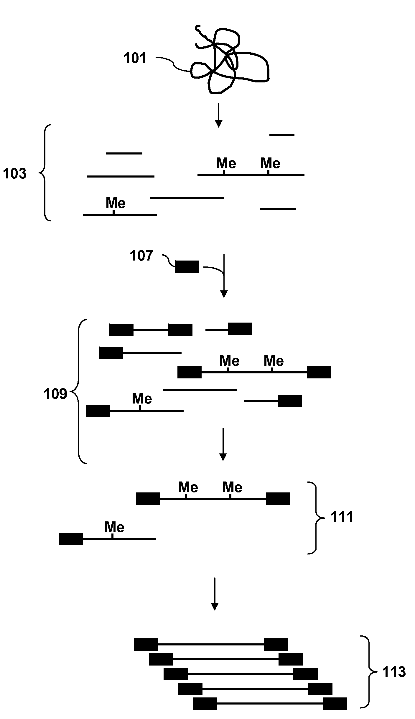
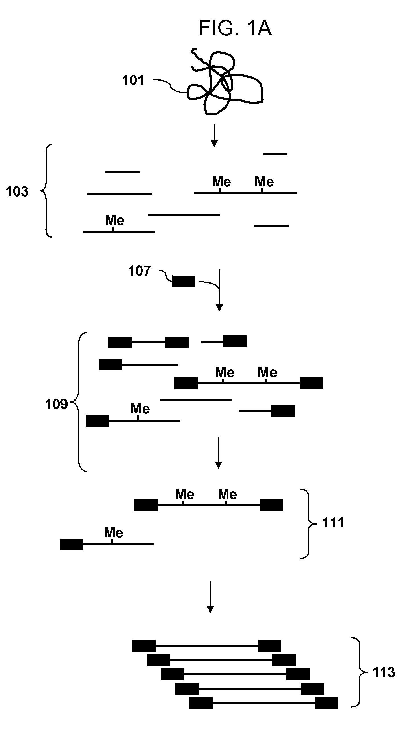
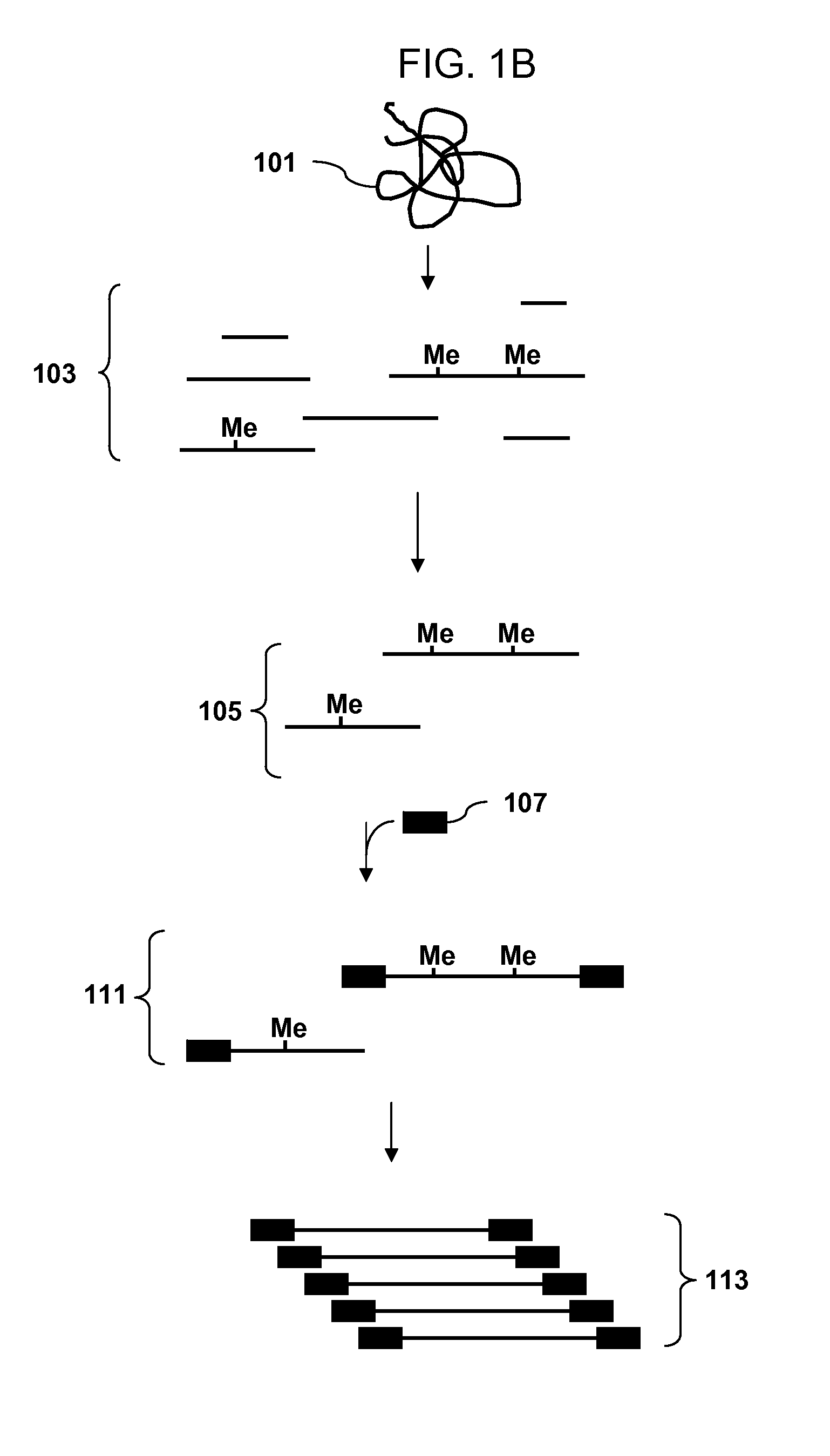
[0007]Methods for analyzing the methylation status of cytosines in genomic DNA are disclosed. In one aspect genomic DNA is fragmented with a restriction enzyme that has at least one degenerate position in the recognition site, adaptors are ligated to the fragments, methylated fragments are affinity purified and a subset of the fragments are amplified. The amplified subset is enriched relative to the genomic DNA sample for fragments that were methylated in the genomic sample. The enriched sample has a complexity that is reduced relative to the genomic sample, there are fewer different sequences present but of those fragments that are present most were methylated in the genomic sample.

Problems solved by technology

Alterations in the normal methylation process have also been shown to be associated with genomic instability (Lengauer et al., Proc. Natl. Acad. Sci.

Method used


Image

  • Analysis of methylation using selective adaptor ligation
  • Analysis of methylation using selective adaptor ligation
  • Analysis of methylation using selective adaptor ligation

Examples


example 1

[0107]A recommended protocol for IP using ab1884 (Abcam). Use 0.5 to 1 μg of fragmented genomic DNA. Dilute fragmented DNA to 100 μl for a final concentration of 0.15% SDS, 1% triton x-100, 150 mM NaCl, 1 mM EDTA pH8.0, 0.5 mM EGTA pH8.0, mM Tris pH8.0, 0.1% BSA, 7 mM NaOH, anti-5mC (up to 30 ug of antibody for saturating conditions), and Prot A / G beads. Rotate overnight at 4° C. Wash 2× with 0.1% SDS, 0.1% DOC, 1% triton, 150 mM NaCl, 1 mM EDTA pH8.0, 0.5 mM, EGTA pH8.0, 10 mM Tris pH8.0. Wash 1× with 0.1% SDS, 0.1% DOC, 1% triton, 500 mM NaCl, 1 mM EDTA pH8.0, 0.5 mM, EGTA pH8.0, 10 mM Tris pH8.0. Wash 1× with 0.25 M LiCl, 0.5% DOC, 0.5% NP-40, 1 mM EDTA pH8.0, 0.5 mM, EGTA pH8.0, 10 mM Tris pH8.0. Wash 2× with 1 mM EDTA pH8.0, 0.5 mM EGTA pH8.0, 10 mM Tris pH8.0. Elute in 1% SDS, 100 mM NaHCO3. Purify the DNA and use the isolated DNA for PCR. Analyze the amplicons by hybridization to an array.

example 2

[0108]Obtain a genomic DNA sample. Fragment the sample with Dde I in NEBuffer 3 at 37° C. Heat inactivate the enzyme at 65° C. for 20 minutes. Ligate an adaptor to the fragments. The adaptor has a primer binding site and a single stranded overhang that is 3′-ATT-5′ and will ligate efficiently to the fragment overhangs that have a 5′ TAA-3′ overhang generated by cleavage, but not to the fragment overhangs that have TTA, TCA or TGA. Immunoprecipitate fragments that contain 5-meC using an antibody to 5-meC. Clean up the immunoprecipitated fragments and amplify by PCR using a primer complementary to the primer binding site in the adaptor. Do the PCR in the presence of dUTP so that uracil is incorporated into the DNA. Treat the amplified DNA with uracil DNA glycosylase and APE 1 to fragment the PCR amplicons. End label using terminal deoxynucleotidyl transferase and Affymetrix′ biotin-labeled DNA labeling reagent (DLR). Hybridize the labeled fragments to an array, stain with biotinylated...


PUM

PropertyMeasurementUnit
temperatureaaaaaaaaaa

Abstract

Methods of analyzing DNA to identify regions of the genome that are methylated in a genomic sample are disclosed. In one aspect genomic DNA is fragmented using a restriction enzyme with a degenerate recognition site, methylated restriction fragments are separated from unmethylated fragments by affinity purification. The complexity of the methylated fragments is reduced by amplification of a subset of the fragments using adaptors that ligate to a subset of the fragments. The amplified product is fragmented, labeled and hybridized to an array of probes. The hybridization pattern is analyzed to determine methylation status of cytosines.

Description

RELATED APPLICATIONS[0001]The present application claims priority to U.S. Provisional Application No. 60 / 774,705, filed Apr. 12, 2006, the entire disclosure of which is incorporated herein by reference.FIELD OF THE INVENTION[0002]The present invention relates to arrays and methods for detecting methylation of nucleic acids.BACKGROUND OF THE INVENTION[0003]The genomes of higher eukaryotes contain the modified nucleoside 5-methyl cytosine (5-meC). This modification is usually found as part of the dinucleotide CpG. Cytosine is converted to 5-methylcytosine in a reaction that involves flipping a target cytosine out of an intact double helix and transfer of a methyl group from S-adenosylmethionine by a methyltransferase enzyme (Klimasauskas et al., Cell 76:357-369, 1994). This enzymatic conversion is the only epigenetic modification of DNA known to exist in vertebrates and is essential for normal embryonic development (Bird, Cell 70:5-8, 1992; Laird and Jaenisch, Human Mol. Genet. 3:1487...

Claims


Application Information

Patent Timeline
Patent Type & Authority Applications(United States)
IPC IPC(8): C12Q1/68C12P19/34
CPCC12Q1/6837C12Q2537/164C12Q1/6809C12Q1/683C12Q2525/191C12Q2537/159
Inventor SHAPERO, MICHAEL H.NAUTIYAL, SHIVANI
Owner AFFYMETRIX INC
Learn More