
Dual-radiation type mammography apparatus and breast imaging method using the mammography apparatus

a mammography and apparatus technology, applied in mammography, medical science, diagnostics, etc., can solve the problems of deteriorating image resolution, long time required for image taking, and increased exposure dose, so as to facilitate examination of breasts, maximize treatment efficiency, and accurately determine the location and size of lesion

Inactive Publication Date: 2008-04-17
WONKWANG UNIV CENT FOR IND ACAD COOPERATION
View PDF2 Cites 12 Cited by
  • Summary
  • Abstract
  • Description
  • Claims
  • Application Information

AI Technical Summary

Benefits of technology

[0015] An object of the present invention is to provide a dual-radiation type mammography apparatus that can easily examine the breast by taking an image by scanning the breast using a plurality of fan beams that are formed from beams radiated from an x-ray generating unit and radiated at different angles and reforming the image as a three-dimensional breast image and can accurately determine a location and size of lesion, thereby enabling the data collection for maximizing treatment efficiency, minimizing the exposure dose to a patient by reducing the image taking time, and minimizing the patient's pain.

Problems solved by technology

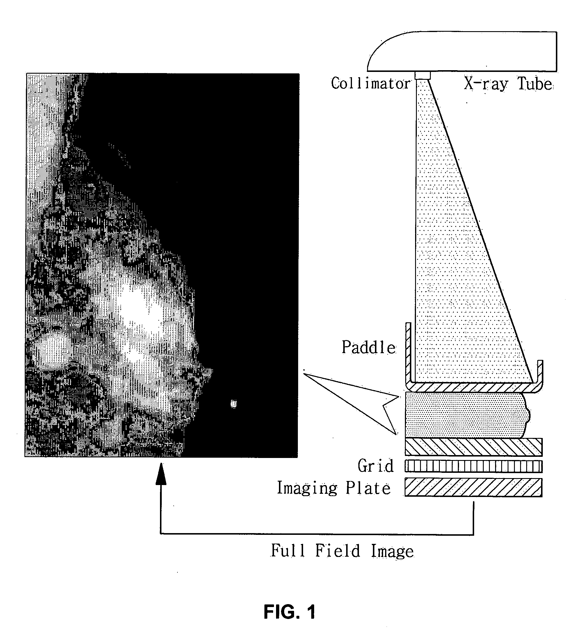
However, since the photon flux is too weak, the image must be taken for a long time.
However, since the x-rays of the lower energy band (8-15 keV) are absorbed in the breast, the exposure dose increases and the resolution of the image is deteriorated.
Therefore, it is difficult to accurately identify the locations and sizes of the lesions.
However, the combining process of the images is complicated.
However, in order to perform the above-describe method, the patient's breast are pressed for a long time.
This exacerbates the patient's pain and increases the exposure dose, thereby inducing a secondary accident.
Furthermore, since the two-dimensional image is used to identify the locations and sizes of the lesions, this method cannot assist the user to accurately identify the locations and sizes of the lesions.

Method used


Image

  • Dual-radiation type mammography apparatus and breast imaging method using the mammography apparatus
  • Dual-radiation type mammography apparatus and breast imaging method using the mammography apparatus
  • Dual-radiation type mammography apparatus and breast imaging method using the mammography apparatus

Examples


Embodiment Construction

[0026] Reference will now be made in detail to the preferred embodiments of the present invention, examples of which are illustrated in the accompanying drawings. Wherever possible, the same reference numbers will be used throughout the drawings to refer to the same or like parts.

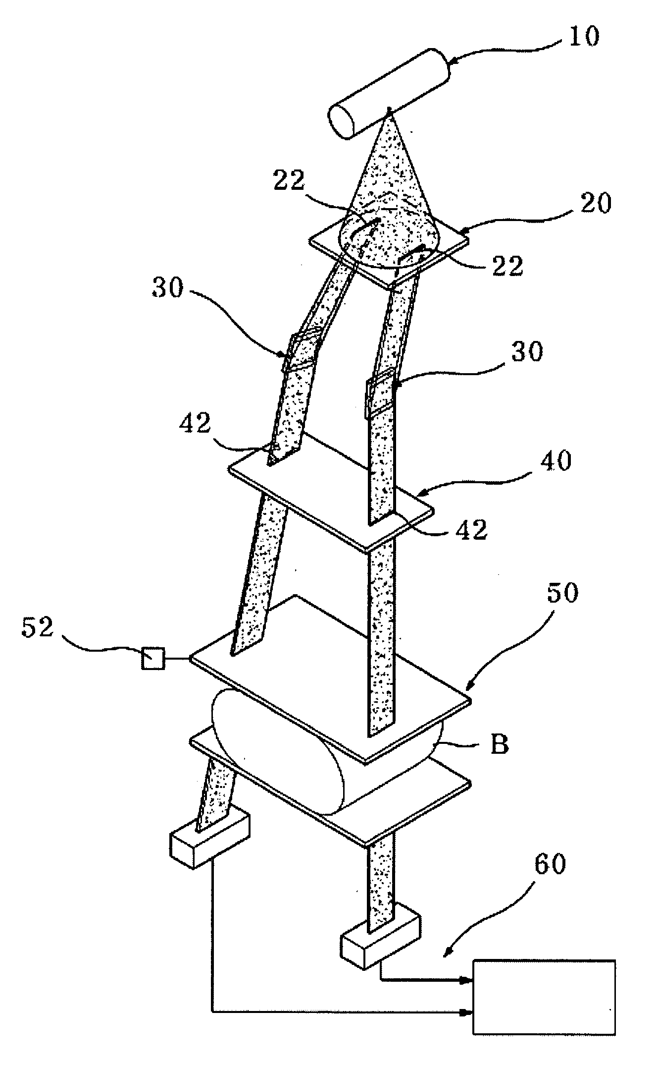
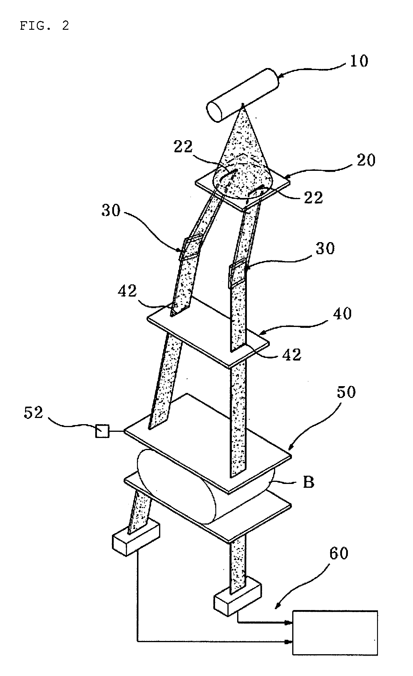
[0027]FIG. 2 is a schematic perspective view of a dual radiation type mammography apparatus according to an embodiment of the present invention, and FIG. 3 is a conception view of the dual radiation type mammography apparatus of FIG. 2.

[0028] Referring to FIGS. 2 and 3, a mammography apparatus includes a first collimator 20 radiating a plurality of beams from solid angle beams generated from an x-ray generating unit 10, a plurality of multi-layered filter units 30 radiating the plurality of beams from the first collimator 20 at different angles, a second collimator 40 for uniformly maintaining a width of the beams radiated at different angles through the multi-layered filter units 30, a breast fixing unit...


PUM


Abstract

A dual-radiation type mammography apparatus for examining a breast by taking a breast image using x-ray beams is provided. The mammography apparatus includes an x-ray generating unit generating the x-ray beams, a first collimator provided with transmission holes through which a solid angle beam generated from the x-ray beams radiated from the x-ray generating unit, in the course of which the solid angle beam is changed into a plurality of beams each having a predetermined size, a plurality of multi-layered filter units that filters off some of the beams passing through the holes of the first collimator to deflect only beams in a first specific energy band, a breast fixing unit fixing the breast B that will be scanned by the beams deflected by the multi-layered filter units, and a plurality of image output units provided for the respective beams scanning the breast fixed by the breast fixing unit and reforming two-dimensional images formed by the beams deflected at different angles into a three-dimensional image.

Description

BACKGROUND OF THE INVENTION [0001] 1. Field of the Invention [0002] The present invention relates to a dual-radiation type mammography, and more particularly, to a dual-radiation type mammography apparatus that can easily examine the breast by taking a breast image by scanning the breast using a plurality of fan beams that are formed from beams radiated from an x-ray generating unit and radiated at different angles and by reforming the image as a three-dimensional breast image and that can accurately determine a location and size of lesion. [0003] 2. Description of the Related Art [0004] Generally, a mammography apparatus (or referred to as “breast cancer examining apparatus”) uses a rotating target type x-ray generating device. Molybdenum or rhodium is usually used as a target material. A maximum tube voltage applied to an anode of the x-ray generating device is 40 kVp. Electron beams radiated from a filament of an x-ray generating unit collide with the anode, by which x-ray beams ...

Claims


Application Information

Patent Timeline
Patent Type & Authority Applications(United States)
IPC IPC(8): A61B6/04
CPCA61B6/4035A61B6/502A61B6/482A61B6/00
Inventor YOON, KWON-HA
Owner WONKWANG UNIV CENT FOR IND ACAD COOPERATION
Learn More