Method for detecting coronary endothelial dysfunction and early atherosclerosis
- Summary
- Abstract
- Description
- Claims
- Application Information
AI Technical Summary
Benefits of technology
Problems solved by technology
Method used
Image
Examples
example 1
Clinical Evaluation of Myocardial Perfusion Heterogeneity Quantified by Markovian Analysis of PET Images
[0036]In this paradigm, the resting perfusion image serves as a baseline for comparison with the stress perfusion image for identifying discrete regional perfusion abnormalities due to flow-limiting coronary artery stenosis, myocardial scar, or hibernating myocardium. The present example analyzes and quantifies the distinctly different diffuse patchy heterogeneity of resting myocardial perfusion as a marker of coronary endothelial dysfunction associated with coronary atherosclerosis, independently from and around these traditional discrete regional myocardial perfusion defects caused by flow-limiting stenosis or myocardial scar.
[0037]A mathematic technique from Markovian homogeneity analysis is used to provide precise, objective, automated quantification of resting perfusion heterogeneity in 1,034 subjects, its normal limits in 50 healthy reference subjects, and its close associat...
example 2
Protocol for Demonstrating Improvement of Myocardial Perfusion Heterogeneity with Administration of a Selective ETA-Receptor Antagonist
[0076]FIG. 6 is a schematic flow diagram illustrating the drug and placebo administration protocol for a study to demonstrate that myocardial perfusion heterogeneity, quantified by Markovian Homogeneity analysis of cardiac PET perfusion images, will improve after treatment with a selective endothelin receptor A antagonist (ETA-receptor antagonist), compared to treatment with placebo.
[0077]The visually apparent heterogeneity of resting myocardial perfusion and / or its improvement after dipyridamole or adenosine stress on high quality, non-invasive PET images is proposed as one manifestation of coronary arteriolar endothelial dysfunction. Positron emission tomography (PET) is necessary for imaging this pattern of heterogeneous perfusion without the attenuation artifacts and poor depth dependent resolution of standard single photon emission tomography (S...
example 3
Demonstration of Endothelin-Induced Myocardial Perfusion Defects
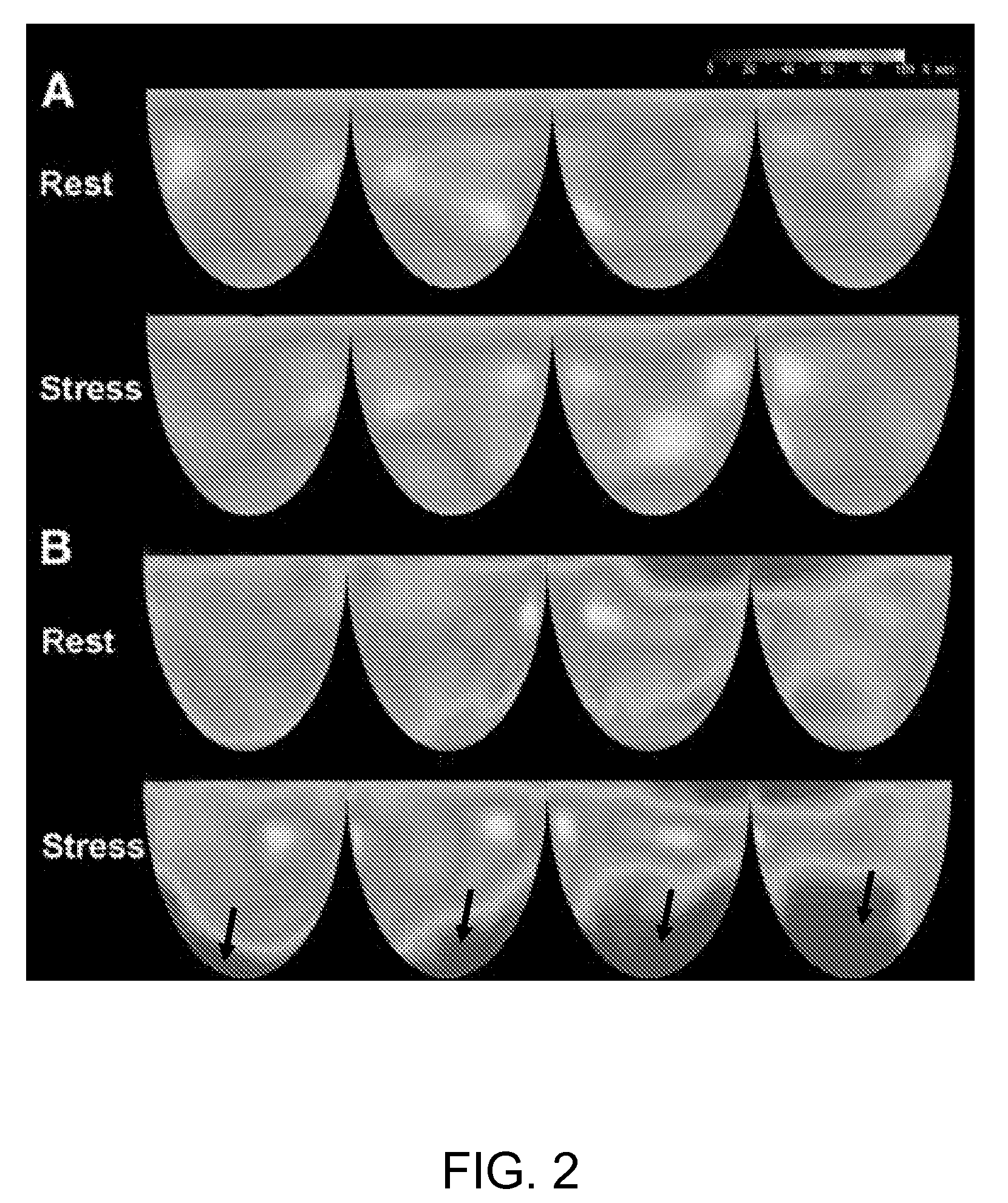
[0097]In diagnostic myocardial perfusion imaging, the resting perfusion image serves as a baseline for comparison to the exercise or pharmacological stress image where a new or worsening stress-induced perfusion abnormality indicates flow-limiting stenosis. This paradigm of rest-stress perfusion imaging is based on the concept of coronary flow reserve and perfusion imaging during pharmacological stress for assessing coronary artery stenosis. In the absence of attenuation artifacts as with positron emission tomography (PET), a persisting fixed perfusion defect is clinically interpreted as myocardial scar or hibernating myocardium due to flow-limiting stenosis.
[0098]However, resting myocardial perfusion defects that improve or disappear during dipyridamole stress in the absence of myocardial scar or flow-limiting stenosis have also been described. In Example 1, it was demonstrated that myocardial perfusion heterogeneity a...
PUM
 Abstract
Description
Claims
Application Information
 - R&D
- Intellectual Property
- Life Sciences
- Materials
- Tech Scout
- Unparalleled Data Quality
- Higher Quality Content
- 60% Fewer Hallucinations
Browse by: Latest US Patents, China's latest patents, Technical Efficacy Thesaurus, Application Domain, Technology Topic, Popular Technical Reports.
© 2025 PatSnap. All rights reserved.Legal|Privacy policy|Modern Slavery Act Transparency Statement|Sitemap|About US| Contact US: help@patsnap.com


