
Animal Model of Prostate Cancer and Use Thereof

a prostate cancer and animal model technology, applied in the field of mammals, can solve the problems of difficult to prove the malignant potential of patients, inability to determine the prognosis, and behind clinicians and basic researchers, and achieve the effect of less evident lobular formation

Inactive Publication Date: 2008-02-28
CHUNG SHAN MEDICAL UNIVERSITY
View PDF0 Cites 103 Cited by
  • Summary
  • Abstract
  • Description
  • Claims
  • Application Information

AI Technical Summary

Benefits of technology

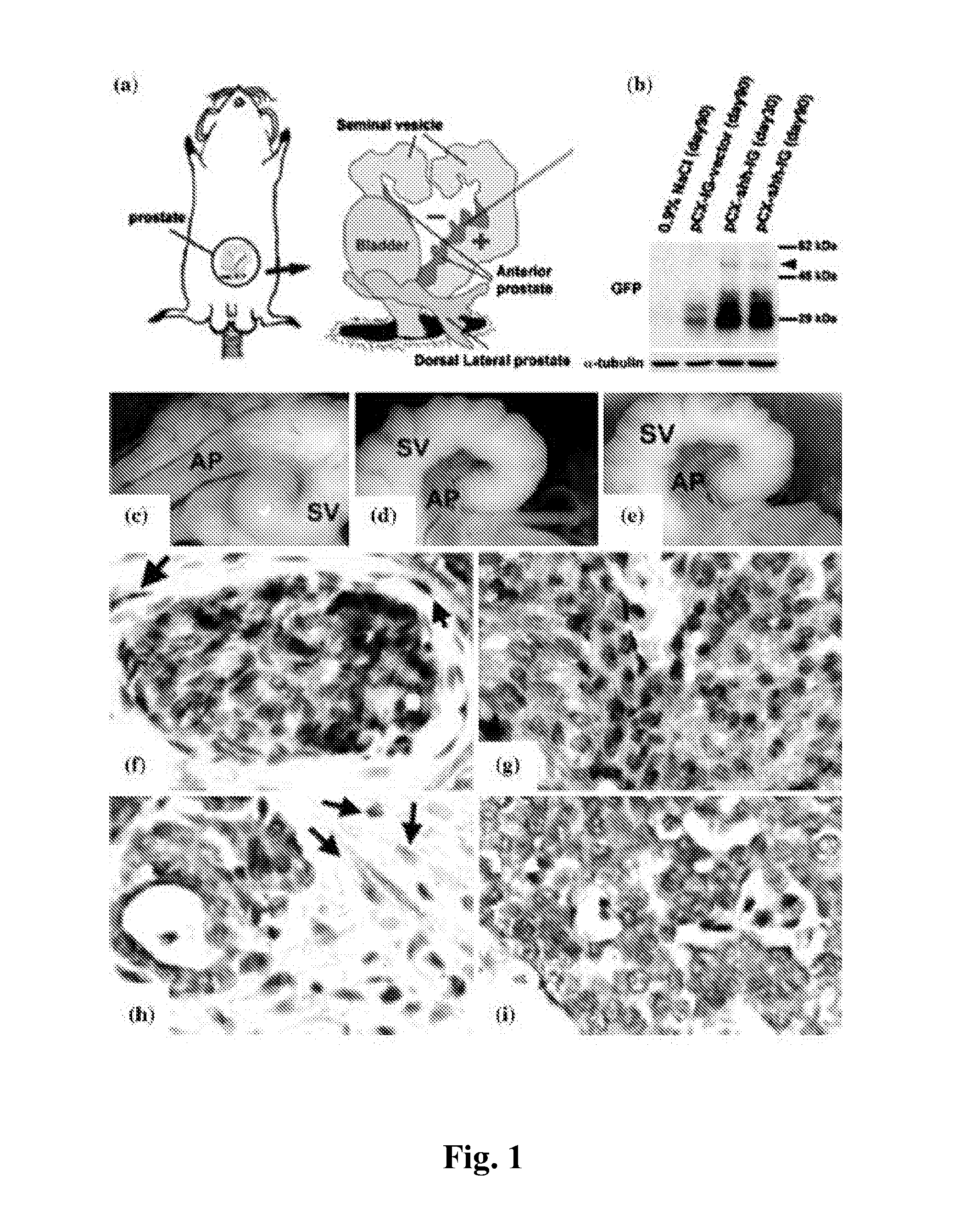
[0046]Gross morphological effects were examined at days 7, 20, 30, and 90 after the manipulation and the most prominent changes were found at day 30 so far. By day 30 after the injection, the anterior prostates (AP) of the pCX-shh-IG injections exhibited overgrowth (FIG. 1c), in contrast to those seen in the pCX-IG vehicle controls (FIG. 1d) or in the 0.9% NaCl blank controls (FIG. 1e). We assumed that the overgrowth in the pCX-shh-IG-injected prostates was due to the effects of Hedgehog overexpression, since no comparable result was found in the two control groups. In addition, seminal vesicles (SV) in the pCX-shh-IG-injected prostates showed less evident lobular formation as compared to those in the pCX-IG and the 0.9% NaCl injections (FIG. 1c vs. FIGS. 1d and e). The size of seminal vesicles appeared to enlarge with pCX-shh-IG injections. Furthermore, h...

Problems solved by technology

Despite the increasing incidence, prostate cancer presents some obstacles that hind clinicians and basic researchers from understanding its pathogenesis. prostate cancer is characterized by slow clinical progression, involvement of multiple genetic and epigenetic events, multifocal and heterogeneous nature of tumorigenesis, and inability to determine prognosis for disease progression (A. M. De Marzo et al., Urology 62, 55 (2003); C. Abate-Shen, M. M. Shen, Trends Genet 18, S1 (2002)).
The non-SV40-Tag models displayed high proportions of atypical epithelial lesions representing different degrees of PIN without frequent progression into invasive carcinoma, which makes it difficult to prove their malignant potential (J. H. Park et al., Am J Pathol 161, 727 (2002)).

Method used


Image

  • Animal Model of Prostate Cancer and Use Thereof
  • Animal Model of Prostate Cancer and Use Thereof
  • Animal Model of Prostate Cancer and Use Thereof

Examples


example

Example 1

Construction of a Mouse Expressing Hedgehog Protein

[0043]The Shh expression and vehicle vector, pCX-shh-IG and pCX-IG, were provided by Dr. Kerby C. Oberg, Loma Linda University. Male outbred FVB strain mice aged 8 to 10 weeks purchased from National Laboratory Animal Center, Academia Sinica, Taipei, were used for the injections. The mice were anesthetized with phenobarbital and exposed of their prostate glands by surgery (FIG. 1). For each injection, 10 μl of pCX-Shh-IG at 0.1 μg / μl in 0.9% NaCl was injected into the anterior lobe alone or into both the anterior and the dorsolateral lobes. Parallel injections with 10 μl of pCX-IG in 0.9% NaCl or 0.9% NaCl alone were performed as controls. After injections, the prostate lobes were subjected to electric stimulations (electroporation) for 10 seconds set at 20 volts, 1 to 2 pulses per second, using a Digitimer DS7 Stimulator (Digitimer, Hertfordshire, England). After electroporation, the mice were caged and maintained until us...

example 2

Immunohistochemical Detection

[0044]To confirm the efficiency of the prostate cancer formation in the mouse model, immunohistochemical detection was conducted as stated below. Tissue were dissected, fixed in 4% paraformaldehyde in PBS (Sigma), and processed to obtain 7 μm thick sections following standard histological preparations. For Gli-1, Gli-2, Gli-3, Hip detections, the sections were processed through citrate buffer (pH 2.0) for 4 min, followed by another citrate buffer (pH9.0) for 5 min and thorough washes before antibody binding. For CK14, p63, GFP, E-cadherin, N-19, 5E1, Ptc-1, Fgfr-2, and Fgf-2, the sections were treated with citrate buffer (pH 6) for 10 min before antibody binding. For Fgfr-1, Fgf-7, and Fgf-10, proteinase K (10 μg / ml) treatment was performed on ice for 5 min before processing through citrate buffer (pH 8.0) and antibody binding. The slides were then covered with antibody solutions at 4° C. overnight, then further processed with biotinylated secondary anti...

example 3

Confirmation of Hedgehog Overexpression in the pCX-shh-IG Injections

[0045]To solidify any data obtained as a result of Hedgehog overexpression, we examined Hedgehog expression status after the manipulation. We first examined the presence of GFP signals in wholemount preparations and in tissue sections. When both methods showed no convincing signal, Western analysis was used as a double check. In wholemounts (data not shown), GFP signals were detected in 15 prostates in a total of 25 pCX-shh-IG injections and in 8 of the 10 pCX-IG injections, but in none of the 0.9% NaCl blank controls. With immunohistochemistry, 23 of the 25 pCX-shh-IG injections and 8 of the 10 pCX-IG injections exhibited definite signals for GFP, with no positive signal detected in the 0.9% NaCl injections (Table 1). The two pCX-shh-IG injections without definite GFP signal in tissue sections were double checked using Western analyses (FIG. 1b) and RT-PCR (data not shown); both were found positive for GFP. We then...


PUM

PropertyMeasurementUnit
Levelaaaaaaaaaa

Abstract

The present invention relates to an adult mammal which exhibits growth or replication of abnormal cells in a target tissue or organ by over-expressing Hedgehog protein in such target tissue or organ. The present invention also relates to a method of preparing an adult animal model of prostate cancer. The invention further relates to a method of evaluating an agent for treating prostate cancer.

Description

FIELD OF THE INVENTION[0001]The present invention relates to a mammal susceptible to prostate cancer, a method of preparing a animal model of prostate cancer, and a method of evaluating an agent for treating prostate cancer.BACKGROUND OF THE INVENTION[0002]Prostate cancer, apart from skin cancer, is the most common male malignancy and the second leading cause of cancer deaths in men in the United States (R. T. Greenlee, T. Murray, S. Bolden, P. A. Wingo, CA Cancer J Clin 50, 7 (2000)). It had surpassed lung cancer in 1990 and was estimated to cause 31,000 deaths in 2002 in the United States alone (W. Isaacs, A. De Marzo, W. G. Nelson, Cancer Cell 2, 113 (2002)). Despite the increasing incidence, prostate cancer presents some obstacles that hind clinicians and basic researchers from understanding its pathogenesis. prostate cancer is characterized by slow clinical progression, involvement of multiple genetic and epigenetic events, multifocal and heterogeneous nature of tumorigenesis, ...

Claims


Application Information

Patent Timeline
IPC IPC(8): A01K67/027
CPCA01K2207/15A01K2217/00C12N15/8509A01K2267/0331C07K14/46A01K2227/105
Inventor LIN, PEI-CHENGCHANG, HAN-HSINCHEN, BO-YIE
Owner CHUNG SHAN MEDICAL UNIVERSITY
Learn More