
Tumor-targeted optical contrast agents

a technology of optical contrast and tumors, applied in the field of tumor visualization and detection, can solve the problems of hepatobiliary toxicity, limited scope in the ability to induce large changes in the absorption and emission properties of dyes, and various attempts to obviate this problem have not been very successful, so as to enhance tumor detection and preserve the fluorescence efficiency of dye molecules.

Inactive Publication Date: 2008-01-10
MALLINCKRODT INC
View PDF10 Cites 5 Cited by
  • Summary
  • Abstract
  • Description
  • Claims
  • Application Information

AI Technical Summary

Benefits of technology

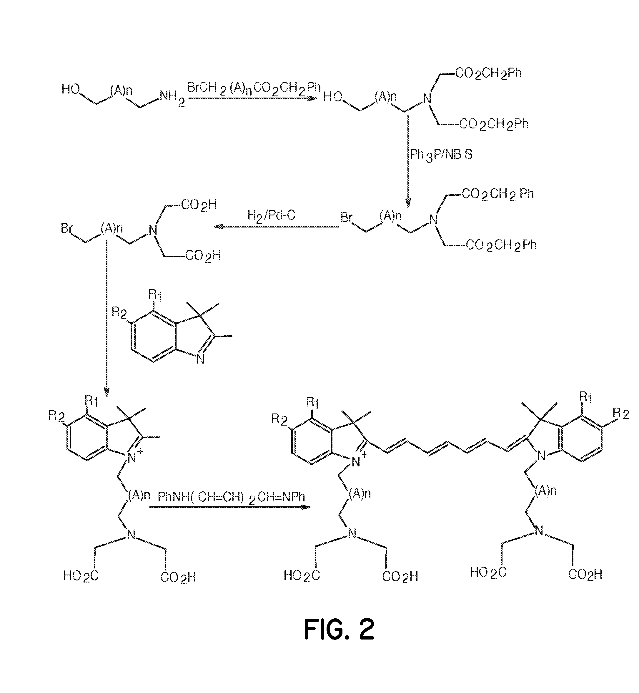
[0009] The invention is directed to compositions, and methods of preparing the compositions, of low molecular weight biomolecule-dye conjugates to enhance tumor detection. The inventive compositions preserve the fluorescence efficiency of the dye molecules, do not aggregate in solution, form starburst dendrimers, are capable of absorbing or omitting light in the near infrared region (beyond 800 mm), and can be rendered tissue-specific.

Problems solved by technology

A major drawback in the use of cyanine dye derivatives is the potential for hepatobiliary toxicity resulting from the rapid clearance of these dyes by the liver (G. R. Cherrick, et al., Indocyanine green: Observations on its physical properties, plasma decay, and hepatic extraction.
Various attempts to obviate this problem have not been very successful.
Another major difficulty with current cyanine and indocyanine dye systems is that they offer a limited scope in the ability to induce large changes in the absorption and emission properties of these dyes.
Further, most cyanine dyes do not have the capacity to form starburst dendrimers, which are useful in biomedical applications.
For the purpose of tumor detection, many conventional dyes are useful for in vitro applications because of their highly toxic effect on both normal and abnormal tissues.
Other dyes lack specificity for particular organs or tissues and, hence, these dyes must be attached to bioactive carriers such as proteins, peptides, carbohydrates, and the like to deliver the dyes to specific regions in the body.
However, this approach does have some serious limitations in that the diffusion of high molecular weight bioconjugates to tumor cells is highly unfavorable, and is further complicated by the net positive pressure in solid tumors (R. K. Jain, Barriers to Drug Delivery in Solid Tumors, Scientific American 1994, 271:58-65.
Furthermore, many dyes in general, and cyanine dyes in particular, tend to form aggregates in aqueous media that lead to fluorescence quenching.

Method used


Image

  • Tumor-targeted optical contrast agents
  • Tumor-targeted optical contrast agents
  • Tumor-targeted optical contrast agents

Examples


example 1

Synthesis of Bis(ethylcarboxymethyl)indocyanine Dye

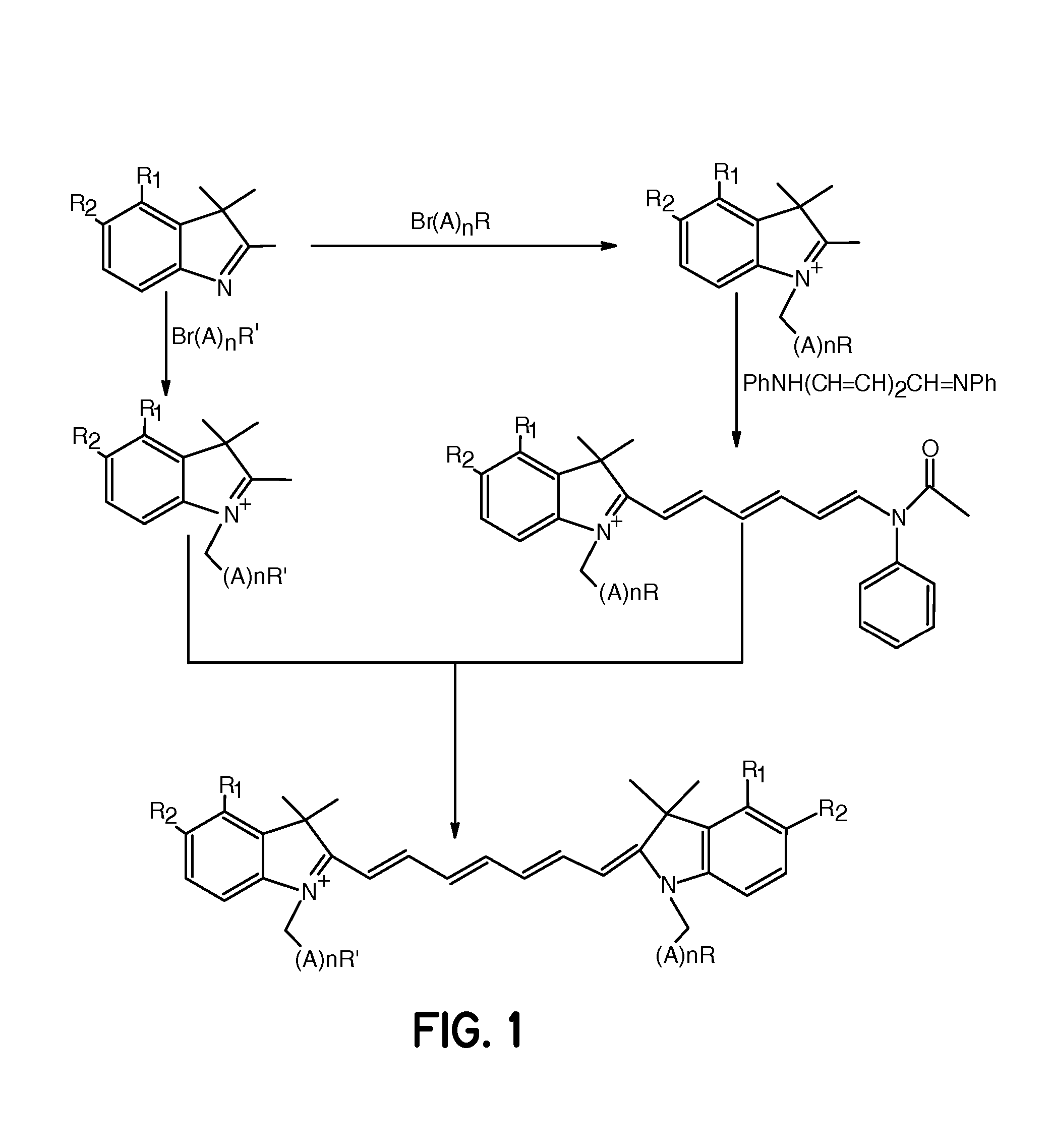
FIG. 1, R1, R2=Fused Phenyl; A=CH2, n=1 and R═R′═CO2H

[0062] A mixture of 1,1,2-trimethyl-[1H]-benz[e]indole (9.1 g, 43.58 mmoles) and 3-bromopropanoic acid (10.0 g, 65.37 mmoles) in 1,2-dichlorobenzene (40 mL) was heated at 110° C. for 12 hours. The solution was cooled to room temperature and the red residue obtained was filtered and washed with acetonitrile:diethyl ether (1:1) mixture. The solid obtained was dried under vacuum to give 10 g (64%) of light brown powder. A portion of this solid (6.0 g; 16.56 mmoles), glutaconaldehyde dianil monohydrochloride (2.36 g, 8.28 mmoles) and sodium acetate trihydrate (2.93 g, 21.53 mmoles) in ethanol (150 mL) were refluxed for 90 minutes. After evaporating the solvent, 40 mL of 2 N aqueous HCl was added to the residue. The mixture was centrifuged and the supernatant was decanted. This procedure was repeated until the supernatant became nearly colorless. About 5 mL of water:acetonitrile (3:2...

example 2

Synthesis of Bis(pentylcarboxymethyl)indocyanine Dye

FIG. 1, R1, R2=Fused Phenyl; A=CH2, n=4 and R═R′═CO2H

[0063] A mixture of 1,1,2-trimethyl-[1H]-benz[e]indole (20 g, 95.6 mmoles) and 6-bromohexanoic acid (28.1 g, 144.1 mmoles) in 1,2-dichlorobenzene (250 mL) was heated at 110° C. for 12 hours. The green solution was cooled to room temperature and the brown solid precipitate formed was collected by filtration. After washing the solid with 1,2-dichlorobenzene and diethyl ether, the brown powder obtained (24 g, 64%) was dried under vacuum at room temperature. A portion of this solid (4.0 g; 9.8 mmoles), glutaconaldehyde dianil monohydrochloride (1.4 g, 5 mmoles) and sodium acetate trihydrate (1.8 g, 12.9 mmoles) in ethanol (80 mL) were refluxed for 1 hour. After evaporating the solvent, 20 mL of 2 N aqueous HCl was added to the residue. The mixture was centrifuged and the supernatant was decanted. This procedure was repeated until the supernatant became nearly colorless. About 5 mL ...

example 3

Synthesis of Bisethylcarboxymethylindocyanine Dye

FIG. 1, R1═R2═H; A=CH2, n=1 and R═R′═CO2H

[0064] This compound was prepared as described in Example 1 except that 1,1,2-trimethylindole was used as the starting material.


PUM

PropertyMeasurementUnit
of wavelengthaaaaaaaaaa
temperatureaaaaaaaaaa

Abstract

Cyanine dye bioconjugates useful for diagnostic imaging and therapy are disclosed. The conjugates include several cyanine dyes with a variety of bis- and tetrakis (carboxylic acid) homologs. The compounds may be conjugated to bioactive peptides, carbohydrates, hormones, drugs, or other bioactive agents. The small size of the compounds allows more favorable delivery to tumor cells as compared to larger molecular weight imaging agents. The various dyes are useful over the range of 350 to 1300 nm, the exact range being dependent upon the particular dye. The use of dimethylsulfoxide helps to maintain the fluorescence of the compounds. The inventive compounds are useful for diagnostic imaging and therapy, in endoscopic applications for the detection of tumors and other abnormalities, for localized therapy, for photoacoustic tumor imaging, detection and therapy, and for sonofluoresence tumor imaging, detection and therapy.

Description

RELATED APPLICATIONS [0001] This application is a Division of co-pending U.S. patent application Ser. No.10 / 654,033 filed Sep. 3, 2003; which is a Continuation-In-Part of U.S. Pat. No. 6,641,798 filed May 23, 2001; which is a Continuation-In-Part of U.S. Pat. No. 6,180,087 filed Jan. 18, 2000, each of which is expressly incorporated by reference herein in its entirety.FIELD OF INVENTION [0002] This invention relates generally to compositions of cyanine dye bioconjugates with bioactive molecules for diagnosis and therapy, and particularly for visualization and detection of tumors. BACKGROUND OF THE INVENTION [0003] Several dyes that absorb and emit light in the visible and near-infrared region of electromagnetic spectrum are currently being used for various biomedical applications due to their biocompatibility, high molar absorptivity, and / or high fluorescence quantum yields. The high sensitivity of the optical modality in conjunction with dyes as contrast agents parallels that of nu...

Claims


Application Information

Patent Timeline
Patent Type & Authority Applications(United States)
IPC IPC(8): A61K49/00C07K7/08C07D419/02C07D417/02C07D413/02A61K41/00
CPCA61K41/0033A61K49/0032A61K49/0056C07K14/655C07D405/06C07K1/13C07D209/14
Inventor ACHILEFU, SAMUEL I.RAJAGOPALAN, RAGHAVANDORSHOW, RICHARD B.BUGAJ, JOSEPH E.PERIASAMY, MUTHUNADAR P.
Owner MALLINCKRODT INC
Learn More