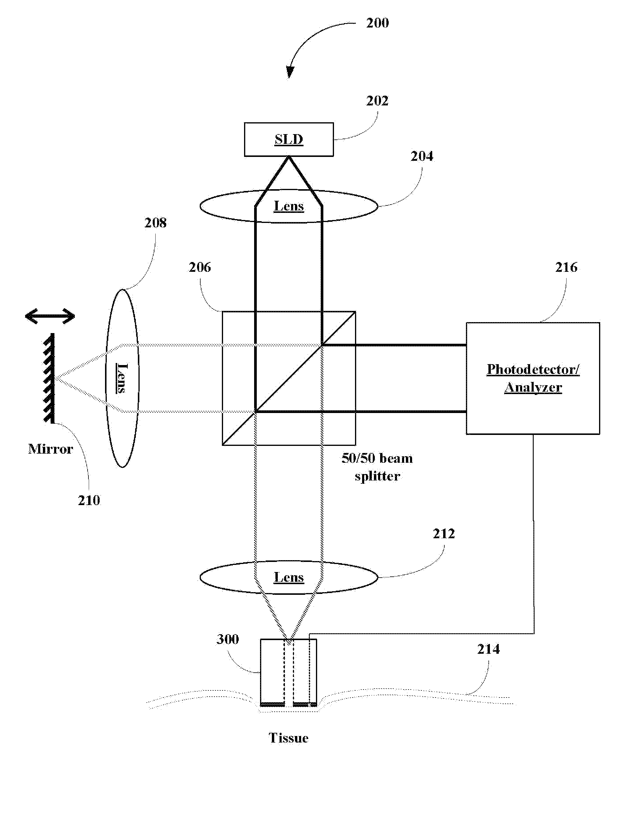
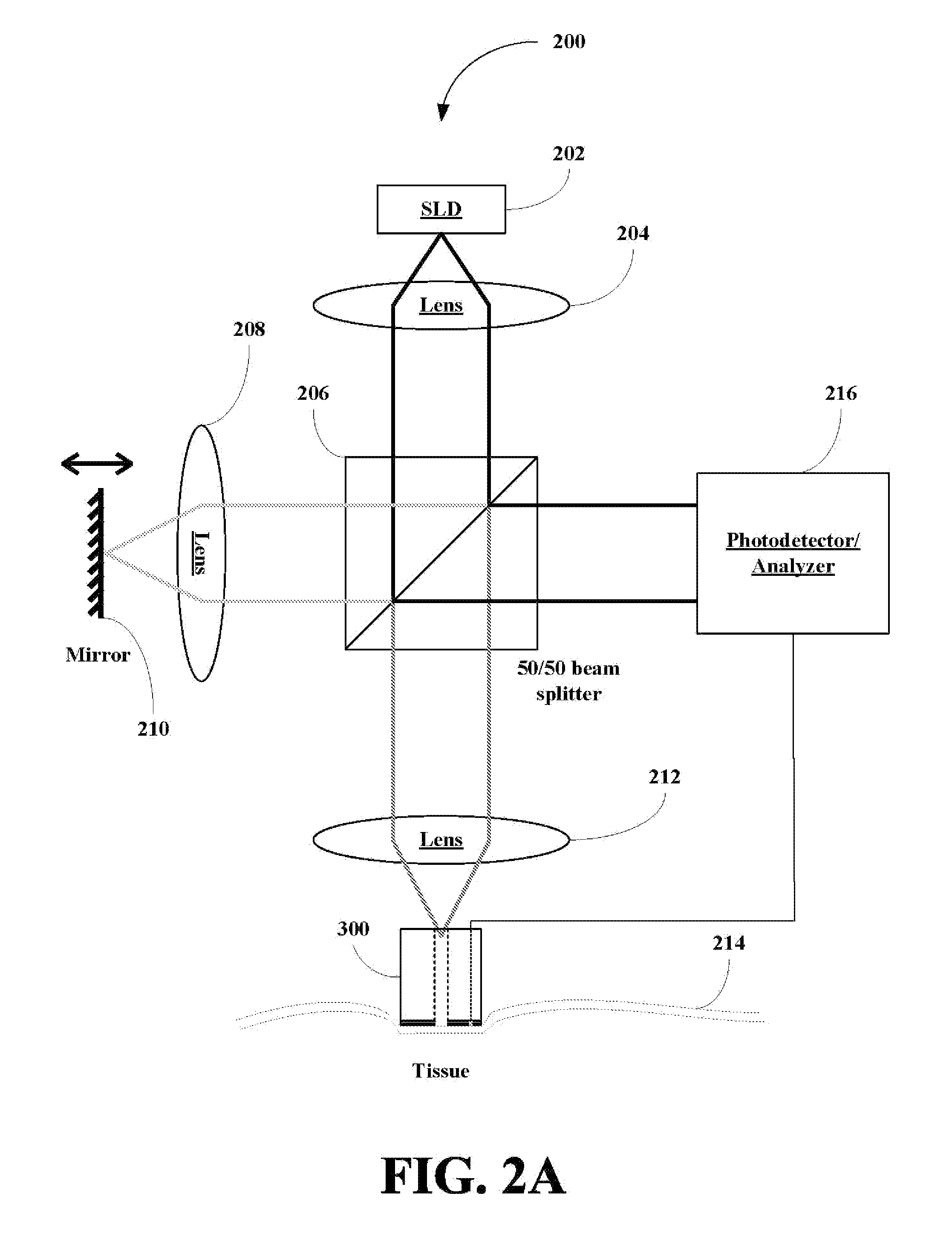
[0012] The present invention also provides a method for continuous noninvasive glucose monitoring in an animal including an human using an OCT based glucose
monitoring system. The method includes the step of generating
radiation. A first portion of radiation is directed onto a
single site of a tissue site or an area of a tissue site to generate backscattered and / or reflected radiation, where the tissue site is maintained at a desired temperature with a temperature variation of less than or equal to 1° C. during the OCT scan. A second portion of the radiation is directed to a reflector to generate reference radiation. The backscattered and / or reflected radiation and the reference radiation are then combined and forwarded to a detected and detected to produce
optical coherence tomography signals. A glucose concentration is then calculated on a continuous basis or periodic basis using a single OCT slope or a composite OCT slope of the
optical coherence tomography signals over the surface, where the number of signals is sufficient to improve the
signal-to-
noise ratio of a composite OCT
signal improving the OCT derived glucose concentration. The method can also include the step of using glucose concentration values obtained from invasive samplings of blood (routinely used in
critically ill patients) to calibrate the OCT-based sensor and improve OCT glucose concentration accuracy. The method is especially well suited for patients undergoing
cardiac surgery, where careful control of glucose level leads to a substantial reduction in mortality and morbidity of in such patients. In certain embodiments, the tissue site is warmed to a desired elevated temperature and held constant at the temperature with a temperature variation of less than or equal to 1° C.
[0013] The present invention also provides a method for continuous noninvasive glucose monitoring in
critically ill patients. The method includes the step of generating radiation. A first portion of radiation is directed to a single location of a mucosa or a plurality of locations of a mucosa such as an
oral mucosa of the patient to generate backscattered and / or reflected radiation, where the tissue site is maintained at a desired temperature with a temperature variation of less than or equal to 1° C. during the OCT scan. A second portion of the radiation is directed to a reflector to generate reference radiation. The backscattered and / or reflected radiation and the reference radiation are then detected to produce optical coherence
tomography signals. A glucose concentration is then calculated on a continuous basis or periodic basis using a single OCT slope or a composite slope of the optical coherence
tomography signals, where the number of signals is sufficient to improve the
signal-to-
noise ratio of a composite OCT signal improving the OCT derived glucose concentration. The method can also include the step of using glucose concentration values obtained from invasive samplings of blood (routinely used in
critically ill patients) to calibrate the OCT-based sensor and improve OCT glucose concentration accuracy. The method is especially well suited for patients undergoing
cardiac surgery, where careful control of glucose level leads to a substantial reduction in mortality and morbidity of in such patients. The inventors believe that probing of mucosa may provide more accurate glucose monitoring due to better blood
perfusion and glucose transport compared in the mucosa as compared to
skin tissue. In certain embodiments, the tissue site is warmed to a desired elevated temperature and held constant at the temperature with a temperature variation of less than or equal to 1° C.
[0015] The present invention provides a computer readable media containing program instructions for measuring glucose concentration of a plurality of 1-D scan of a tissue area. The computer readable media including instructions for storing a plurality of 1-D optical coherence
tomography (OCT) signals in memory. The computer readable media also includes instructions for combining the signals into a
composite signal with an improved signal-to-
noise ratio. The computer readable media also includes instructions for determining the glucose concentration using the
composite signal. The instructions for determining the glucose concentration include determining a slope of the composite OCT signal and determining an OCT glucose concentration using the slope. The computer readable media can also include instructions to identify structures within the tissue area at a given depth in the tissue which improve the OCT glucose concentration value relative to the actual blood glucose concentration. The computer readable media also includes instructions for maintaining a temperature of the tissue site at a desired temperature with no more than a 1° C. temperature variation during the scanning. The computer readable media can also include instructions for
data filtering and / or
smoothing of the OCT data to improve an accuracy of OCT glucose concentration measurements and to improve a correlation between [GluOCT] and [Glub].
[0016] The present invention provides a computer readable media containing program instructions for continuously measuring glucose concentration of a plurality of 1-D scan of a tissue area. The computer readable media includes instructions for storing a plurality of 1-D optical coherence tomography (OCT) signals in memory, instruction of forming a composite OCT signal from the plurality of 1-D scans and instructions for determining the glucose concentration within the tissue using the
composite signal. The instructions for determining the glucose concentration include instructions for correlating a change in the slope with an optical or morphological change in the tissue. The computer readable media can also include instructions to identify structures within the tissue area at a given depth in the tissue which improve the OCT glucose concentration value relative to the actual blood glucose concentration in the tissue. The computer readable media also includes instructions for maintaining a temperature of the tissue site at a desired temperature with no more than a 1° C. temperature variation during the scanning. The computer readable media can also include instructions for warming a tissue site and maintaining a temperature of the tissue site at a desired temperature with no more than a 1° C. temperature variation during the scanning. The computer readable media can also include instructions for
data filtering and / or
smoothing of the OCT data to improve an accuracy of OCT glucose concentration measurements and to improve a correlation between [GluOCT] and [Glub].
[0023] The present invention also provides multi-
wavelength OCT, where one or more wavelengths (single
wavelength or narrowly banded
wavelength-narrow wavelength bandwidth) are used in OCT scanning. The scanning method can include performing a first 1-D scan at a location at a first frequency and then a second 1-D scan at the same location at a second frequency. The method can include making additional 1-D scans at other frequencies as well, but generally the inventors believe that two wavelength are sufficient if judiciously selected. Alternatively, the method can include scanning a portion or all of a tissue area at a first wavelength and then scanning the same or different portion or all of the tissue area with a second wavelength. The wavelength are selected from the
electromagnetic spectrum between about 700 and about 2000 nm. In certain embodiments, the first wavelength is a longer wavelength generally between about 1300 nm and about 2000 nm and the second wavelength is a shorter wavelength generally between about 700 nm and 1300 nm. The longer wavelength data correlates with water contributions to the OCT signal and the longer wavelength data is thus used to correct the OCT data at shorter wavelength, which generally correlates between glucose contributions to the OCT signal. The longer wavelength OCT signals are more water specific allowing efficient removal of water contributions, while shorter wavelength improve contrast. The combination of the two signal types can be used to enhance glucose specificity by better accounting for artifacts do to water. Alternatively, the OCT scan can be collected at one or more glucose specific wavelengths, but currently no
light source are commercially available that generate light at those wavelengths. The two wavelength specific signals can be combined using an acceptable mathematical technique such as ratiometric analysis. In certain embodiments, the tissue site is warmed to a desired elevated temperature and held constant at the temperature with a temperature variation of less than or equal to 1° C.


