
Suppression of xenotransplant rejection

a technology for transplanting organs and suppressing grafts, applied in the direction of fusion polypeptides, dna/rna fragmentation, unknown materials, etc., can solve the problems of xenogeneic organ rapid rejection, poor knockout effect, and inability to prevent har alone, so as to improve the effect of knockou

Inactive Publication Date: 2007-07-05
IMPERIAL INNOVATIONS LTD
View PDF0 Cites 16 Cited by
  • Summary
  • Abstract
  • Description
  • Claims
  • Application Information

AI Technical Summary

Benefits of technology

This approach effectively prolongs graft survival by reducing leukocyte adhesion and infiltration, potentially leading to indefinite graft survival and donor-specific tolerance without compromising the recipient's immune system.

Problems solved by technology

However, the limited supply of donor organs means that many patients have little or no chance of receiving a transplanted organ and thus die before a suitable organ is found.
One problem associated with xenografting is that xenogeneic organs are rejected rapidly upon re-vascularisation by a humoral process called hyperacute rejection (HAR).
This recognition triggers the complement cascade which in turn leads to rejection.
However, it is becoming clear that preventing HAR alone is unlikely to be sufficient to prevent rejection of xenogeneic organs.
Beyond DXR there is the problem of T-lymphocyte mediated rejection.
It has been demonstrated (Dorling et al., 1994) that the T-cell response to porcine xenografts is at least equivalent to the response against allografts, but is likely to be more aggressive and may be difficult to control with standard doses of systemic immunosuppressive drugs.
However due to the high on / off rate of these selectin-carbohydrate interactions, this class of receptors cannot support firm adhesion of leukocytes.
This is likely to promote trafficking of human lymphocytes and monocytes into a porcine xenograft and trigger rejection.
It might be possible to target porcine VCAM-1 specifically with monoclonal antibodies, but it is thought that repeated administration of these antibodies would result in sensitization of the recipient and a decline in efficiency of blockade.
Systemic administration of agents such as the VCAM-Ig fusion protein, cyclic peptide antagonists naturally-occurring fungal cyclopeptolides mentioned above would result in blockade not only of the pVCAM-VLA-4 interaction within the graft but also in other tissues of the recipient and may impair the ability of the recipient to fight infections.

Method used


Image

  • Suppression of xenotransplant rejection
  • Suppression of xenotransplant rejection
  • Suppression of xenotransplant rejection

Examples


example 1

Identification of VCAM-Specific sFv from a Phage-Display Library

[0075] A phage-display antibody library was used to generate an sFv specific for VCAM-1. The library used contains >108 clones generated using a bank of 50 cloned human VH gene segments with a random nucleotide sequence encoding CDR3 lengths of 4-12 residues (Richardson et al., 1993). This library has already been used to isolate specific single-chain antibodies to a variety of antigens including haptens, foreign and self antigens. However selection has depended upon the availability of purified or recombinant antigen. We developed a novel screening strategy to overcome the lack of recombinant porcine VCAM.

[0076] cDNA for porcine VCAM was used to generate stable cell lines that exhibit high levels of surface porcine VCAM expression as assessed by flow cytometry. VCAM positive cells were incubated with 3 μM CellTracker™ Green CMFDA (5-chloromethylfluorescein diacetate, (Molecular Probes, Oregon) for 1 hour at 37° C. On...

example 2

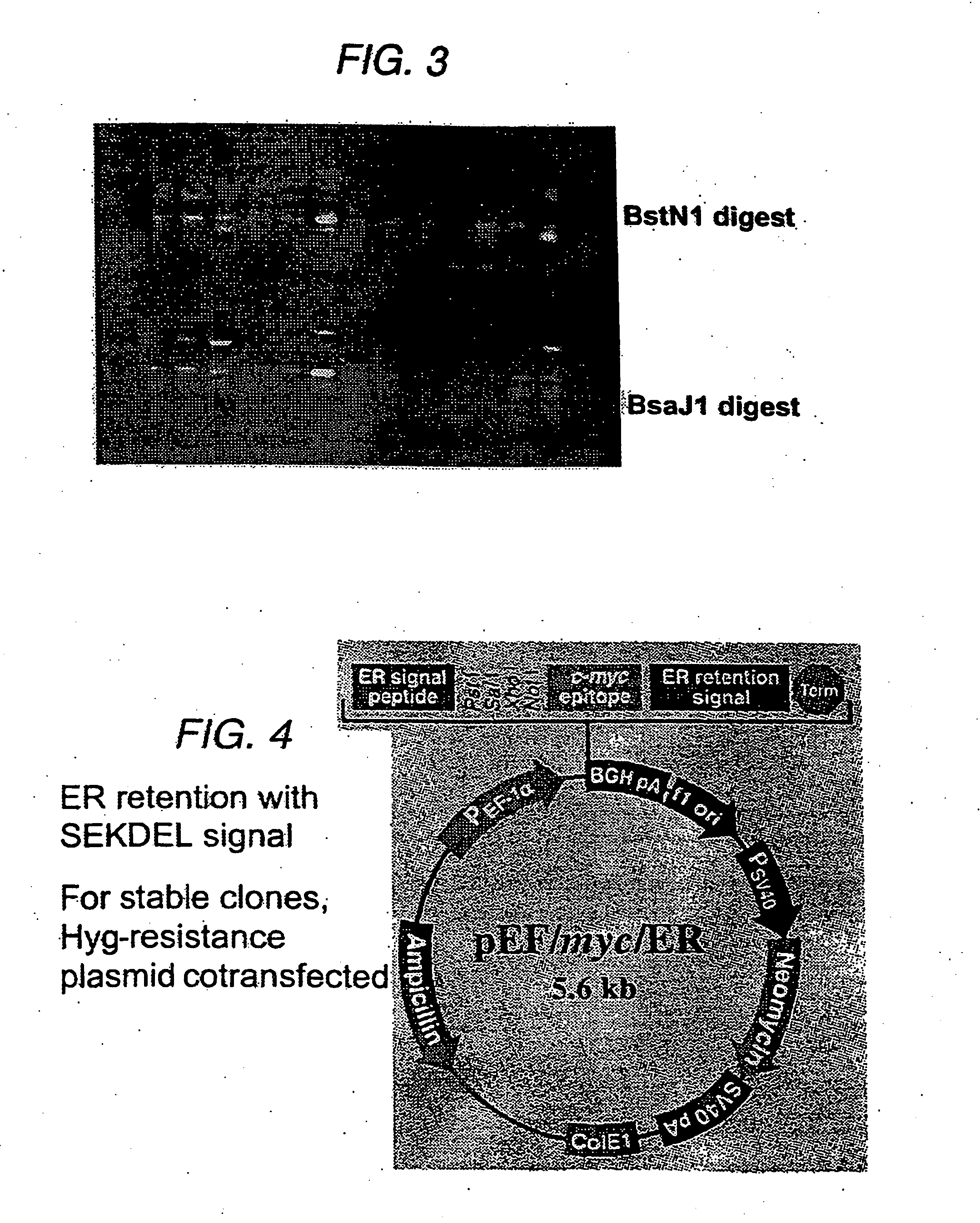
Subcloning of sFv for Targeted Intracellular Expression

[0079] Our strategy was to engineer the VCAM-specific sFv to be retained within the ER, so that providing that the sFv-VCAM interaction was of sufficient affinity, both molecules would be retained within the ER and degraded, thereby reducing cell-surface VCAM levels. Initially, the sFv has been expressed using a constitutively active promoter, the promoter from the human elongation factor 1α-subunit (EF-1α).

[0080] The sFv was amplified from the phagemid vector by PCR (30 cycles, annealing temperature 55° C., 1.5 μM Mg2+) using the primers:

(5′)CAGTCTATGCGGCCCCATTCA(3′);and(5′)TCCACAGGCGCGCACTCCCAGCCGGGCATGGCCCAGGT(3′).

[0081] The resulting fragment was subcloned into BssHII / Notl sites in pEF / myc / ER (Invitrogen, BV). The sFv was directed to the ER by incorporation of the sequence of a signal peptide from a mouse VH chain at the 5′-end of the sFv gene; this peptide is cleaved upon translocation into the ER. The sFv is retained b...

example 3

Effect of sFv Constructs on VCAM Expression

Co-transfection of sFv / ER with REF / GFP / ER

[0082] Functional analysis of the constructs was carried out by transfection into an immortalized porcine endothelial cell line A9. These were generated by microinjection of pZipSVU19 DNA into primary aortic endothelial cells (Dorling et al., 1996). The immortalized cells retain the characteristics of endothelial cells but unlike primary ECs, demonstrated constitutive expression of VCAM. Cytokine treatment of the cells increased VCAM expression marginally (RMFI increase from 38.2 to 66.3 at 72 hours).

[0083] Transfection of DNA was carried out with the liposome formulation LipofectAMINE (Life Technologies) using a modification of the manufactures protocol. LipofectAMINE reagent is a 3:1 (w / w) formulation of the polycationic lipid 2,3-dioleyloxy-N-[2(sperminecarboxamido)ethyl]-N,N-dimethyl-1-propanaminium trifluoroacetate (DOSPA) and the neutral lipid dioleoyl phosphatidtlethanolamine (DOPE) in wat...


PUM

PropertyMeasurementUnit
temperatureaaaaaaaaaa
volumeaaaaaaaaaa
final volumeaaaaaaaaaa

Abstract

This invention relates to the suppression of graft rejection, particularly to the suppression of xenograft rejection. In particular, the invention relates to biological tissues that contain endothelial cells that may be induced to generate a compound which down-regulates the expression of a cell adhesion molecule in these cells.

Description

[0001] The present invention relates to the suppression of graft rejection, particularly to the suppression of xenograft rejection. [0002] The success of allogeneic organ transplantation has become well established in the last few decades. However, the limited supply of donor organs means that many patients have little or no chance of receiving a transplanted organ and thus die before a suitable organ is found. One potential solution to this problem is “xenografting”, or the use of organs from a non-human (“xenogeneic”) animal donor. [0003] Porcine donor organs are particularly suitable candidates for transplantation because pigs are anatomically and physiologically similar to humans) are in abundant supply and are relatively free of pathogens that are capable of causing infections in humans. Furthermore, transgenic technology affords the possibility of genetically modifying the donor tissue to abrogate the rejection response. [0004] One problem associated with xenografting is that ...

Claims


Application Information

Patent Timeline
Patent Type & Authority Applications(United States)
IPC IPC(8): A01K67/027C07H21/04C12P21/06A61K48/00C07K14/705A61K35/12A61L27/00C07K14/47C07K16/28C07K19/00C12N5/07C12N5/078C12N5/10C12N15/09C12N15/113
CPCA01K2217/05A61K48/00A61K2035/122C07K14/47C07K2319/04C07K2317/21C07K2317/622C07K2317/81C07K2319/00C07K16/2836
Inventor RAMRAKHA, PUNIT SATYAVRATGEORGE, ANDREW JOHN TIMOTHYHASKARD, DORIANLECHLER, ROBERT IANDORLING, ANTHONY
Owner IMPERIAL INNOVATIONS LTD
Learn More