Method for enhanced ocular drug penetration

a drug penetration and enhanced technology, applied in the field of enhanced ocular drug penetration, can solve the problems of drug diffusion through the pupil, drug availability in aqueous humor does not ensure availability at more remote target tissues, drug diffusion is very low, etc., and achieve the effect of enhancing ocular drug penetration

Inactive Publication Date: 2007-05-24
INSERM TRANSFERT
View PDF2 Cites 8 Cited by
  • Summary
  • Abstract
  • Description
  • Claims
  • Application Information

AI Technical Summary

Benefits of technology

[0031] In the method of the present invention, the co-administration of placental growth factor (PlGF) with model-drug, either simultaneously or at different administration times, was evidenced to cause a temporary leak in the orbital, episcleral / scleral and choroidal vessels and the consequent release of the model-drug cleared by these vessels at sites with intimate proximity to the target tissues. Furthermore, the same PlGF intervention was evidenced to lead to increased permeability of the RPE barrier and consequently to enhanced model-drug penetration towards the retina.
[0032] Therefore, in view of the limitations of existing ocular drug delivery systems, it is an object of the present invention to provide a method for enhanced ocular drug penetration that allows for effective concentrations of ocular therapeutics in intraocular tissues.
[0033] It is an object of the present invention to provide a method and composition that will improve and enhance the process of drug penetration to ocular and periocular tissues in a unique and novel way that overcomes the obstacle of ocular surface barriers and blood-ocular barriers.
[0038] The object of the present invention can be accomplished by either simultaneous or non-simultaneous co-administration of the ocular therapeutic agent and the factor that can increase the permeability of ocular surface and blood-ocular barriers, as dictated by the time table that leads to an optimal effectiveness.

Problems solved by technology

In any case, drug availability in aqueous humor does not ensure availability at more remote target tissues like the ciliary body and the retina.
However, drug diffusion through the pupil is very low due to the narrow and apparently impermeable iris-lens canal, the counter-flow of the aqueous humor from the posterior to anterior chamber, the relatively impermeable lens, and the dense zonular diaphragm that connects the lens with the ciliary processes of the ciliary body.
Furthermore, the bulk of the vitreous is a dilutional and to some extent metabolic obstacle that faces drugs on their way from the anterior vitreous border to the posterior retina.
In the prior art, the RPE together with the scleral / episcleral and choroidal vasculature were recognized as a significant obstacle that critically limits the permeation of drugs to the retina when these drugs were administered extraocularly, either topically or subconjunctively.
Nevertheless, the role of the ocular vascular beds in delivering or diverting topically applied drugs to or away from the target tissue remained debatable.
However, the existing approaches for delivering drugs to intraocular tissues suffer from significant and well recognized limitations.
Therefore, the need for effective and minimally invasive method for intraocular drug penetration remained unmet.

Method used


Image

  • Method for enhanced ocular drug penetration
  • Method for enhanced ocular drug penetration

Examples


example 1

[0073] Intervention: PlGF was administered to the vitreous cavity and fluorescent labeled dextran solution was injected intravenously. In the control group animals, the same intervention was done but with the administration of buffered physiological solution (BPS) instead of PlGF.

[0074] Results: Special attention was made to obtain similar sections through the central retinal blood vessels for both of the PlGF treated and control animals. While the drug was seen to be confined to the lumen of blood vessels in the control group, the same retinal vessels have show significant leak of drug in the PlGF treated group, with subsequent drug penetration to the retina.

example 2

[0075] Intervention: PlGF was administered topically on the ocular surface followed by topical administration of the fluorescent-labeled dextran solution. In the control group animals, the same intervention was done but with the administration of BPS instead of PlGF.

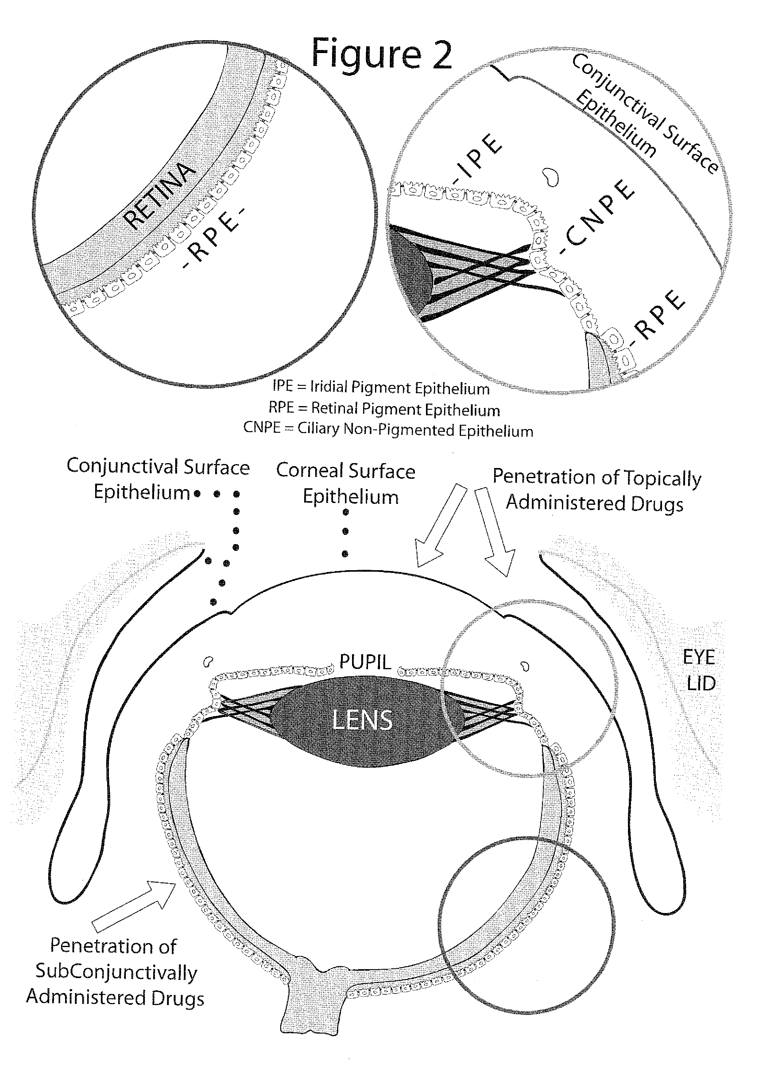
[0076] Results: In the control group animals there was no drug penetration through the surface epithelium of the cornea and conjunctiva. After the PlGF intervention numerous sites of drug penetration between adjacent cells of the ocular surface epithelium were observed. Consequently, there was a remarkable drug penetration to the corneal stroma, deep penetration through the conjunctiva and penetration to the subconjunctival space. Furthermore, collecting vessels loaded with drug were leaking and releasing the drug at periocular, episcleral / scleral and choroidal sites, with drug penetration into the ciliary body and anterior retina.

example 3

[0077] Intervention: PlGF was administered to the anterior subconjunctival space followed by administration of the fluorescent-labeled dextran solution to the anterior subconjunctival space. In the control group animals, the same intervention was done but with the administration of BPS instead of PlGF.

[0078] Results: Compared to minimal intraocular drug penetration in the control group animals, there was a massive intraocular penetration in the group with PlGF intervention with remarkable drug accumulation in the ciliar body, anterior retina and corneal endothelial cells. Blood vessels loaded with the drug and leaking were seen again in the PlGF treatment group.


PUM

PropertyMeasurementUnit
thicknessaaaaaaaaaa
volumeaaaaaaaaaa
pore diameteraaaaaaaaaa

Abstract

Provided is a method for enhanced intraocular drug penetration that comprises the co-administration of ocular therapeutics with agents that increases the permeability of ocular and periocular vessels and ocular epithelial barriers. Due to its unique and novel concept and additive nature, the method of the present invention can be used in combination with previous methods for enhancement of ocular drug penetration.

Description

FIELD OF THE INVENTION [0001] The invention relates generally to methods for enhanced ocular drug penetration and, more specifically, the invention relates to the co-administration of ocular therapeutics with agents that increases the permeability of ocular and periocular vessels and ocular epithelial barriers by unique strategy and thus with additive ocular penetration enhancement effect that allows the utilization of this invention in combination with known methods for ocular drug penetration enhancement. BACKGROUND OF THE INVENTION [0002] The following is a brief overview of eye anatomy that, together with the schematic description in FIGS. 1 and 2, is needed to understand the bellow discussion on obstacles to intraocular drug penetration. [0003] The ocular surface consists of the superficial layers of the cornea and conjunctiva. It is well accepted that one of the main tasks of the ocular surface is to create a defense barrier against penetration from undesired molecules. In hum...

Claims


Application Information

Patent Timeline
Patent Type & Authority Applications(United States)
IPC IPC(8): A61K38/18A61K38/13A61K31/7048A61K31/704A61K31/573A61K31/57A61K31/554A61K31/553
CPCA61K9/0019A61K9/0048A61K31/553A61K31/554A61K31/57A61K31/573A61K31/704A61K31/7048A61K38/1866
Inventor ABDULRAZIK, MUHAMMAD
Owner INSERM TRANSFERT
Why Eureka
Try Eureka