Use of biomolecular targets in the treatment and visualization of brain tumors

Inactive Publication Date: 2007-02-01
MEDAREX INC
View PDF3 Cites 47 Cited by
  • Summary
  • Abstract
  • Description
  • Claims
  • Application Information

AI Technical Summary

Benefits of technology

[0009] The present invention provides methods and reagents for specifically targeting brain tumor neoplastic cells for both therapeutic and imaging purposes, by targeting the brain tumor target protein, DDR1, which is identified as being overexpressed in brain tumors, and thus allow for the selective inhibition of cell function or selective marking for visualization with therapeutic or visualizing compositions which have a specific affinity for these protein targets.
[0011] Agents that bind to, or otherwise inhibit DDR1 function can inhibit the invasiveness of astrocytoma cells, and are effective in preventing the spread of brain tumors through extracellular matrix and basement membrane. Inhibitors can also target matrix metalloproteases that are induced by activation of DDR1, e.g. thiol, alkylcarbonyl, phosponamidate and hydroxamate MMP inhibitor compounds, such as marimastat and prinomastat.

Problems solved by technology

The intracranial space and physical layout of the brain create significant obstacles to treatment and recovery.
Because of the relatively small volume of the skull cavity, minor changes in the volume of tissue in the brain can dramatically increase intracranial pressure, causing damage to the entire organ.
Thus, even small tumors can have a profound and adverse affect on the brain's function.
The cramped physical location of the cranium also makes surgery and treatment of the brain a difficult and delicate procedure.
However, because of the dangers of increased intracranial pressure from the tumor, surgery is often the first strategy of attack in treating brain tumors.
However, it also prevents therapeutic molecules, including many chemotherapeutic agents that are useful in other types of tumors, from crossing into the brain.
However, the amount of radiation necessary to completely destroy potential tumor-producing cells also produce unacceptable losses of healthy brain tissue.
The retention of patient cognitive function while eliminating the tumor mass is another challenge to brain tumor treatment.
Thus, it is impossible to define a true “tumor margin,” unlike, for example, in lung or bladder cancers.
Unlike reproductive (ovarian, uterine, testicular, prostate, etc.), breast, kidney, or lung cancers, the entire organ, or even significant portions, cannot be removed to prevent the growth of new tumors.
This pervasive and variable nature greatly adds to the difficulty of treating brain tumors while preserving the health and function of normal brain tissue.
Although current surgical methods offer considerably better post-operative life for patients, current combination therapy methods (surgery, low-dosage radiation, and chemotherapy) have only improved the life expectancy of patients by one month, as compared to the methods of 30 years ago.
Without effective agents to prevent the growth of brain tumor cells that are present outside the main tumor mass, the prognosis for these patients cannot be significantly improved.
Although some immuno-affinity agents have been proposed and tested for the treatment of brain tumors, see, for example, the tenascin-targeting agents described in U.S. Pat. No. 5,624,659, these agents have not proven sufficient for the treatment of brain tumors.

Method used


Image

  • Use of biomolecular targets in the treatment and visualization of brain tumors
  • Use of biomolecular targets in the treatment and visualization of brain tumors
  • Use of biomolecular targets in the treatment and visualization of brain tumors

Examples


example 1

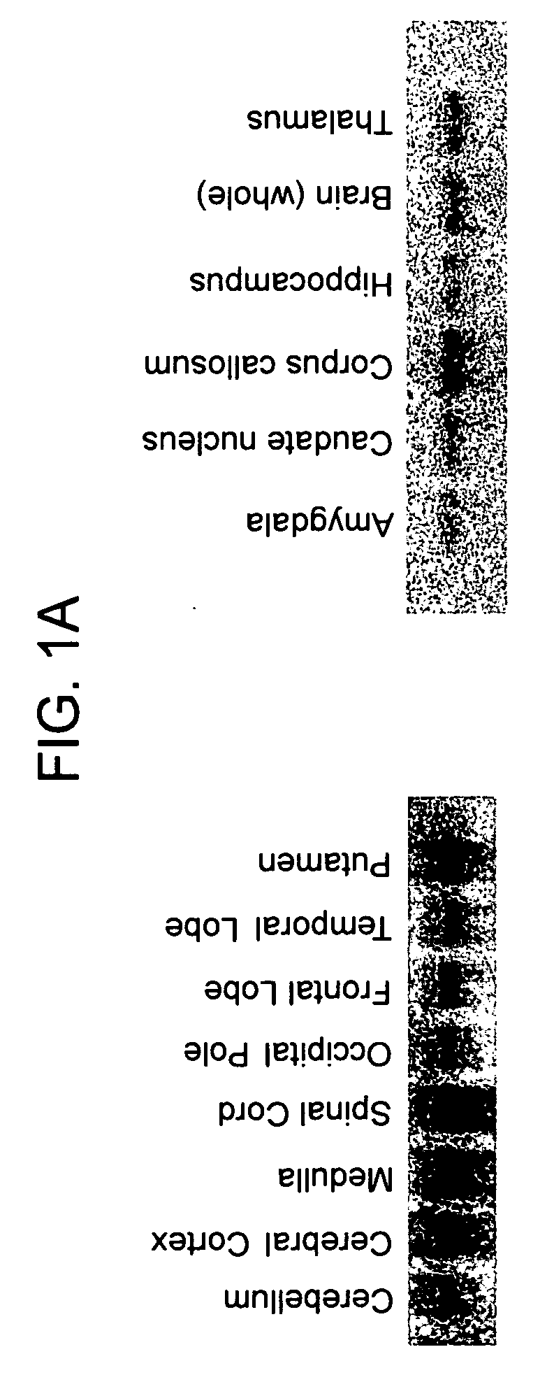
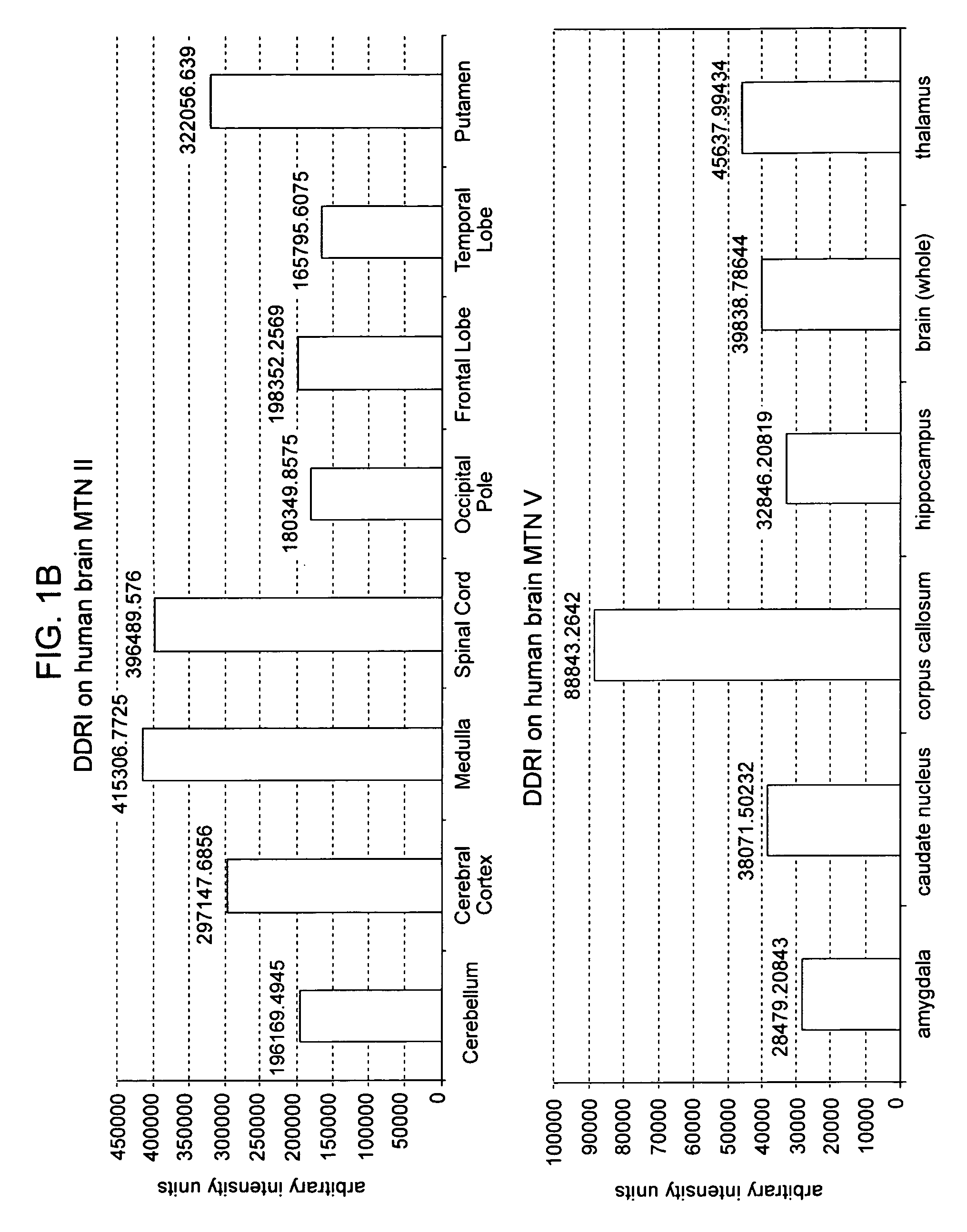
[0208] Brain Tumors: Tumor tissue, confirmed as astrocytoma grade IV by neuropathology, from unknown patients was snap frozen in the operation hall and served as experimental sample. Human whole brain tissue (Clontech Laboratories, Palo Alto, USA) served as control sample. Poly-A+ RNA prepared from the cells was converted into double-stranded cDNA (dscDNA) and normalized as described in co-pending U.S. patent application Ser. No. 09 / 627,362, filed on Jul. 28, 2000. Subtractive hybridization was carried out using the dscDNA from tumors with an excess of dscDNA prepared from the same region of a non-cancerous brain. Differentially expressed gene fragments were cloned into a plasmid vector, and the resulting library was transformed into E. coli cells. Inserts of recombinant clones were amplified by the polymerase chain reaction (PCR). The PCR products (fragments of 200-2000 bp in size) were sequenced using an oligonucleotide complementary to common vector sequences. The resulting seque...

example 2

Discoidin Domain Receptor-1a (DDR1a) Promotes Glioma Cell Invasion and Adhesion in Association with Matrix Metalloproteinase-2

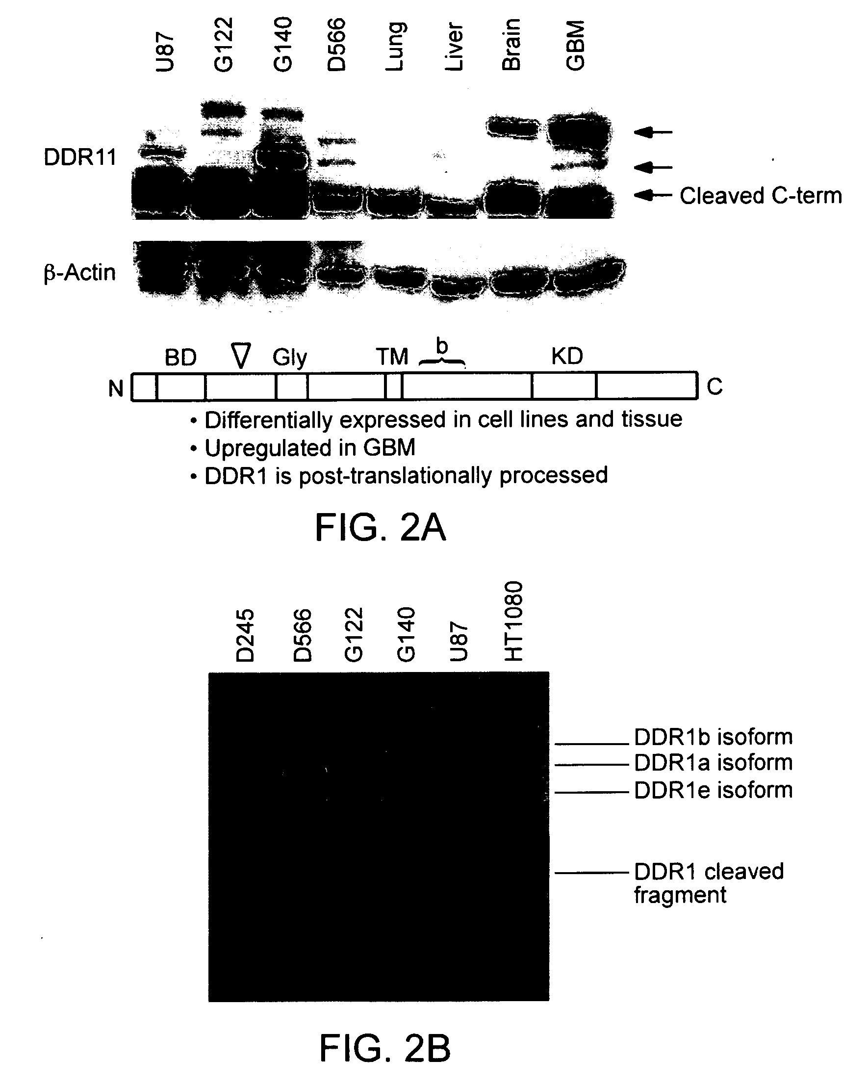
[0243] Invasion of glioma cells involves the attachment of invading tumor cells to extracellular matrix (ECM), disruption of ECM components, and subsequent cell penetration into adjacent brain structures. Discoidin domain receptor 1 (DDR1) tyrosine kinases constitute a novel family of receptors characterized by a unique structure in the ectodomain (discoidin-1 domain). These cell surface receptors bind to several collagens and facilitate cell adhesion. In the following example it is shown that DDR1 is overexpressed in glioma tissues using cDNA arrays, immunohistochemistry and Western blot analysis. Functional comparison of two splice variants of DDR1 (DDR1a and DDR1b) reveal novel differences in cell based glioma models. Overexpression of either DDR1a or DDR1b caused increased cell attachment. However, glioma cells overexpressing DDR1a display enhanced invas...


PUM

PropertyMeasurementUnit
Fractionaaaaaaaaaa
Solubility (mass)aaaaaaaaaa
Stabilityaaaaaaaaaa

Abstract

The present invention relates to the use of proteins that are differentially expressed in primary brain tumor tissues, as compared to normal brain tissues, as biomolecular targets for brain tumor treatment therapies. Specifically, the present invention relates to the use of therapeutic and imaging agents, which specifically bind to one or more of the identified brain tumor protein targets. The present invention also provides compounds and pharmaceutically acceptable compositions for administration in the methods of the invention. Nucleic acid probes specific for the spliced mRNA encoding these variants and affinity reagents specific for the novel proteins are also provided.

Description

BACKGROUND OF THE INVENTION [0001] Among tumors, those of the brain are considered to have one of the least favorable prognoses for long term survival: the average life expectancy of an individual diagnosed with a central nervous system (CNS) tumor is just eight to twelve months. Several unique characteristics of both the brain and its particular types of neoplastic cells create daunting challenges for the complete treatment and management of brain tumors. Among these are the physical characteristics of the intracranial space; the relative biological isolation of the brain from the rest of the body; the relatively essential and irreplaceable nature of the organ mass; and the unique nature of brain tumor cells. [0002] The intracranial space and physical layout of the brain create significant obstacles to treatment and recovery. The brain is primarily comprised of astrocytes, which make up the majority of the brain mass, and serve as a scaffold and support for the neurons, which carry...

Claims


Application Information

Patent Timeline
IPC IPC(8): A61K39/395C12Q1/68G01N33/574
CPCA61K2039/505C07K16/2851C12Q1/6886G01N33/57407C12Q2600/136C12Q2600/158C12Q2600/112
Inventor NAGAVARAPU, USHASHIVAK, DAVID A.CHIN, DANIEL J.FOEHR, ERIK D.GONZALEZ-ZULUETA, MIRELLA
Owner MEDAREX INC
Why Eureka
Try Eureka