
Methods for detecting and confirming minimal disease

Inactive Publication Date: 2007-01-25
HEMATOLOGICS
View PDF6 Cites 55 Cited by
  • Summary
  • Abstract
  • Description
  • Claims
  • Application Information

AI Technical Summary

Benefits of technology

[0049] One advantage of the present invention is that very few cells are needed for genetic analysis to confirm the presence of minimal disease. Thus, in certain embodiments, the number of sorted cells to be analyzed can be about 2000,1500, 1000, 950, 900, 850, 800, 750, 700, 650, 600, 550, 500, 450, 400, 350, 300, 250, 200 or fewer cells.
[0050] Following detection of minimal disease using flow cytometry fluorescence activated cell sorting, the sorted cells are subjected to genetic analysis using any of a variety of reagents and techniques known in the art and described for example, in Current Protocols in Molecular Biology (John Wiley & Sons, NY, N.Y.), or Innis, Ed., PCR Protocols, Academic Press (1990). In this regard, any genetic analysis that confirms that the sorted population of cells represents minimal disease is contemplated for use herein. The genetic analysis to be performed will depend on the disease setting and can be determined by the skilled artisan. One advantage of the present invention is that the confirmatory genetic analysis does not require patient-specific reagents. However, such reagents can be used where available if desired.
[0051] Nucleic acid from the sorted cells is isolated using techniques known in the art such as described in Current Protocols in Molecular Biology (John Wiley & Sons, NY, N.Y.) or using any of a variety of commercially available reagents. The nucleic acid for subsequent analysis may be genomic DNA, RNA, including HnRNA and mRNA, or cDNA.
[0052] Examples of subsequent analysis which may be performed on samples of genetic material isolated from the sorted cell population of interest include, but are not limited to, polymerase chain reaction (PCR), ligase chain reaction (LCR), reverse transcriptase initiated PCR (RT-PCR), DNA or RNA hybridization techniques including restriction fragment length polymorphism (RFLP) and other techniques using genetic probes such as fluorescence in situ hybridization (FISH), DNA analysis by variable number of tandem repeats (VNTR) or short tandem repeats (STR), or other genotype analysis, CpG methylation analysis (see, for example, Cottrell et al., Nucleic Acids Research 2004; Vol. 32, No. 1 e10), genomic sequencing, enzymatic assays, affinity labeling, methods of detection using labels or antibodies and other similar methods.
[0053] The genetic analysis may involve the use of oligonucleotides. As used herein oligonucleotides is used as the term is normally understood in the art, that is, to mean a short string of nucleotides. In this regard, the oligonucleotides can be used as either primers or probes and can be of varying lengths as is appropriate for the molecular technique they are being used for, such as PCR, RT-PCR, hybridization assays, FISH, and the like. Generally oligonucleotides are from about 8-50 nucleotides in length but they can be shorter or much longer. In some embodiments, the oligonucleotides can be 10,11, 12, 13, 14, 15, 16, 17, 18, 19, 20, 21, 22, 23, 24, 25, 26, 27, 28, 29, 30, 31, 32, 33, 34, 35, 36, 37, 38, 39, 40, 41, 42, 43, 44, 45, 46, 47, 48, 49, 50, 51, 52, 53, 54, 55, 56, 57, 58, 59, 60, or more nucleotides in length. In certain embodiments, the oligonucleotides can be 65, 70, 75, 80, 85, 90, 95, 100, 105,110, 115,120, 130, 140, 150, or even 200 nucleotides in length. As would be recognized by the skilled artisan, oligonucleotides can be synthesized or otherwise constructed using techniques well known in the art.
[0054] In one embodiment, the genetic analysis comprises the detection of a translocation event. Illustrative translocation events include, but are not limited to: BCR-ABL; BCL1 / JH t(11;14); BCL2 / JH t(14;18); BCR / ABL t(9;22); PML / RAR t(15;17); translocations involving MLL and any of its translocation partners (e.g., AF4, AF6, AF9, ENL and ELL), the t(10;11)(p12;q23) translocation, which is a recurrent event in acute myeloid leukemias; c-myc (8q24) translocations involving t(8;14) (translocations involving t(8;14) occurs in less than 5% of human multiple myeloma cases, but between 10 to 20% of tumors have genetic abnormalities near this locus (Bergsagel, 1998); Bcl-1 / PRAD-1 / cyclin D1 (11q13). A cluster of translocation events occur in this locus resulting in chronic lymphocytic leukemia (CLL) and lymphoma; FGFR3 and / or MMSET (4p16.3) Approximately 25% of myeloma has translocation of the receptor tyrosine kinase FGFR3 to the IgH locus; MUM1 / IRF4 / ICSAT / PIP / LSIRF (6p25) IRF4 is a member of the interferon regulatory factor family which are know to be involved in B-cell proliferation and differentiation. This recurrent translocation was seen in 2 of 11 MM; Cyclin D3 (6p21) is overexpressed in ˜3% of multiple myeloma cell lines and ˜4% of primary multiple myeloma tumors; c-maf (16q23) Translocation of c-maf into the IgH locus results in overexpression of c-maf in approximately 25% of multiple myeloma cell lines. And other translocation events known in the art. Translocation events can be detected using any of a variety of techniques known in the art, such as by FISH and PCR.

Problems solved by technology

In addition to the cumbersome set-up of patient specific assays, several gene rearrangements generally must be used simultaneously as PCR targets since single rearrangements are unstable and can be lost during clonal transformation and following disease relapse due to continuing gene rearrangements or further gene deletions.
In particular for malignancies demonstrating oligoclonality with multiple subclones present at diagnosis, the likelihood of losing a PCR-target during follow-up is significantly increased.
Thus, conventional detection of residual disease using patient-specific reagent panels suffers from the following limitations:
Thus, there are cases where minimal disease must be detected without a prior diagnostic specimen.

Method used


Image

  • Methods for detecting and confirming minimal disease
  • Methods for detecting and confirming minimal disease
  • Methods for detecting and confirming minimal disease

Examples


example 1

Cell Sorting and Genotype Analysis of Abonrmal B and T Lymphocytes

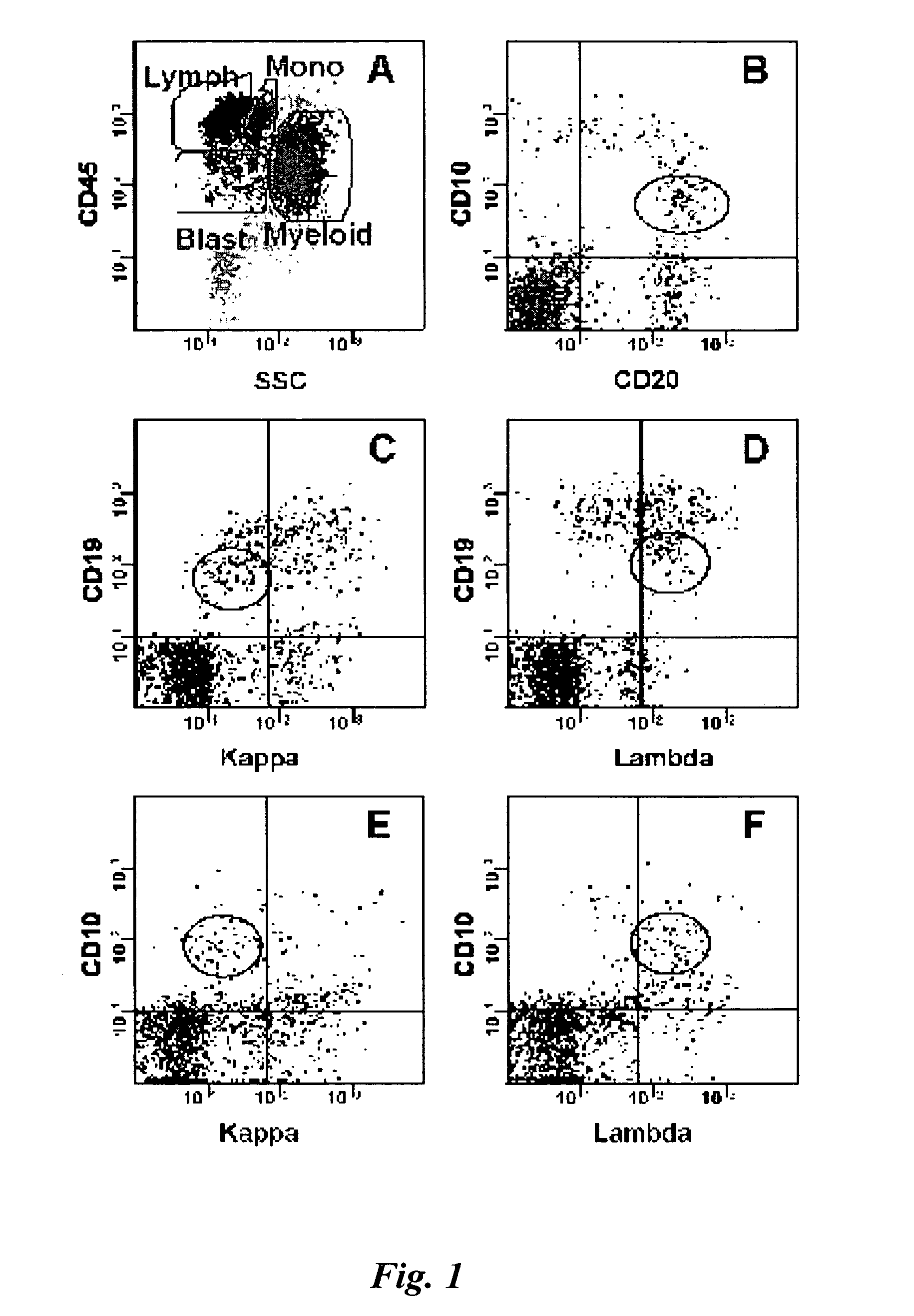
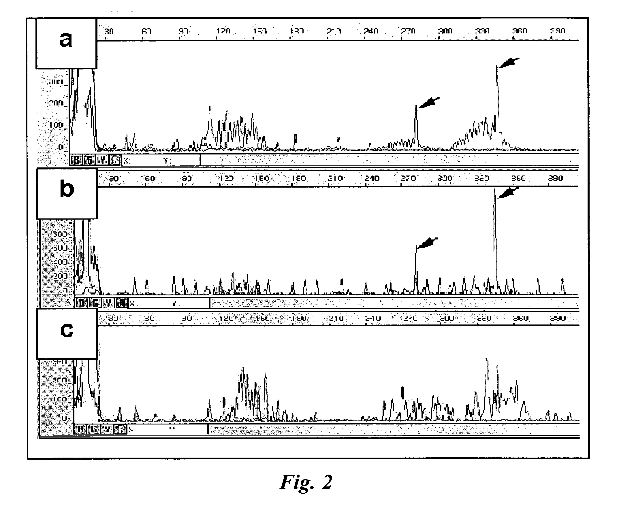
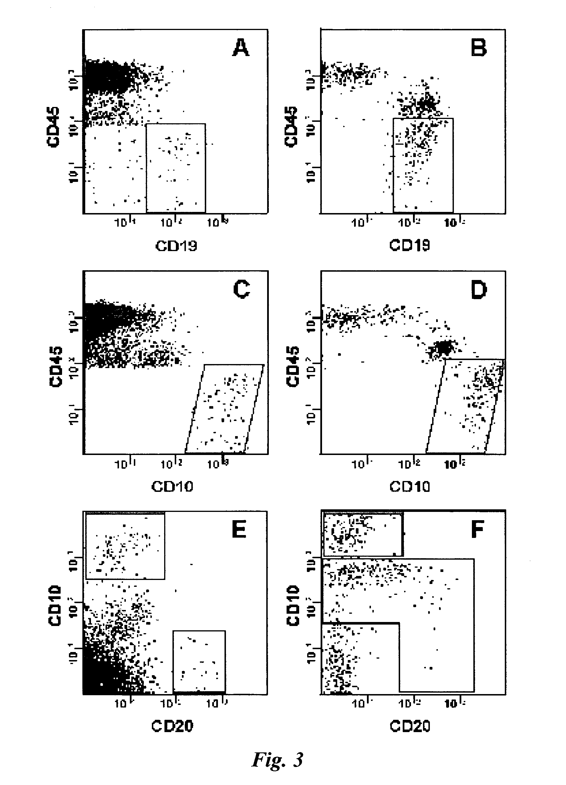
[0066] This example describes a quantitative two-step technique for detecting and confirming low levels of disease by integrating phenotype analysis using standardized flow cytometry panels, cell sorting and genotype analysis using multiplex gene rearrangement PCR thereby confirming the presence of both aberrant phenotype linked to a specific genotype. The feasibility of combining cell sorting with clonality profiling to effectively lower sensitivity limits for disease detection and to provide independent confirmation of the tumor detection without the need for patient specific assay designs is demonstrated. Bone marrow aspirates from three patients with small abnormal B lymphoid populations and one peripheral blood specimen with a small aberrant T cell population as detected by flow cytometry were analyzed by Immunoglobulin heavy chain (IgH) or T cell receptor gamma chain (TCRG) gene rearrangement PCR with and witho...


PUM

PropertyMeasurementUnit
Fractionaaaaaaaaaa
Fractionaaaaaaaaaa
Timeaaaaaaaaaa

Abstract

The present invention provides improved methods for detection of minimal disease. More specifically, the invention provides methods for combining cell sorting with clonality profiling to effectively lower sensitivity limits for disease detection and to provide independent confirmation of the tumor detection without the need for patient specific assay designs.

Description

BACKGROUND OF THE INVENTION [0001] 1. Field of the Invention [0002] The present invention relates generally to improved methods for confirming the presence of minimal disease in cancer patients and, more particularly, to methods useful for initial diagnostics and for monitoring the presence of minimal residual disease following treatment. [0003] 2. Description of the Related Art [0004] The detection of minimal disease can play a significant role not only in monitoring response to therapy but also in the accurate diagnosis of the underlying cause of major clinical signs. Several studies have shown that quantitative detection of minimal residual disease (MRD) in lymphoid malignancies predicts clinical outcome. (Szczepanski T, et al., Lancet Oncol 2001; 2: 409-17; van Dongen J J, et al., Lancet 1998; 352: 1731-8; Bruggemann M, et al., Acta Haematol 2004; 112: 111-9; Cave H, et al., N Engl J Med 1998; 339: 591-8; Coustan-Smith E, et al., Blood 2000; 96: 2691-6; Coustan-Smith E, et al., ...

Claims


Application Information

Patent Timeline
IPC IPC(8): C12Q1/68C12P19/34
CPCC12Q1/6804C12Q1/6827C12Q1/6886C12Q2600/16G01N2333/70503G01N33/57492C12Q2563/131
Inventor LOKEN, MICHAEL R.ZEHENTNER-WILKINSON, BARBARA K.
Owner HEMATOLOGICS
Learn More