
Ultrasound guiding system and method for vascular access and operation mode

Inactive Publication Date: 2006-08-17
RCH SYST
View PDF14 Cites 39 Cited by
  • Summary
  • Abstract
  • Description
  • Claims
  • Application Information

AI Technical Summary

Benefits of technology

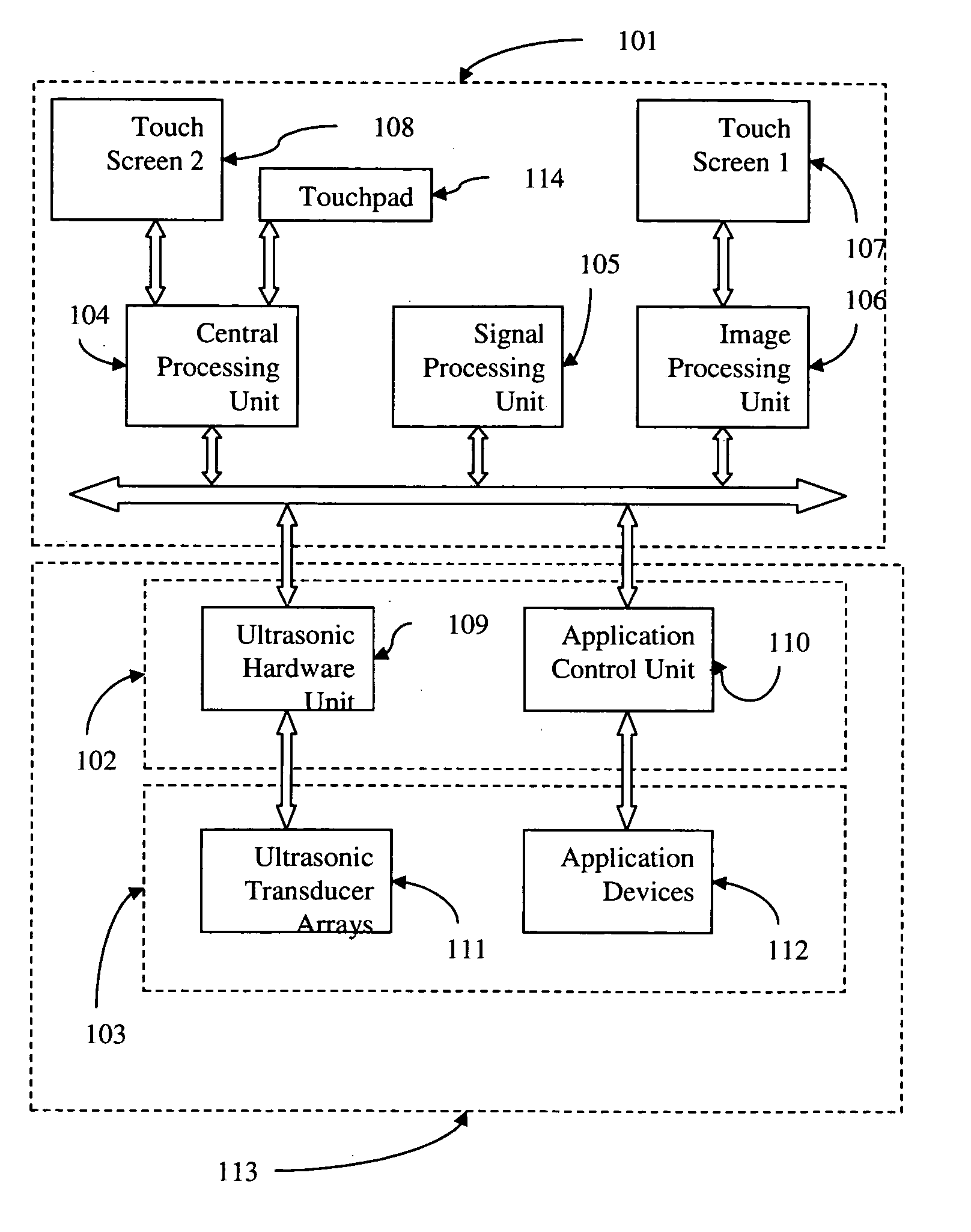
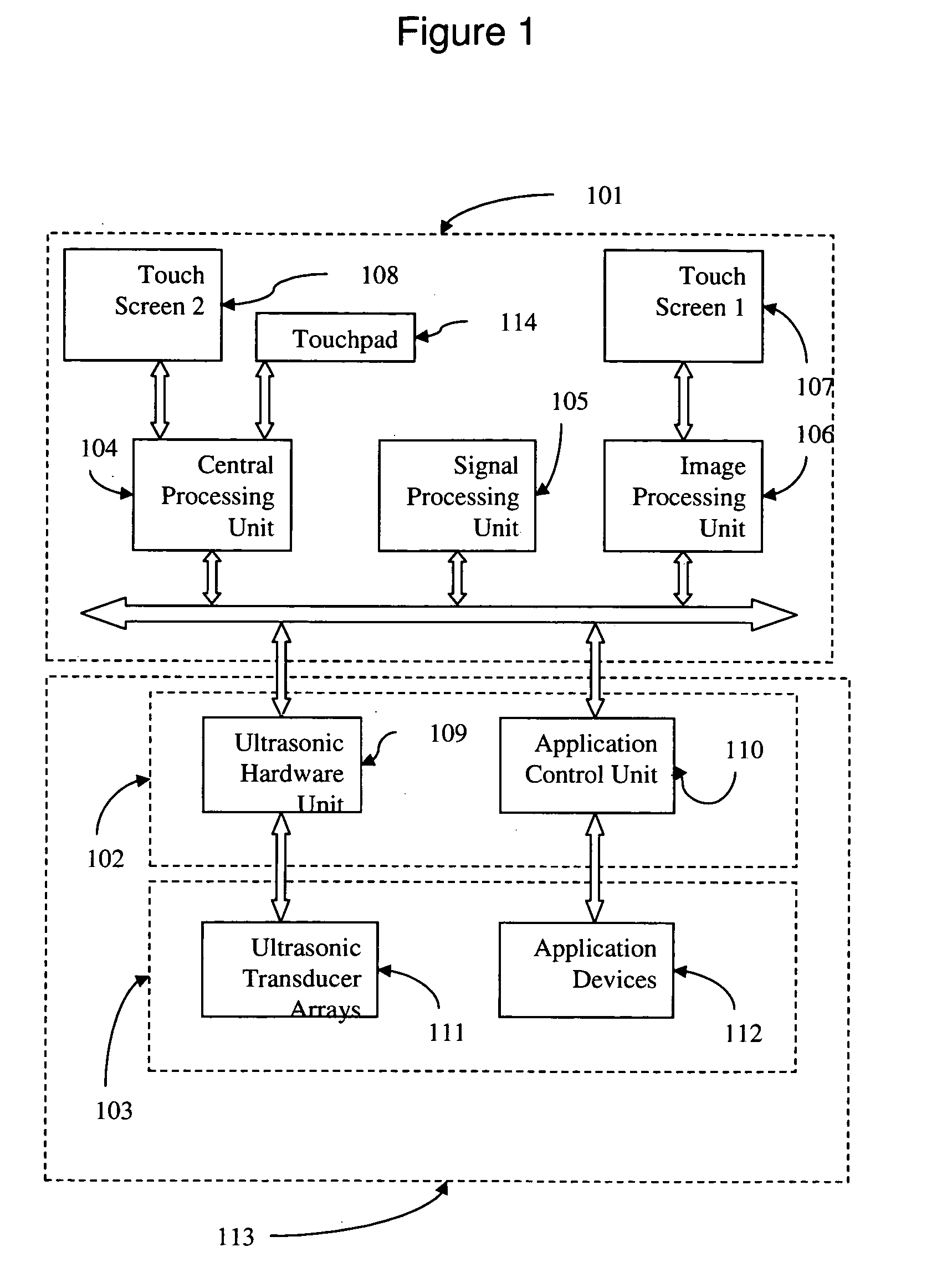
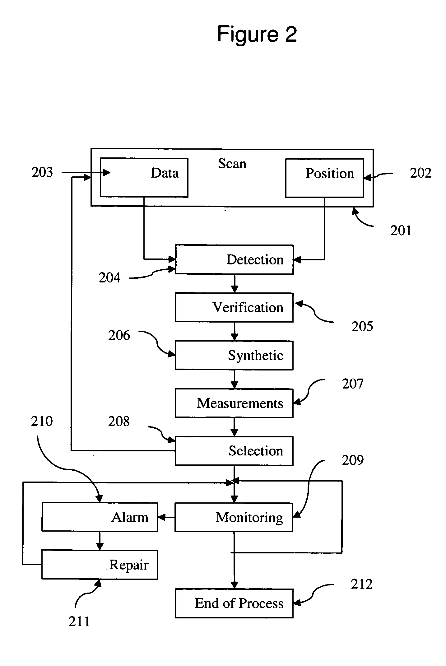
[0014] The present invention comprises of a method for detection, recognizing and classifying of blood vessels and further more calculates the optimal insertion parameters of catheter to the blood vessels. The present invention also exempts the user from handling several procedures simultaneously such as positioning the probe, observing the image or data on the systems screen and locating an accurate location on the surface and thus allowing the user to practice a more accurate procedure.

Problems solved by technology

The operations become rather complex for an insertion of catheters into veins or arteries.
Multiple attempts at penetration may result in extreme discomfort to the patient and loss of valuable time during emergency situations.
For example, while attempting to access the internal jugular vein, the carotid artery may be punctured accidentally, resulting in severe complications or even death.
None of the patents mentioned above address the needs of detecting, recognizing and classifying blood vessels and integrating a display, a pointing device and a tracking device in the probe housing for simpler and easier usage in medical procedures.

Method used


Image

  • Ultrasound guiding system and method for vascular access and operation mode
  • Ultrasound guiding system and method for vascular access and operation mode
  • Ultrasound guiding system and method for vascular access and operation mode

Examples


Embodiment Construction

Introduction

[0033] Ultrasound equipment has developed rapidly over the past 30 years and is now used routinely for numerous medical applications, for example: assessment of arterial stenosis, venous insufficiency and venous thrombosis. Ultrasound images are obtained by holding a probe on the skin surface. An ultrasonic scanner usually has a range of probes with different characteristics, e.g., a linear array probe. This produces a rectangular image which is displayed with the skin surface at the top, the vertical axis showing depth into the body and the horizontal axis showing position along the probe. When imaging blood vessels, the probe can either be placed along the vessel to produce a longitudinal scan or across the vessel to produce a transverse scan. To produce the images, the probe emits short pulses of ultra-sound, and these travel into the body from the probe. Within the soft tissues or at boundaries between them, a small proportion of the ultrasound is scattered or refle...


PUM


Abstract

The present invention provides an ultrasound_medical imaging system with integrated devices and functionalities for use in wide range of medical procedures that require vascular and arterial access. The present invention discloses a method for detection, recognition and classification of blood vessels. Said method further calculates the optimal insertion parameters of catheter to the blood vessels. The present invention also exempts the user from handling several procedures simultaneously such as positioning the probe, observing the image or data on the systems screen and locating an accurate location on the surface and thus allowing the user to practice a more accurate procedure.

Description

CROSS-REFERENCE TO RELATED APPLICATIONS [0001] This application is related to U.S. Provisional Patent Application 60 / 643,168 filed Jan. 12, 2005 and U.S. Provisional Patent Application 60 / 650,969 filed Feb. 9, 2005 whose disclosure is incorporated herein in its entirety by reference.BACKGROUND OF THE INVENTION [0002] The present invention relates to the field of ultrasonic probes and more particularly to an ultrasonic probe for vascular and arterial access procedures. [0003] Diagnosing human organs using ultrasound scanning is a well known procedure. Ultrasonic transducers based probes direct ultrasonic waves, which travel through a selected biological medium. [0004] Reflections are obtained each time the ultrasonic waves encounter impedance variation interfaces in the biological medium, such as fat and muscle. The returned echoes are received and processed by the imaging system that adds up all scanning lines received from the transducer and provides an image. The number of scannin...

Claims


Application Information

Patent Timeline
IPC IPC(8): A61B8/14
CPCA61B5/02007A61B5/489A61B5/7264A61B8/06A61B8/0833A61B8/0841A61B8/0858A61B8/4483A61B8/463A61B8/483A61B8/488
Inventor HAIM, RONENWISCHNIEWSKI, ESTHERPERETS, URISAFRAN, VALERY
Owner RCH SYST
Learn More