Osteopontin-coated surfaces and methods of use

a technology of osteopontin and coating, applied in the field of osteopontin coating surfaces and methods of use, can solve the problems of not being able to make good bone implants, material surfaces that cannot bind macromolecules supporting osteoblast function, etc., and achieves enhanced osseointegration and bone apposition, increased implant placement indications, and improved osseointegration and osseointegration rate

Inactive Publication Date: 2006-05-18
CHILDRENS MEDICAL CENT CORP
View PDF1 Cites 19 Cited by
  • Summary
  • Abstract
  • Description
  • Claims
  • Application Information

AI Technical Summary

Benefits of technology

[0013] Molecules and / or compounds involved in directing the migration or the attachment of cells to matrices of a particular environment are of interest in the presently described invention. The implants coated with these these peptides increase the rate of osseointegration and the percentage of bone apposition. Implant surfaces should have such properties which permit the phenomenology of the relevant cells. The achievement of reproducible biological integration of implants calls for a delineation of the molecular biological events relevant to the morphogenesis of the desired interfacial tissue. Material surfaces that can not bind the macromolecules supportive of osteoblast function, are not likely to make a good bone implant. An enhanced rate of osseointegration and / or augmented percentage of bone apposition around implants or cell recruitment systems of the invention increases implant placement indications, expedites loading time, and opens new fields for research in implant materials.
[0025] In another aspect the invention features a coated implant. The implant includes a coated material which increases the proliferation of osteoblasts by at least about 100% when compared to an uncoated material based on the human osteoblast cell (HOS) proliferation assay.

Problems solved by technology

Material surfaces that can not bind the macromolecules supportive of osteoblast function, are not likely to make a good bone implant.

Method used


Image

  • Osteopontin-coated surfaces and methods of use
  • Osteopontin-coated surfaces and methods of use
  • Osteopontin-coated surfaces and methods of use

Examples


example 1

Coating of Implants

[0126] Titanium, plastic, glass and chromocobalt (CrCo) surfaces were coated with human recombinant OPN. Attachment and proliferation of human osteoblasts by means of matrix formation markers was evaluated using uncoated surfaces as a control. Also the amount of adhesion protein that can be coated to these surface was investigated.

[0127] The human recombinant phosphorylated form of osteopontin (rhOpn) was used as an adhesion molecule. This form of osteopontin migrates on 10% SD S-gels with an apparent molecular weight of 78 Kd, making it easy to differentiate from osteopontin secreted by osteoblasts which migrates in the same gels with an apparent molecular weight of 58 Kd.

[0128] The experiments outlined below investigate the expression and mineralization of extra cellular matrix components in human osteoblasts cultured on titanium disks, plastic, glass and chromocobalt surfaces coated with recombinant osteopontin. The adhesion molecule rhOPN used as a coating ...

example 2

Effect of Ca++ Ions on the Binding of Osteopontin to Ti Disks

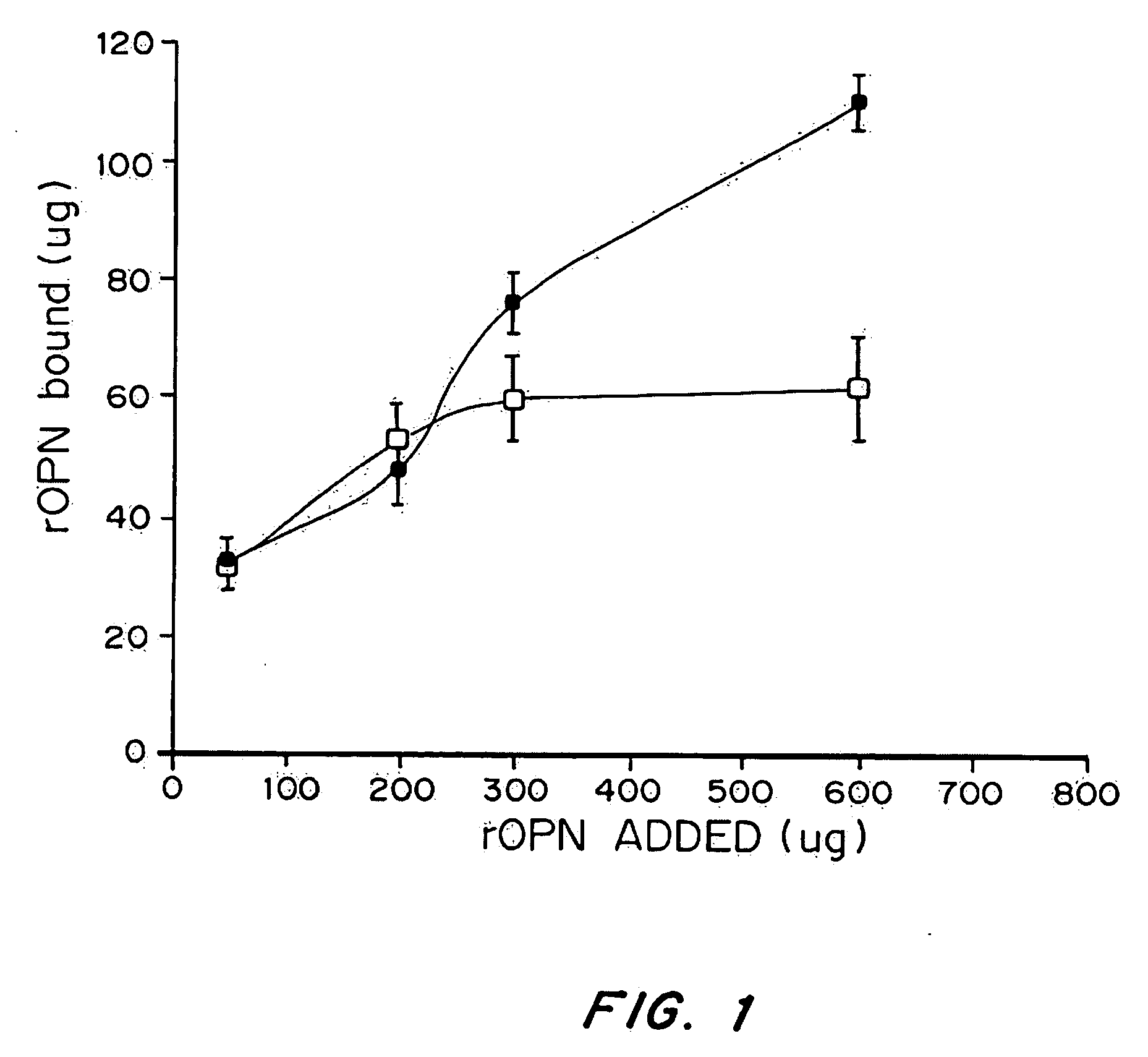
[0142] Increasing concentration of 35S-labeled OPN (60, 200, 400, 600 ug) were incubated with titanium disks either with (▪) or without () CaCl2 at 4° C. After 24 h the unbound protein was removed and the Ti disks were washed with PBS. Bound OPN was extracted from the disks with scintilation fluid and counted. Each experiment was done in triplicates and reported as mean±SEM.

[0143] To investigate whether exogenously added Ca++ had any effect on the binding of rhOPN to Ti, the binding of rhOPN to Ti disks was measured with and without added CaCl2. The results, presented in FIG. 1, demonstrate that in the absence of added CaCl2 the Ti disks saturate at 60 μg of rhOPN, but in the presence of 100 mM CaCl2 the Ti disks can bind more rhOPN saturating at more than 110 μg protein / disks.

example 3

Attachment of HOS Cells to Ti Surfaces Coated with rhOPN

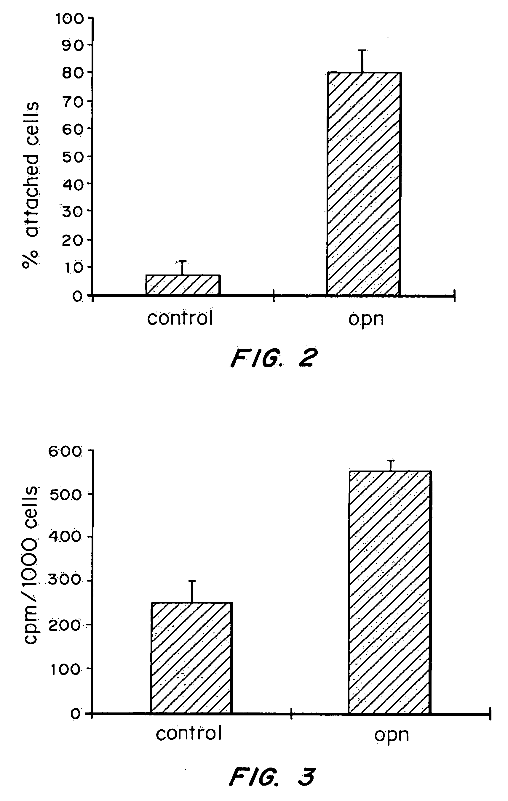
[0144] 5000 cells (total cpm 1000) were plated on either coated or uncoated Ti disks and incubated at 37° C. in a humidified atmosphere (95% air 5% CO2). After 30 min, unattached cells were removed and the disks were washed with PBS. The total number of attached cells was determined for the total cpm released for the disks after the cells were lysed with 10% TCA and solubilized in 5 ml scintillation fluid. All measurements were done in triplicates and graphed as mean±Standard error of the mean.

[0145] The initial events following seeding of cells onto Ti surfaces include the attachment, migration and proliferation of the seeded cells. Coating Ti disks with 50 μg of rhOPN enhanced by 1100% the attachment of HOS cells to Ti disks (FIG. 2), after 30 min. These results are consistent with the role of osteopontin in promoting cell attachment and spreading.


PUM

PropertyMeasurementUnit
molecular weightaaaaaaaaaa

Abstract

An osteopontin containing implant which increases the rate of osseointegration and the percentage of bone apposition is described. In one embodiment, the implant includes osteopontin or an active fragment thereof or an active peptide derived thereform. In another embodiment, the implant includes a material suitable for use in vivo within a subject in combination with a releasable form of osteopontin forming an osteopontin containing implant. The disclosed osteopontin derived peptides bind to various cell types and play important roles in cellular differentiation and / or motility. Many of these interactions are mediated by integrins as disclosed. Antibodies provide a mechanism to abolish or attenuate the activities of the claimed peptides.

Description

[0001] This application is related to U.S. Provisional Application No. 60 / 327,273 filed on Oct. 5, 2001, U.S. Provisional Application No. 60 / 241,248 filed on Oct. 18, 2000.BACKGROUND OF THE INVENTION [0002] The process that leads to successful osseointegration of an implant into the surrounding tissues is a complex one that involves cell migration, attachment, differentiation, proliferation, extracellular matrix synthesis and finally mineralization of that matrix. Implant materials are as biocompatible as their surface chemistry allows for a favorable interaction with the biological molecules relevant for that tissue. [0003] For example, placement of endosseous dental implants has been limited to areas of favorable bone character, and fixtures must remain unloaded after placement for considerable periods of time. The primary challenges faced in the fabrication of new endosseous implants are to increase the rate of osseointegration and the percentage of bone apposition. Histological ...

Claims


Application Information

Patent Timeline
Patent Type & Authority Applications(United States)
IPC IPC(8): A61K38/17C07K14/435A61C8/00A61K38/00A61L27/00A61L27/34A61P43/00C07K14/47C07K14/52C07K16/24
CPCA61K38/00A61L27/34C07K14/51C07K14/52C07K16/24C08L89/00A61P43/00
Inventor ASHKAR, SAMYSALCEDO, JAIRO
Owner CHILDRENS MEDICAL CENT CORP
Why Eureka
Try Eureka