X-ray imaging apparatus and method for mammography and computed tomography

a computed tomography and x-ray imaging technology, applied in the field of x-ray apparatus for medical radiology, can solve the problem that conventional mammography does not detect all cancers, and achieve the effect of effective irradiation of tissu

Active Publication Date: 2006-05-04
ADVANCED X RAY TECH
View PDF4 Cites 13 Cited by
  • Summary
  • Abstract
  • Description
  • Claims
  • Application Information

AI Technical Summary

Benefits of technology

[0010] Thus, it is an object of the present invention to provide an apparatus with a movable, monochromatic, small size, point x-ray secondary source that is movable and can effectively irradiate the tissue to be examined.
[0013] It is also an object of the invention to provide an imaging apparatus that effectively moves the focal spot of the secondary target re-emitting a monochromatic x-ray beam close to the compression paddle of the apparatus, or directly to a desired region of tissue.
[0015] It is yet a further object of the invention is to provide the imaging apparatus with means for smoothly moving a primary x-ray source (x-ray tube) and collimator with a secondary target with particular reference to orthogonal collimator with a secondary target with particular reference to orthogonal coordinates through pre-defined positions.
[0016] The ability to place the secondary target at any desirable point allows one to obtain x-ray images of the patient's tissue from different angles. These images can then be processed for reconstruction of a 3D image.
[0018] The x-ray apparatus includes means for smoothly moving the primary x-ray source and the collimator with the secondary target as a unit. In one embodiment, the collimator also has means for movement of the secondary target with a semilens along the optical axis by a defined distance.

Problems solved by technology

Although mammographic screening for breast cancer has proved invaluable in the early detection and diagnosis of breast cancer, conventional mammography does not detect all cancers.

Method used


Image

  • X-ray imaging apparatus and method for mammography and computed tomography
  • X-ray imaging apparatus and method for mammography and computed tomography
  • X-ray imaging apparatus and method for mammography and computed tomography

Examples


Embodiment Construction

)

1. The Apparatus

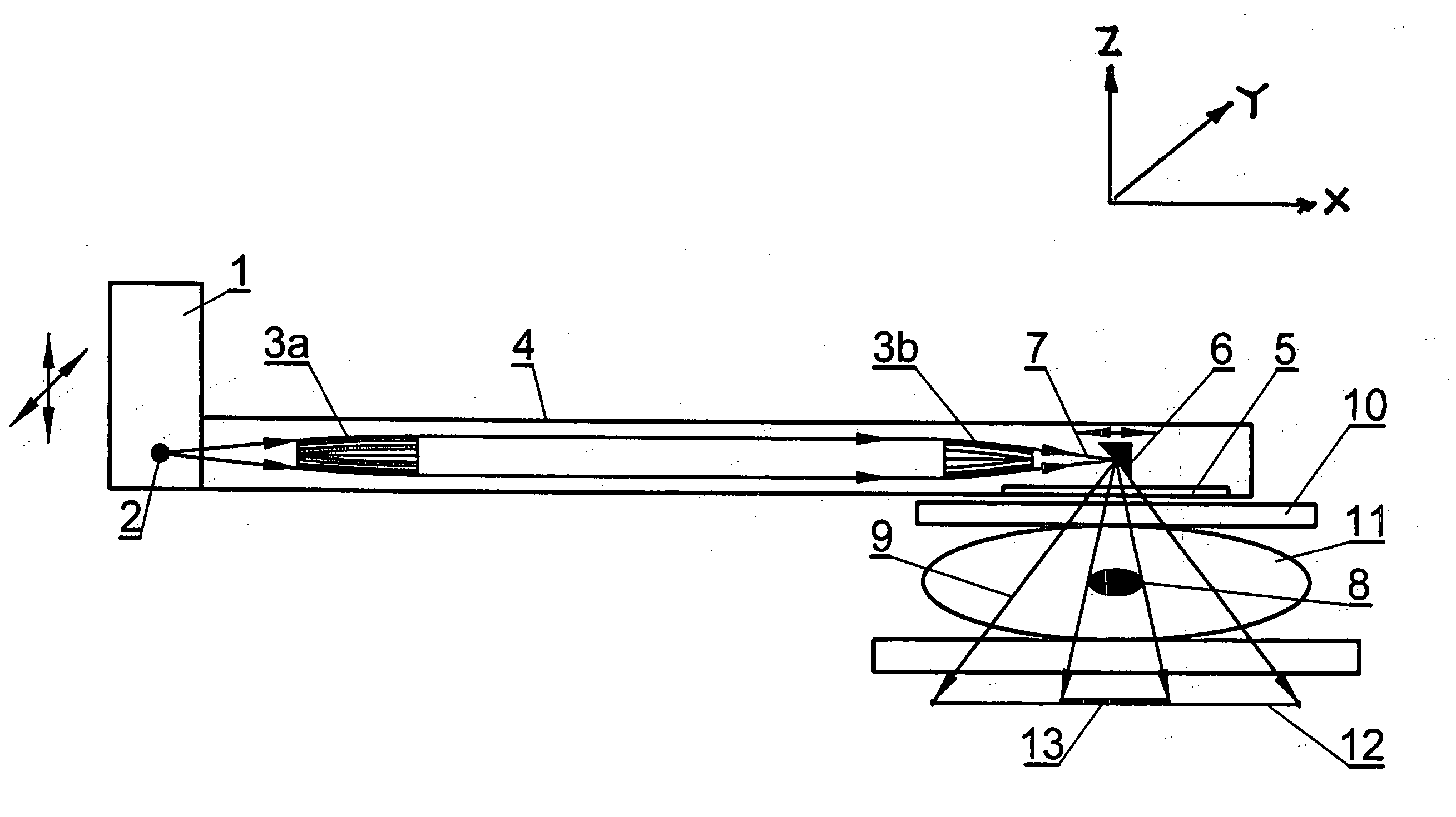
[0029]FIG. 1 shows one embodiment of an x-ray imaging apparatus for mammography. The apparatus has: an x-ray source 1 (x-ray tube with a Rh or Ag anode) with a point focus 2 and a capillary lens, means for shaping, or preferably two semilenses 3a and 3b within a collimator 4 (attached to the x-ray tube 1). The collimator has an output window 5 and a secondary or pseudo-target 6. The pseudo-target 6 or means for monochromatizing is preferably made from a metallic material such as Mo that allows two-stage production of monochromatic x-ray radiation with the highest effectiveness.

[0030] First, a primary x-ray beam 7 generated by an x-ray tube 1 is guided towards the secondary target 6 through the collimator 4. The primary x-ray beam 7 excites the pseudo-target 6 and causes the target to emit x-ray fluorescence that is mainly defined by the characteristic lines of the target material and the parameters of the incident x-ray beam. At least part of the re-emitted fluor...


PUM


Abstract

An x-ray imaging apparatus for mammography. The apparatus places the focal spot of the x-ray source as close as possible to a compression paddle or into a desired region of tissue. The apparatus comprises an x-ray source, a collimator with conditioning optics which focus and direct x-ray radiation to a metal pseudo-target in the collimator or a needle placed along the x-ray beam, paddles for breast compression and a detector. The pseudo-target re-emits monochromatic x-ray radiation with an energy, intensity, and space distribution that are adjustable in accordance with a desired imaging procedure.

Description

CROSS-REFERENCE TO RELATED APPLICATION [0001] This application claims the benefit of U.S. provisional application Ser. No. 60 / 624,411, filed Nov. 2, 2004, which is incorporated herein by reference.BACKGROUND OF THE INVENTION [0002] 1. Field of the Invention [0003] The present invention relates to an x-ray apparatus for medical radiology. More specifically, the invention relates to an x-ray apparatus for mammography, including receiving tomosynthesis data and obtaining radiological images in galactography (see, e.g., U.S. Pat. No. 6,480,566 which is incorporated by reference herein) and angiography. This invention also can be used in computer tomography. [0004] 2. Background Art [0005] Mammography is a tool for diagnosing breast cancer. Images of a tumor can be viewed on film or (if it is used a digital detector) on a computer screen. Successful treatment of breast cancer depends on early diagnosis. Mammography plays a central part in early detection because it can show changes in th...

Claims


Application Information

Patent Timeline
IPC IPC(8): A61B6/04
CPCA61B5/6848A61B6/08A61B6/4429A61B6/502A61B2562/17
Inventor GUTMAN, GEORGE
Owner ADVANCED X RAY TECH
Why Eureka
Try Eureka