Liposomal vectors
a technology of liposomes and vectors, applied in the field of liposome vectors, can solve the problems of reducing treatment effectiveness, and achieve the effects of less inflammation, less inflammation, and more productive gene transfer
- Summary
- Abstract
- Description
- Claims
- Application Information
AI Technical Summary
Benefits of technology
Problems solved by technology
Method used
Image
Examples
example 1
Preparation of Lipoplexes and Safeplexes
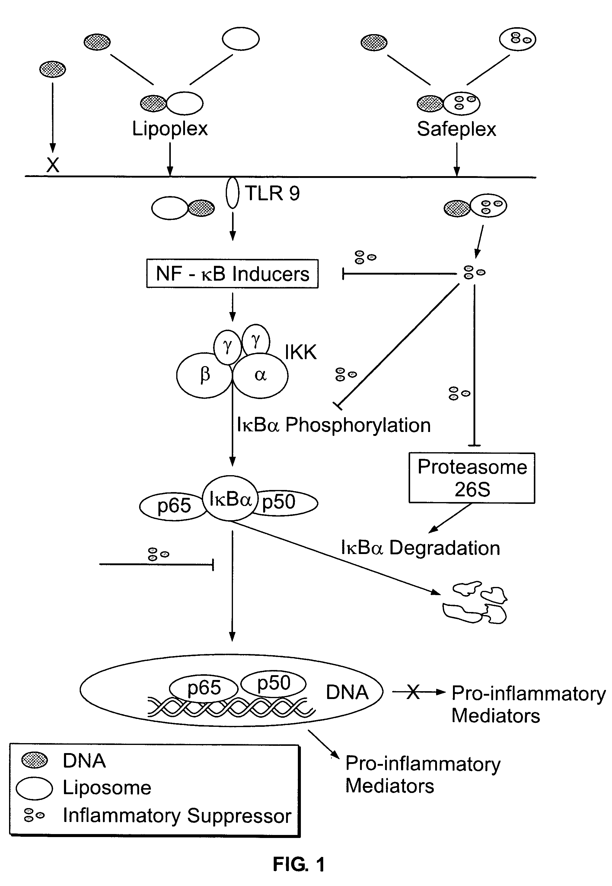
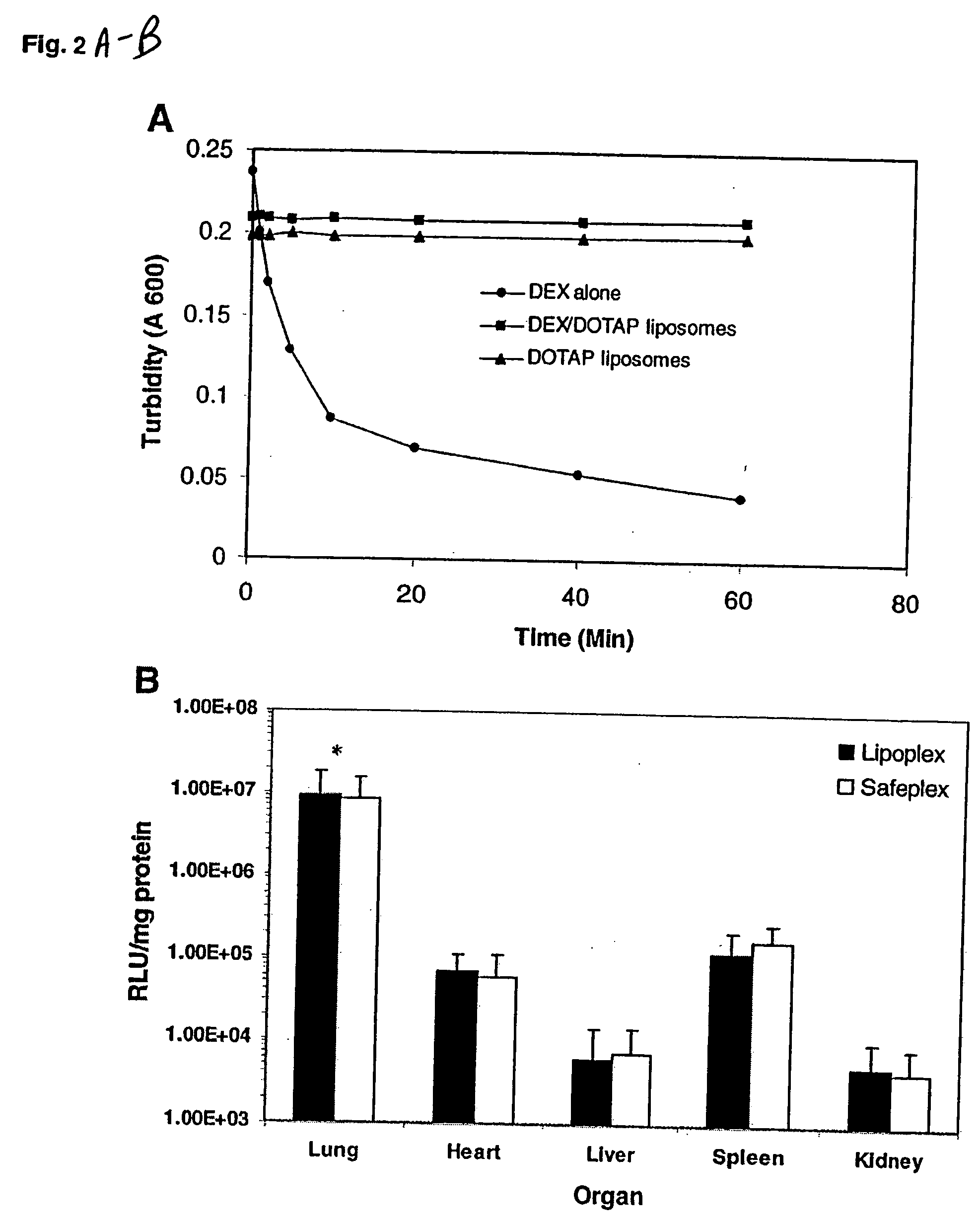
[0062] All inflammatory suppressors were dissolved in organic solvents with concentration of 10 mg / ml (dexamethasone in methyl alcohol, prednisone in methyl alcohol and chloroform (1:1), indomethacin, tetrandrine and gliotoxin in chloroform). DOTAP in chloroform was added with (for the preparation of the safeplexes) or without (for the preparation of the lipoplexes) an inflammatory suppressor, and then was placed under a stream of nitrogen to evaporate the solvent until a thin lipid film formed at the bottom of a glass tube. It was further vacuum-desiccated for 1 h and then hydrated in 5% of dextrose solution to a final concentration of 10 mg DOTAP / ml (4 mg DOTAP / ml for tetrandrine and gliotoxin liposomes). The lipid suspension was briefly sonicated and then sequentially extruded through polycarbonate membrane of pore size of 0.2 μm. The DOTAP liposomes, suppressor / DOTAP liposomes, and pNGVL-3 luciferase plasmid DNA were separately diluted wi...
example 2
Evaluation of In Vivo Transgene Expression
[0063] The luciferase plasmid was used as a reporter gene. CD1 female (18-20 g) mice were injected intravenously with the lipoplexes or safeplexes with a charge ratio of 12 to 1 (+ / −) and sacrificed 6 h after the injection. Each lung was collected and placed in 1 ml of ice-cold lysis buffer and homogenized with a tissue tearor (BioSpec Products, Bartlesville, Okla.) for 20 s at the highest speed. The homogenates were then centrifuged at 14,000 g for 5 min at 4° C. Ten microliters of the supernatant was analyzed with the luciferase assay system (Promega, Madison, Wis.) using an automated LB953 luminometer (Berthod Bad Wildbad, Germany). The protein content of the supernatant was measured with the Bio-Rad Protein Assay System (Bio-Rad, Hercules, Calif.). Luciferase activity was expressed as relative light units per milligram protein (RLU / mg protein).
example 3
Cytokine Assay in the Blood and Organs
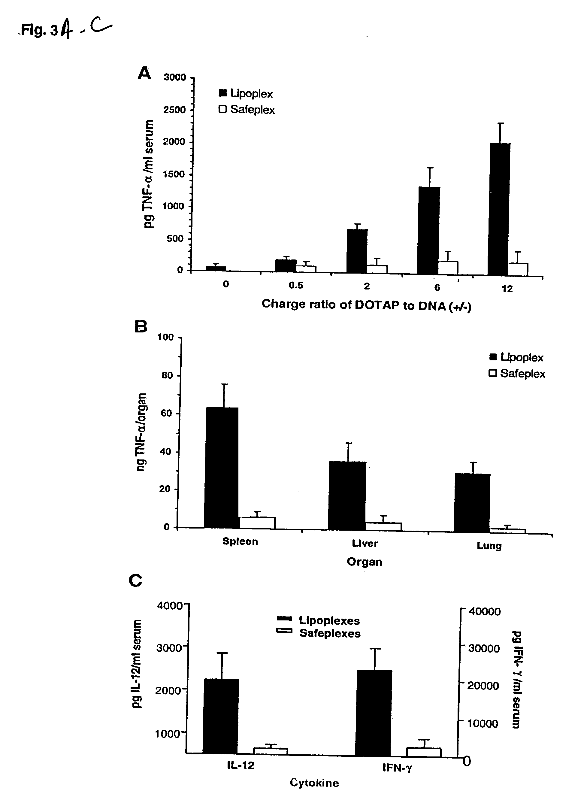
[0064] At indicated time points (2 h for TNF-α, 6 h for IL-12 and IFN-γ) following the injection of a tested sample containing 25 μg of pDNA, the blood and organs (the liver, lung and spleen) were collected. The blood was allowed to clot on ice for at least 4 h and then centrifuged at 3000 g for 20 min at 4° C., and serum was collected for the cytokine assay. For the organ assay, 50 μg of each organ was added with 0.5 ml PBS buffer and homogenized for 20 s at the highest speed. The homogenates were frozen (in liquid nitrogen) and thawed (in 37° C. water bath) three times and then centrifuged at 14,000 g for 5 min. The supernatants were collected for the cytokine assay. The concentrations of cytokines (TNF-α, IL-12 and IFN-γ) were determined with mouse cytokine immunoassay kits (R&D System, Minneapolis, Minn.). Data were analyzed by the paired Student's t test using Excel. Data were considered statistically significant if P<0.01.
PUM
| Property | Measurement | Unit |
|---|---|---|
| Immunogenicity | aaaaa | aaaaa |
Abstract
Description
Claims
Application Information
 - R&D
- Intellectual Property
- Life Sciences
- Materials
- Tech Scout
- Unparalleled Data Quality
- Higher Quality Content
- 60% Fewer Hallucinations
Browse by: Latest US Patents, China's latest patents, Technical Efficacy Thesaurus, Application Domain, Technology Topic, Popular Technical Reports.
© 2025 PatSnap. All rights reserved.Legal|Privacy policy|Modern Slavery Act Transparency Statement|Sitemap|About US| Contact US: help@patsnap.com


