
Robotic microscopy apparatus for high throughput observation of multicellular organisms

a multicellular organism and robotic technology, applied in the field of robotic microscopy apparatus for high throughput observation of multicellular organisms, can solve the problems of limiting the throughput of any screening method used with any model organism, affecting the economic or workforce level of high-throughput analyses with model organisms, and affecting the economic and workforce level. , to achieve the effect of reducing costs

Inactive Publication Date: 2006-03-09
ELEGENICS
View PDF27 Cites 40 Cited by
  • Summary
  • Abstract
  • Description
  • Claims
  • Application Information

AI Technical Summary

Benefits of technology

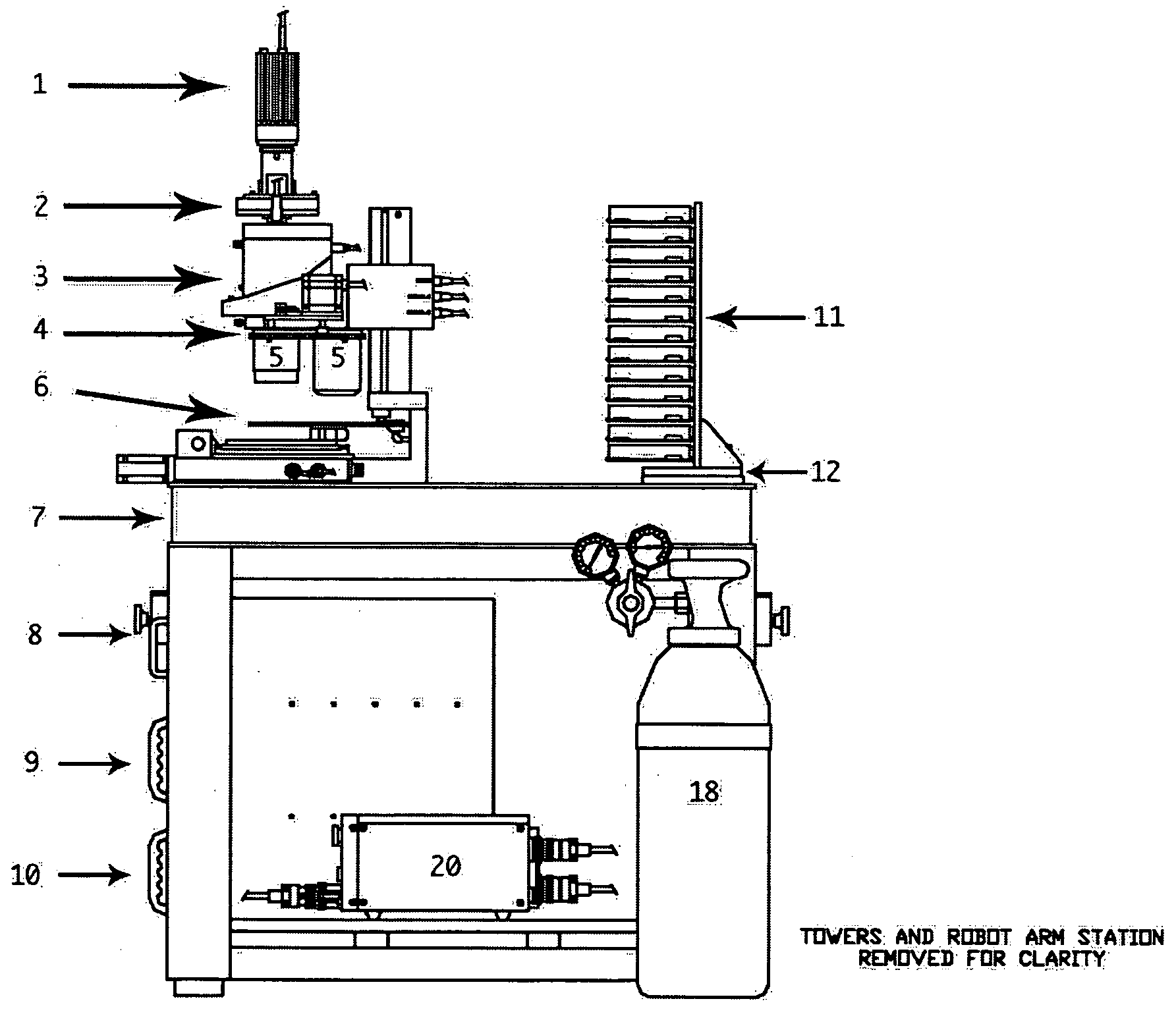
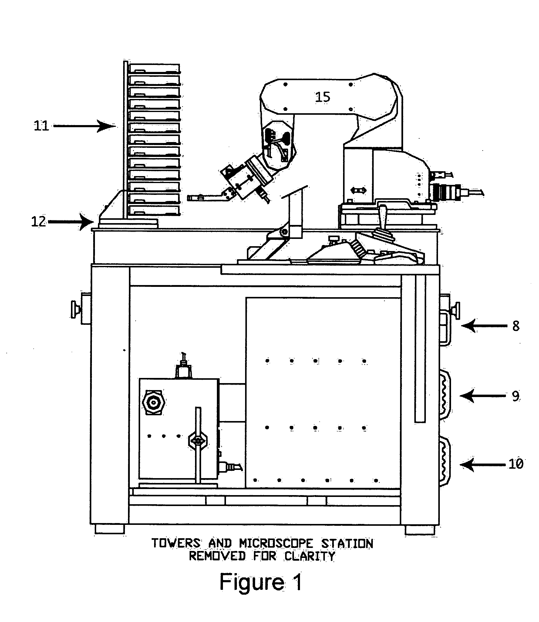
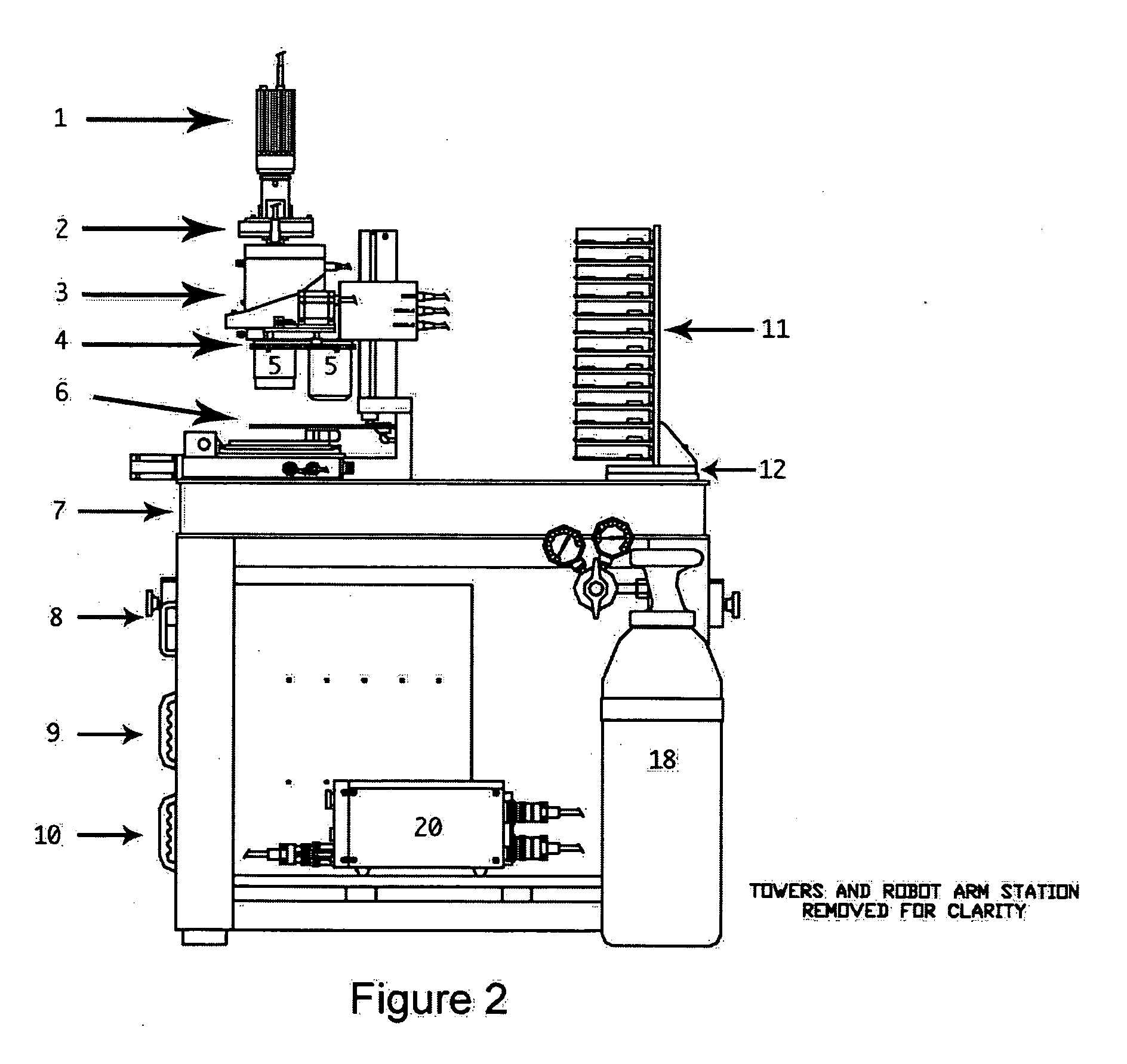
"The present invention is a robotic microscopy apparatus that can screen, detect, count, and image whole multicellular organisms, tissues, individual cells, and groups of cells on or embedded within agar, collagen, or other defined matrix. The apparatus has a motorized system for plate handling and a stereo-optic system with motorized magnification changer for automated and high throughput screening. The apparatus can detect and image optical characteristics of biological specimens in a plate using a microscope and a robotic system. The biological specimens can be immobilized using a gas and the apparatus can contain multiple light sources, optical filters, and software for processing image data. The apparatus can also contain an automated immobilizing compound injection system and a robotic plate handling system for holding multiple plates. The technical effects of the invention include automation, high throughput screening, and improved accuracy and efficiency in biological research."

Problems solved by technology

Screening methods with model organisms is extremely labor intensive, with experimental personnel required to inspect each plate or well containing a specimen(s) or organism(s).
This clearly limits the throughput of any screening method used with any model organism.
With the limitation imposed by these present human-based screening procedures, high-throughput analyses with model organisms are not practical at the economical or workforce level.
However, each of the currently available apparatuses has limitations.
These limitations singly or in combination include detector, sample, sample holder (e.g., plate versus slide) and most importantly the working distance between the apparatus and sample and the ability to view a sample from the upright position rather than an inverted position.
This short working distance reduces the types of experiments that can be carried out, and the quality of images that can be obtained with the currently available apparatuses when viewing whole organisms.
Additionally, despite these methods and apparatuses, no available apparatus effectively provides for automated imaging of whole organisms.
Further, no apparatus known to the applicant is able to do so in a multi-well format or high throughput manner.
And still further no apparatus known to the applicant is able to temporarily immobilize whole, live organisms to obtain high resolution images.

Method used


Image

  • Robotic microscopy apparatus for high throughput observation of multicellular organisms
  • Robotic microscopy apparatus for high throughput observation of multicellular organisms
  • Robotic microscopy apparatus for high throughput observation of multicellular organisms

Examples


example 1

[0181] Acquire brighffield and fluorescent images of individual objects within a well of multiwell plate. Each well of a 12-well plate is processed with the following steps:

[0182] 1. In order to arrest nematode locomotion, the HIDI control system applies carbon dioxide gas to the well under operation using a proprietary gas applicator

[0183] 2. A brightfield full well image of a well is acquired using a software based auto-focus algorithm

[0184] 3. The full well image is analyzed to identify individual nematodes.

[0185] 4. Five centrally located full sized worms are chosen for observation

[0186] 5. The system magnification is increased so that an acquired image fits one full sized nematode

[0187] 6. The XY imaging stage is moved until the 1st of the five chosen nematodes is centered in the field of view

[0188] 7. A sequence of images are acquired using brightfield, YFP and RFP filter sets

[0189] 8. The XY imaging stage is moved to center the 2nd of the five chosen nematodes in the ...

example 2

[0195] Imaging a full well, and counting and analyzing objects in the well. Each well of a 12-well plate is processed with the following protocol:

[0196] 1. A brightfield full well image of a well is acquired using a software based auto-focus algorithm

[0197] 2. The full well image is processed within a user definable area of interest

[0198] 3. The area of interest is analyzed to identify individual nematodes.

[0199] 4. The identified nematodes are measured in size and counted.

[0200] 5. Counts are parsed into user configurable bins according to nematode size.

[0201] 6. The full well image is stored on disk.

[0202] 7. A count results file is generated containing a table of all well counts for the experiment run.


PUM


Abstract

The present invention relates to a robotic microscopy apparatus that is able to screen, detect, count and image in an automated and high throughput fashion whole multicellular organisms, tissues, individual cells and groups of cells on or embedded within agar, collagen or other defined matrix. To achieve this, the robotic apparatus of the invention images the samples from the top using a microscope with a long working distance. The invention provides robotic systems for plate handling, biological sample immobilization and microscopic examination. The invention also provides for automatic image acquisition, image storage and display, and image analysis.

Description

CROSS-REFERENCE TO RELATED APPLICATIONS [0001] This application claims priority under 35 U.S.C. § 119(e) to U.S. Provisional Application Ser. No. 60 / 607,221 filed on Sep. 2, 2004 and to U.S. Provisional Application Ser. No. 60 / 649,727 filed on Feb. 2, 2005; the disclosures of which are herein incorporated by reference.INTRODUCTION [0002] 1. Background of the Invention [0003] Discovery and technological advances continues to surround high throughput analyses in academic, biotechnological and pharmaceutical settings. Thus far, high throughput analyses have focused primarily on the use of cells as biological entities or targets for testing. [0004] High throughput experimentation has been made possible with the generation of roboticized apparatuses and chemical reagents including, but hot limited to, chemical libraries derived from combinatorial and medicinal chemistry, and biological reagents derived from genome sequencing efforts (e.g. siRNA). [0005] In addition to cells, model multic...

Claims


Application Information

Patent Timeline
Patent Type & Authority Applications(United States)
IPC IPC(8): G02B21/26
CPCG01N15/1463G02B21/365G02B21/16G01N2015/1497G01N15/1433
Inventor HOUSTON, EDWARD S.HOWARD, J. ALBERT JR.ROY, PETER J.JOHANSON, KARL
Owner ELEGENICS
Learn More