
Drug discovery method

a discovery method and drug technology, applied in the field of drug discovery methods, can solve the problems of time and a tremendous commitment of company resources, and achieve the effects of reducing the average number of cycles, rapid availability of new pharmaceuticals, and reducing the cost of drug developmen

Inactive Publication Date: 2006-01-05
VERTEX PHARMA INC
View PDF9 Cites 33 Cited by
  • Summary
  • Abstract
  • Description
  • Claims
  • Application Information

AI Technical Summary

Benefits of technology

[0015] Presently, it takes between three and five years to bring new potentially therapeutic compounds from the early discovery to preclinical development (in vivo animal testing). This lead time requires a tremendous commitment of company resources. Understandably, a technique that could predict the efficacy of the multitude compounds that arrive at or near the initial synthesis stage of drug discovery would have an enormous impact on the efficiency with which new drugs are identified by eliminating, early in the drug discovery process, compounds that have an unfavorable efficacy profile. An immediate expected benefit of more powerful, early stage in vitro effcicacy testing is the reduction of the number of these three to five year drug discovery cycles that result in failures, and thus reduce the average number of cycles required to develop successful new therapeutics. Related benefits include reduced costs for drug development and more rapid availability of new pharmaceuticals to the medical community.
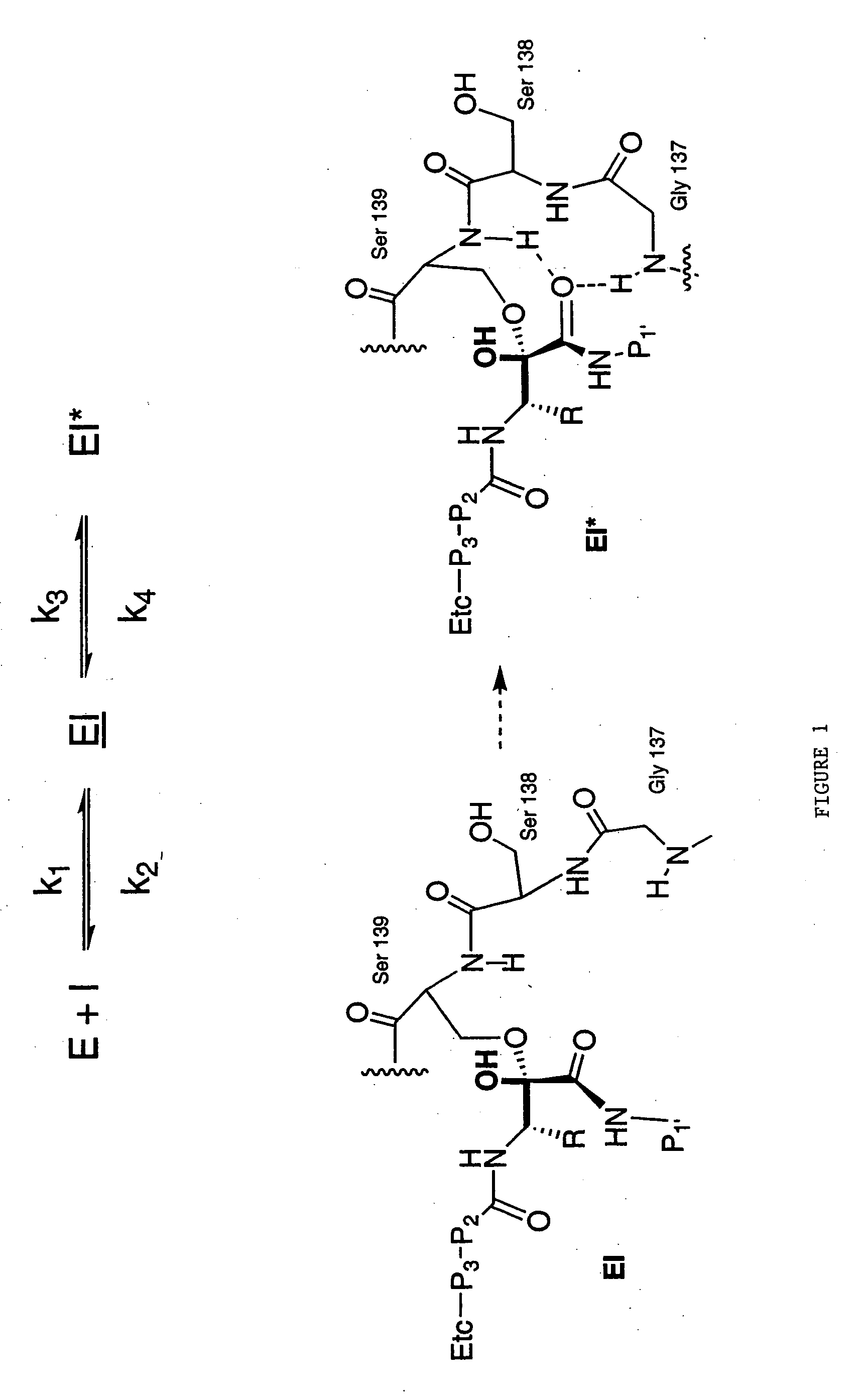
[0016] The present invention provides methods for prioritizing new chemical entities within a class for further development or for identifying such compounds as appropriate lead compounds that will be effective as therapeutic agents early in the drug development and discovery process. As such, these methods can be used to prioritize large numbers of new compounds for further drug development. In addition, the methods greatly increase the probability that an identified agent will be successful in preclinical efficacy testing. The adaptability of these in vitro methods for high-throughput analysis makes them an economical and cost-effective addition to a drug discovery program. In particular, the present invention relates to the kinetics of inhibitors of infectious agents and is directed to a drug discovery method comprising comparing a time a reversible inhibitor is bound to a target of an infectious agent; and a replication time of an infectious agent, or a life-cycle time of the infectious agent. Using such determinations, the present invention teaches methods for selecting a compound having activity against an infectious agent, comprising selecting a target in the agent; identifying a compound that binds reversibly with the target; determining the half life of the compound on the target; determining the half life of the replication cycle of the infectious agent; and selecting a compound that has a half life on the target of at least about 25% of the time of an average life cycle for the infectious agent.

Problems solved by technology

This lead time requires a tremendous commitment of company resources.

Method used


Image

  • Drug discovery method
  • Drug discovery method
  • Drug discovery method

Examples


example 1

[0111] The rates of onset of slow binding inhibition were determined by a modification of the method for measurement of progress curves described in Narjes et. al. (2000).

[0112] Enzyme activity was measured using a continuous assay which monitored the increase of fluorescence which resulted from cleavage of the internally quenched fluorogenic depsipeptide (FRET substrate): [0113] Ac-Glu-Asp-Glu-(Edans)-Asp-Asp-Aminobutyrl-ψ[COO]-Ala-Ser-Lys-(Dabcyl)-NH2

[0114] Two hundred nanomolar NS3 (protease domain) was pre-incubated for 10-15 minutes at room temperature in 50 mM HEPES, pH 7.5, 25 μM KK4A and 5 mM dithiothreitol (DTT). An aliquot of this mixture was then added to 50 mM HEPES, pH 7.5, 15% v / v glycerol, 25 μM KK4A and 5 mM dithiothreitol containing 4 μM to 8 μM FRET peptide substrate (4-8×Km) and inhibitor dissolved in DMSO or DMSO alone as control (1% v / v final concentration; the total assay volume was 1.0 mL and the final enzyme concentration was 1.0 nM. The increase in fluores...

example 2

2-Day HCV Replicon Assay

[0125] Cells containing subgenomic HCV RNA (replicon) are maintained in DMEM containing 10% fetal bovine serum (FBS), 0.25 mg / ml of G418. On the day prior to the assay, 104 HCV replicon cells were plated in each well of a 96-well plate in the presence of 10% FBS but no G418 to allow the cells to attach and to grow overnight (˜16 h). On the day of the assay, the culture media were removed and replaced with media with serially diluted compounds in the presence of 2% FBS and 0.5% DMSO. The replicon cells were treated with the compound for 48 hours, then the reduction of HCV RNA in the cells was determined by quantitative RT-PCR (Taqman) and the cytotoxicity of the compound was determined by MTS-based cell viability assay. The IC50 and CC50 of the compound were calculated from these assays using 4-parameter curve fitting. IC50 represents the concentration of the compound at which the HCV RNA level in the replicon cells is reduced by 50%. CC50 represents the conc...

example 3

9-Day Viral Clearance (VC) Assay

[0126] The replicon cells were plated at a very low density (500 cells per well) in a 96-well plate so that they won't reach confluence after 9 days in culture. Compounds were serially diluted to concentrations at multiples (2×, 5×, etc.) of their respective IC50's in media containing 10% FBS and 0.2% DMSO. The media containing compounds were replaced every three days. The cells were treated with compounds for 3, 6 or 9 days. At the end of experiment, cell numbers were determined in an MTS-based assay with an established standard curve, the level of HCV RNA in the cells was measured by quantitative RT-PCR (Taqman), and then the copy number of HCV replicon RNA per cell in each sample was calculated.


PUM

PropertyMeasurementUnit
Fractionaaaaaaaaaa
Fractionaaaaaaaaaa
Timeaaaaaaaaaa

Abstract

The present invention relates to drug discovery methods, particularly antiviral drug discovery methods.

Description

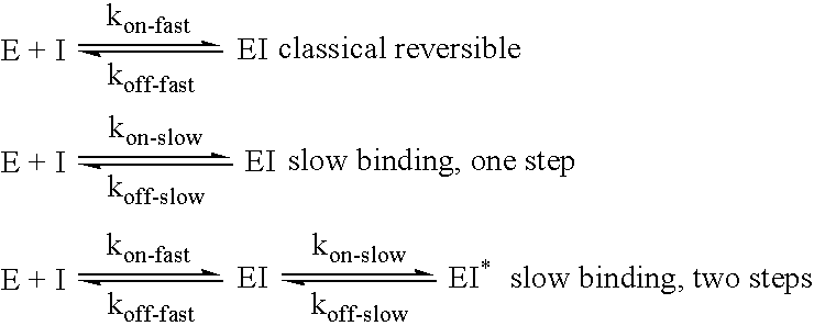
BACKGROUND [0001] The present application claims the benefit of priority of U.S. Provisional Application No. 60 / 515,009 filed Oct. 27, 2003; and U.S. Provisional Application No. 60 / 526,346 filed Dec. 1, 2003. The entire text of each of the foregoing applications is specifically incorporated herein by reference in its entirety.FIELD OF THE INVENTION [0002] The present invention relates to methods of identifying compounds that inhibit infective agents. BACKGROUND OF THE RELATED ART [0003] As noted by Morrison and Walsh (1988), enzyme inhibitors can be categorized based on potency, reversibility, and rates of inhibitor interaction. Classical reversible, and non-time dependent inhibitors exhibit fast steps for binding and release from the enzyme. Equilibria for classical inhibitors are established quickly. For example, a classical inhibitor with a Ki value in the nanomolar range may have rates of binding of 106-108 M−1sec−1 and rates of release from enzyme-inhibitor complex at 1-100 sec...

Claims


Application Information

Patent Timeline
IPC IPC(8): C12Q1/70A61K38/21A61K38/55C12Q1/18G01N
CPCA61K38/212A61K38/215A61K38/217A61K38/553C12Q1/18G01N2333/18A61K2300/00
Inventor PERNI, ROBERT B.GATES, CYNTHIATHOMSON, JOHN
Owner VERTEX PHARMA INC
Learn More