
Non-invasive measurement of blood glucose using retinal imaging

a retinal imaging and blood glucose technology, applied in the field of blood glucose in vivo measurement, can solve the problems of high cost of glucose testing supplies, blood drawn for analysis, and many patients not testing their blood as often as recommended, so as to achieve accurate blood glucose determination

Inactive Publication Date: 2005-11-03
FOVIOPTICS INC
View PDF4 Cites 22 Cited by
  • Summary
  • Abstract
  • Description
  • Claims
  • Application Information

AI Technical Summary

Benefits of technology

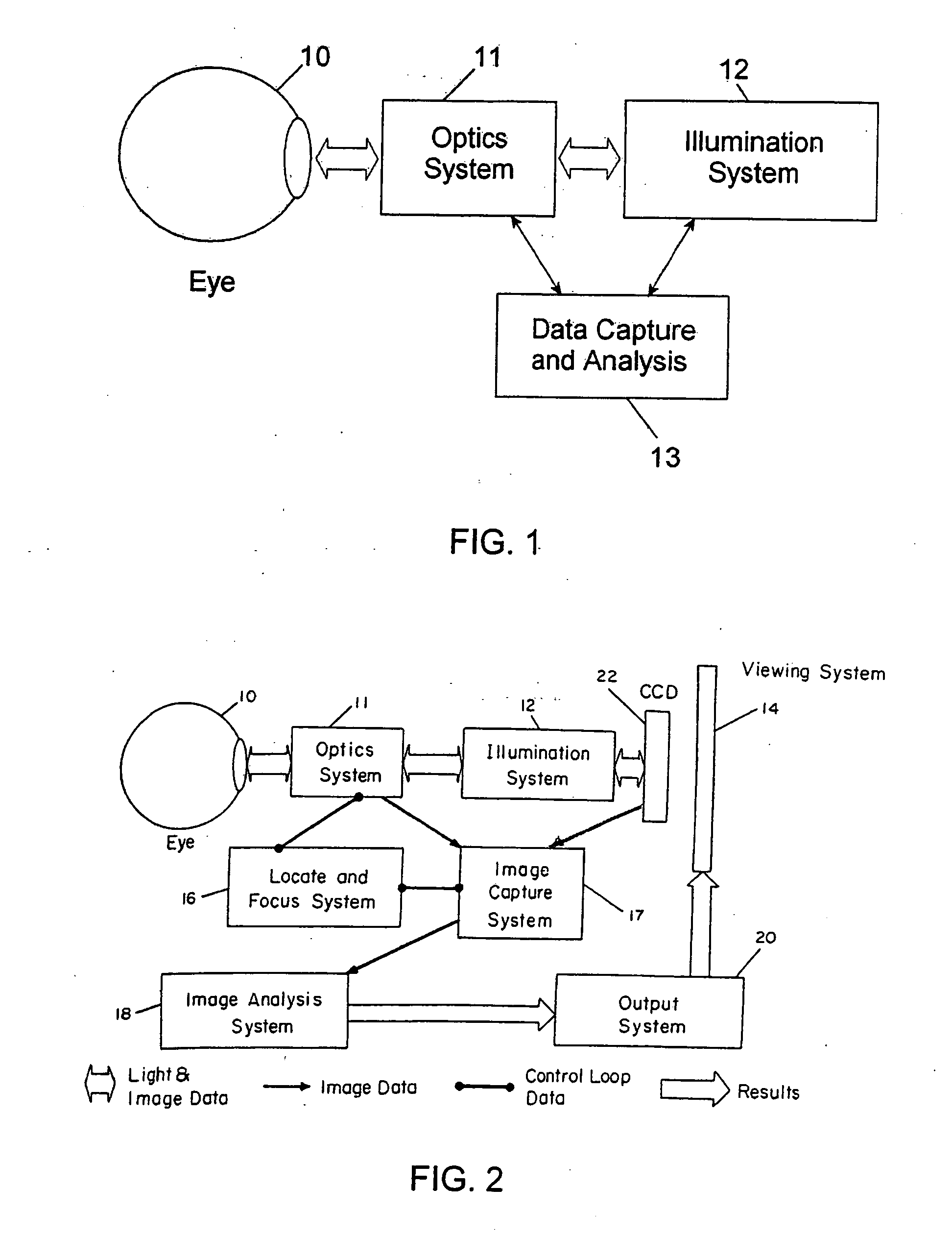
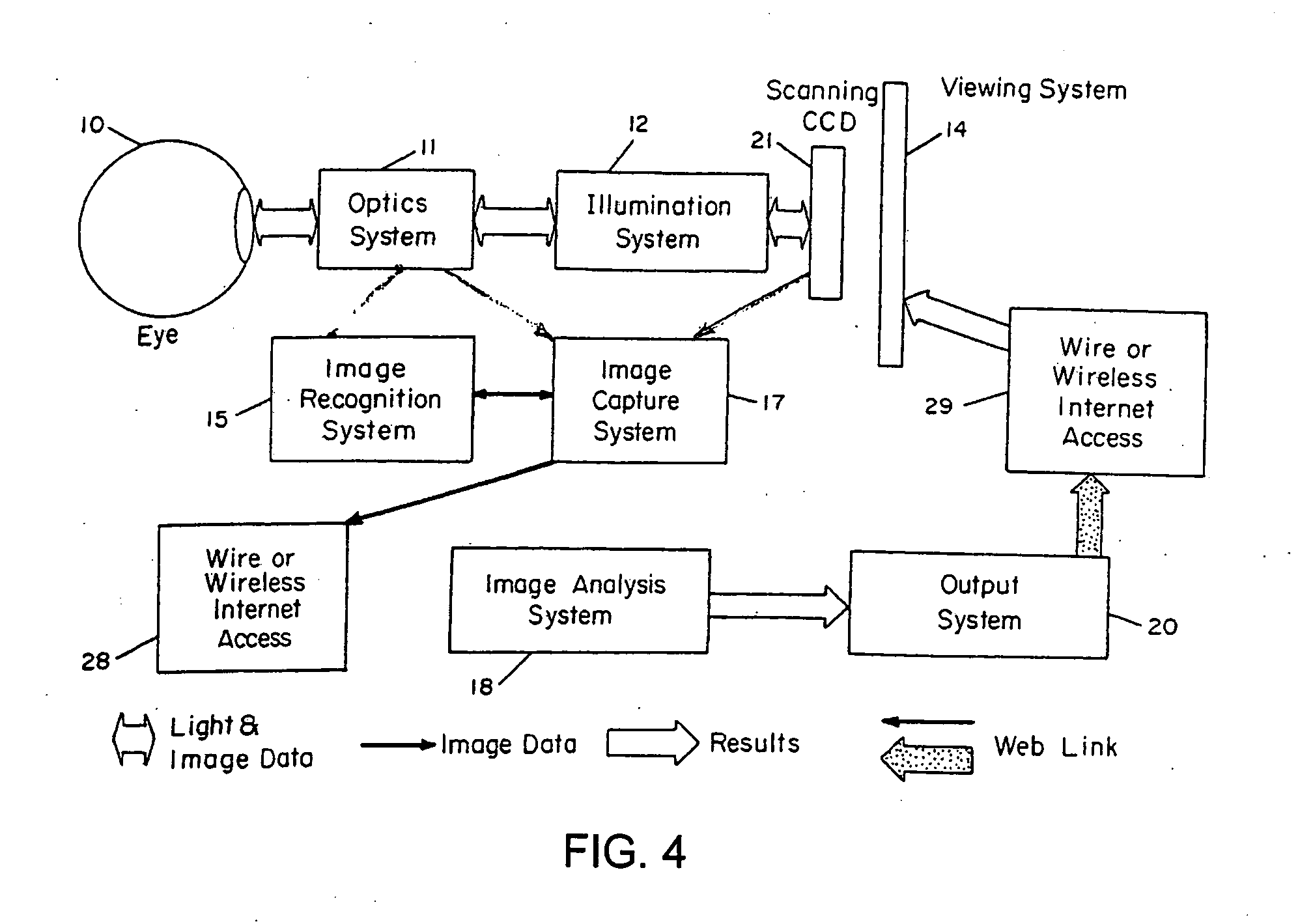
[0006] The present invention carries out measurements of blood glucose in a repeatable, non-invasive manner by measurement of the rate of consumption of glucose, or the rate of production of another substance which is dependent on the glucose concentration of the individual, as an indication of the individual's glucose concentration. The rate of consumption of glucose (or the rate of production of a second glucose concentration-dependent substance) can be the result of the consumption of glucose by a specific organ or part of the body, or by a specific biochemical process in the body. One such process is the rate of regeneration of retinal visual pigments, such as cone visual pigments. The rate of regeneration of visual pigments is dependent upon the blood glucose concentration, by virtue of the glucose concentration limiting the rate of production of a cofactor, NADPH, which is utilized in the rate-determining step of the regeneration of visual pigments. Thus, by measuring the visual pigment regeneration rate, blood glucose can be accurately determined. One preferred embodiment of this invention exposes the retina to light of selected wavelengths at selected times and analyzes the reflection (as color or darkness) from a selected portion of the exposed region of the retina, preferably from the fovea. In addition, since the rate of glucose consumption, or of the production of a glucose-concentration dependent substance can be indicative of illnesses, pathologies or other clinically-significant conditions of the health of the individual, embodiments of this invention can be used to screen for or to diagnose those conditions.

Problems solved by technology

The need to draw blood for analysis is undesirable for a number of reasons, including discomfort to the patient, the high cost of glucose testing supplies, and the risk of infection with repeated skin punctures which results in many patients not testing their blood as frequently as recommended.
As a result of the discomfort, many of these patients do not test as often as is recommended by their physician, with the consequence of poor blood glucose control.
This poor control has been shown to result in increased complications from this disease.
These efforts have been largely unsuccessful primarily due to the variability of absorption and scatter of the light waves in the tissues.
These approaches, which have generally attempted to measure glucose concentration by detecting extremely small optical signals corresponding to the absorbance spectrum of glucose in the infrared or near-infrared portion of the electromagnetic spectrum, have suffered from the size requirements of instrumentation necessary to separate the wavelengths of light for this spectral analysis.
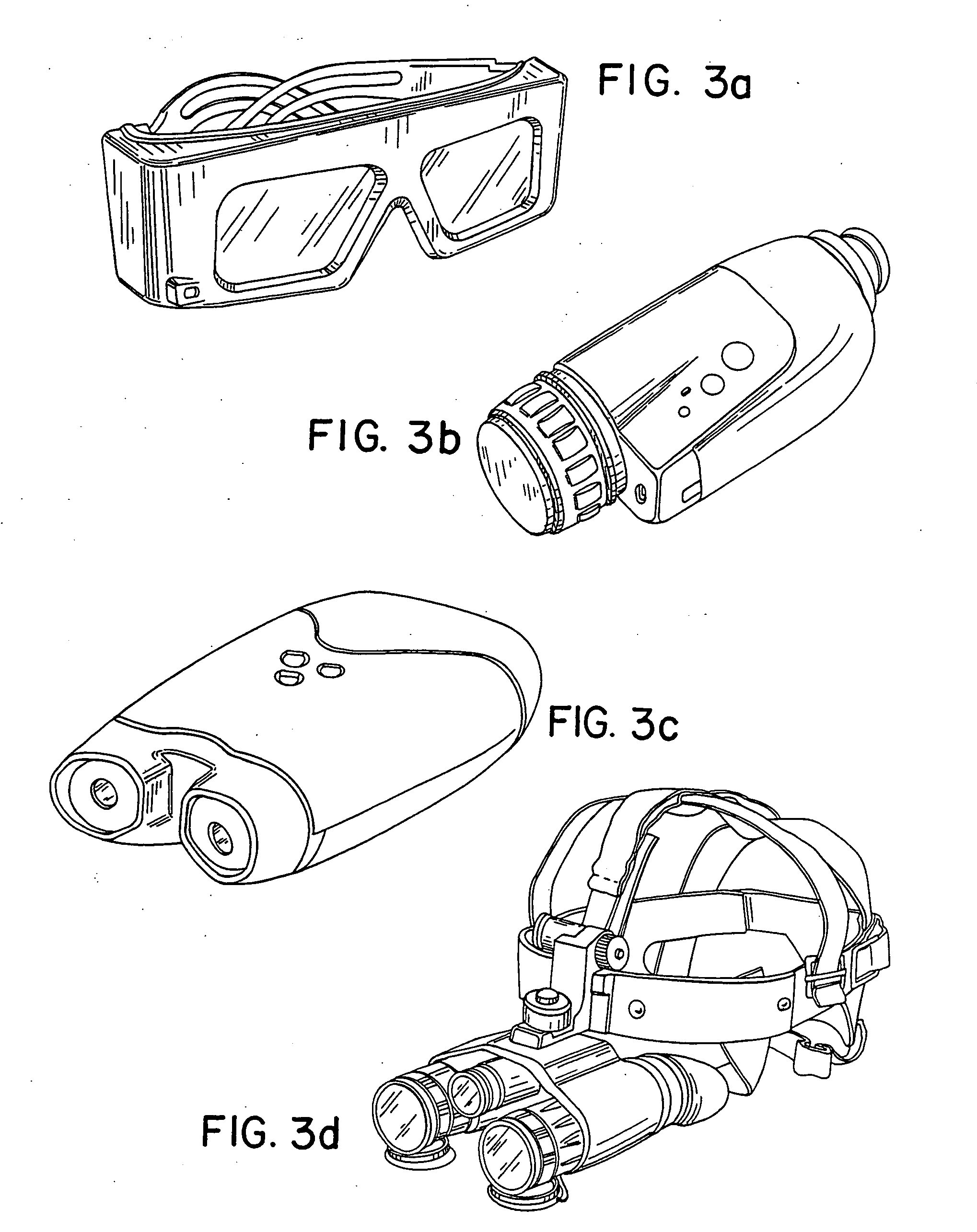
Regardless of approach, the physical size and weight of the instruments described have made it impractical for such a device to be hand-held or worn on the body as a pair of glasses.

Method used


Image

  • Non-invasive measurement of blood glucose using retinal imaging
  • Non-invasive measurement of blood glucose using retinal imaging
  • Non-invasive measurement of blood glucose using retinal imaging

Examples


Embodiment Construction

[0036] Rhodopsin is the visual pigment contained in the rods (that allow for dim vision) and cone visual pigment is contained in the cones of the retina (that allow for central and color vision). The outer segments of the rods and cones contain large amounts of visual pigment, stacked in layers lying perpendicular to the light incoming through the pupil. As visual pigment absorbs light, it breaks down (bleaches) into intermediate molecular forms and initiates a signal that proceeds down a tract of nerve tissue to the brain, allowing for the sensation of sight. During normal vision this bleaching process occurs continuously. Light that reacts with the visual pigments causes a breakdown of those pigments. This phenomenon is termed bleaching, since the retinal tissue loses its color content when a light is directed onto it. In addition, regeneration of the visual pigments occurs at all times, even during the bleaching process. Rod visual pigment absorbs light energy in a broad band cen...


PUM


Abstract

An apparatus carries out measurements of blood glucose in a repeatable, non-invasive manner by measurement of the rate of regeneration of retinal visual pigments, such as cone visual pigments. The rate of regeneration of visual pigments is dependent upon the blood glucose concentration, and by measuring the visual pigment regeneration rate, blood glucose concentration can be accurately determined. This apparatus exposes the retina to light of selected wavelengths in selected distributions and subsequently analyzes the reflection (as color or darkness) from a selected portion of the exposed region of the retina, preferably from the fovea.

Description

CROSS-REFERENCE [0001] This application is a continuation of Ser. No. 10 / 863,619, filed Jun. 8, 2004, which claims the benefit of U.S. Provisional Application No. 60 / 477,245 filed Jun. 10, 2003, which is incorporated herein by reference in its entirety.FIELD OF THE INVENTION [0002] This invention pertains to the field of non-invasive in vivo measurement of blood analytes. BACKGROUND OF THE INVENTION [0003] The measurement of blood glucose by diabetic patients has traditionally required the drawing of a blood sample for in vitro analysis. The blood sampling is usually done by the patient himself as a finger puncture, or in the case of a young child, by an adult. The need to draw blood for analysis is undesirable for a number of reasons, including discomfort to the patient, the high cost of glucose testing supplies, and the risk of infection with repeated skin punctures which results in many patients not testing their blood as frequently as recommended. [0004] Many of the estimated th...

Claims


Application Information

Patent Timeline
Patent Type & Authority Applications(United States)
IPC IPC(8): A61B3/12A61B5/00
CPCA61B5/14532A61B3/10A61B5/6821A61B5/1455
Inventor WOODS, JOE W.SMITH, JOHN L.RICE, MARK J.ROUTT, WILSONMESSERSCHMIDT, ROBERT G.OU, JUNLI
Owner FOVIOPTICS INC
Learn More