
Method to ameliorate osteolysis and metastasis

a technology of osteolysis and metastasis, applied in the field of cancer metastasis prevention and treatment, can solve the problems of systemic elevation of blood calcium levels, no evidence of connecting pth-rp to bone resorption, and add to the uncertainty of the mechanism

Inactive Publication Date: 2005-11-03
XENOTECH CALIFORNIA
View PDF9 Cites 13 Cited by
  • Summary
  • Abstract
  • Description
  • Claims
  • Application Information

AI Technical Summary

Benefits of technology

The invention provides a pharmaceutical formulation for the treatment of localized effects of metastasized cancer cells on bone. The formulation contains an antibody and a bisphosphonate, which can inhibit the growth and osteolysis effects of cancer cells in bone. The treatment is carried out by administering the antibody and bisphosphonate in an injectable formulation. The invention also provides methods to prevent cancer metastasis to bone and inhibit the growth of cancer cells in bone. The protein antibodies and bisphosphonates used in the invention are relatively non-toxic and have minimal side effects.

Problems solved by technology

Both of these result in a systemic elevation of blood calcium levels.
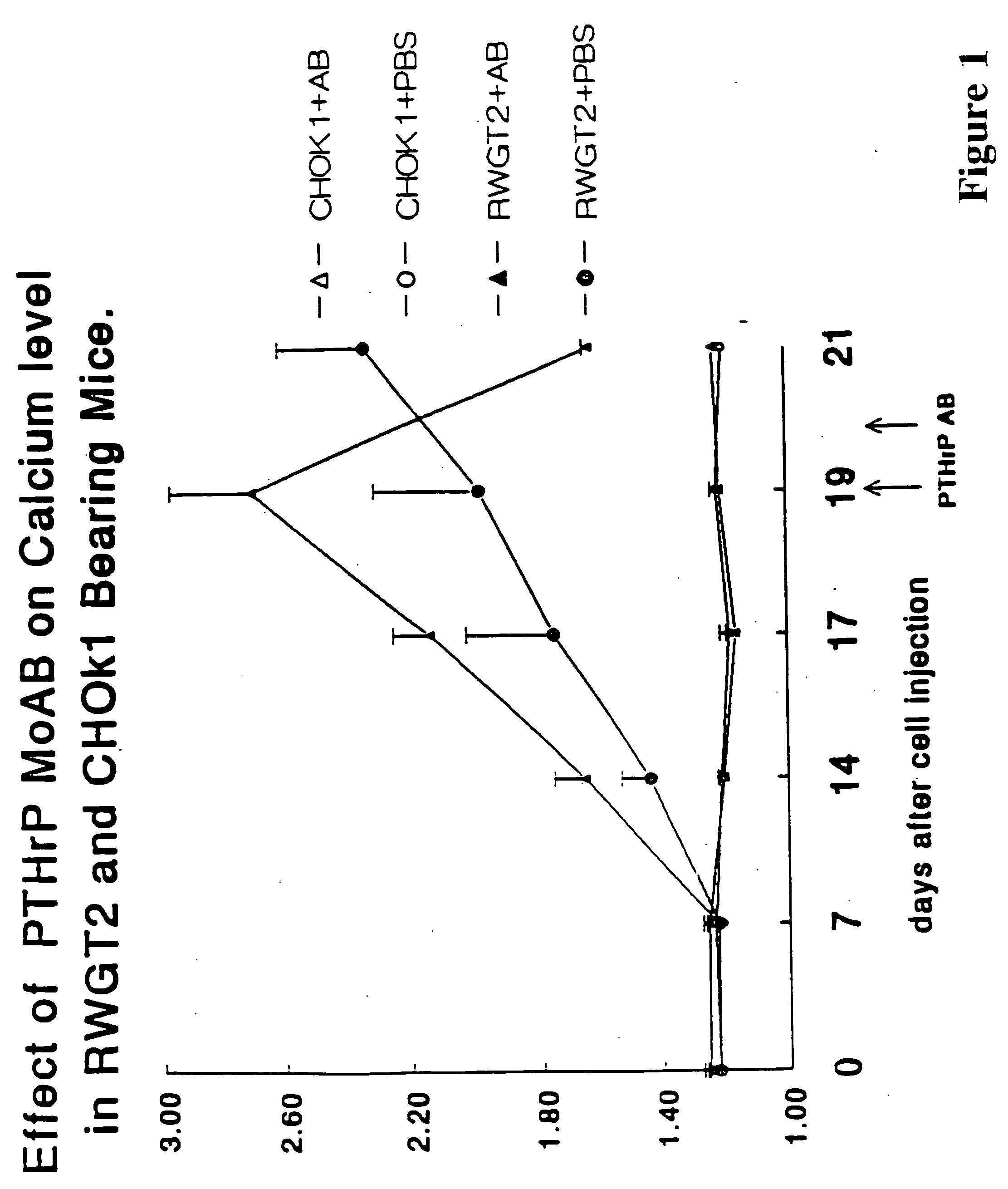
Thus, it is logical that where PTH-rp is circulating, inhibitors of the PTH-like action of PTH-rp would interfere with these biological events, including calcium reabsorption from urine to blood at the kidney tubules and systemic bone resorption.
There is no available evidence to connect PTH-rp to bone resorption mediated by osteoclasts.
The presence of additional factors is particularly important and adds to the uncertainty of the mechanism.
However, the observations related only to the staining of cancer tissues by anti-PTH-rp and are not quantitative.
Thus, the results do not show a causal relationship between the presence of PTH-rp and any acceleration of bone metastasis or growth enhancers.

Method used


Image

  • Method to ameliorate osteolysis and metastasis
  • Method to ameliorate osteolysis and metastasis
  • Method to ameliorate osteolysis and metastasis

Examples


example 1

Effect of Anti-PTH-rp on Bone Resorption Stimulated by Various Agents

[0106] In this example, the percentage of labeled calcium (45Ca) from fetal rat long bone was used as a measure of bone resorption. To label the bones, pregnant female rats at the 18th day of gestation were injected subcutaneously with 50 μCi of labeled calcium salt. The embryos were harvested the next day, at 19 days gestation, and long bones were obtained from the fetuses. The bones were cultured in standard media for 120 hours in the presence or absence of various putative bone resorption stimulation factors and in the presence and absence of the anti-PTH-rp (1-34) antibody used in Preparation A. The results are shown in FIG. 2.

[0107] As shown, untreated bones released roughly 20% of the labeled calcium into the medium over the 120-hour time frame. The addition of 10 ng / ml of PTH-rp to the culture medium results in release of almost 90% of labeled calcium; however, addition of anti-PTH-rp antibodies diminishes...

example 2

Effect of Anti-PTH-rp on Long Bone Lesions

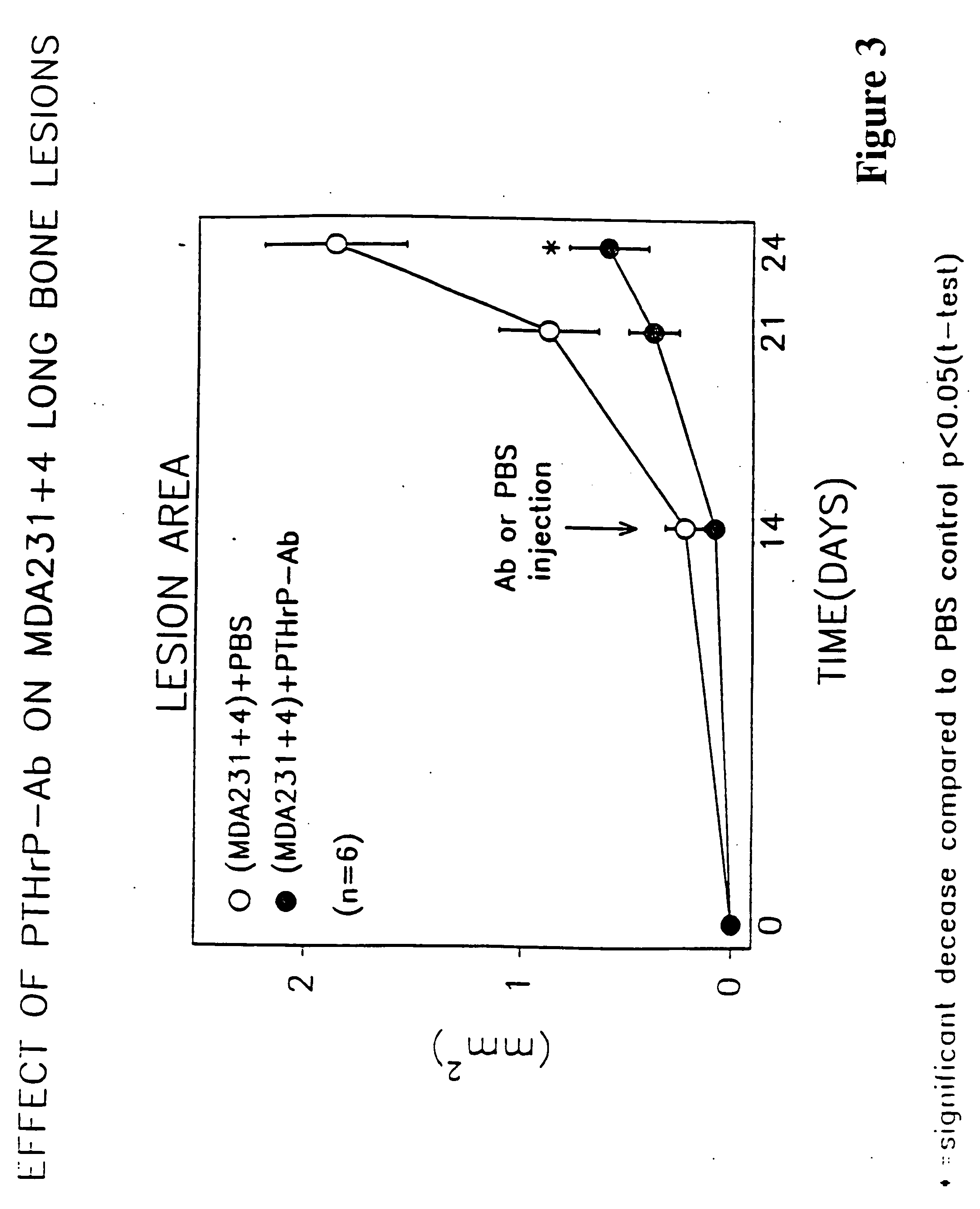
[0108] The osteolytic / metastatic model of Nakai, M. et al. Cancer Res (1992) 52:5395-5399 was used. MDA231±4 cells were injected into the left ventricle with the mice under anesthesia with 0.05 mg / g pentobarbital on day 0. On days 14, 17 and 20, 0.3 ml anti-PTH-rp (1-34) (as per Preparation A described above) containing 75 μg protein or 0.3 ml PBS was injected subcutaneously. The area of bone lesions was estimated on radiographs; the mice were anesthetized deeply, laid down in prone position against the films (22×27 cm X—O Mart AR Kodak, Rochester, N.Y.) and exposed with an x-ray at 35 kvp for 6 seconds using a Faxtron Radiographic Inspection Unit (Model 8050-020, Field Emission Corporation, Inc., McMinnville, Oreg.). Films were developed using an RPX-O Mart Processor (Model M8B, Kodak). All radiographs were evaluated extensively by three different individuals. Metastatic foci, recognized as demarcated radiolucid lesions in bones, were enum...

example 3

[0110] Four week-old female nude mice (Balb c Nu / Nu, Harlan Sprague-Dawley, Houston, Tex.) received the PTH-rPAb (MAb) 7 days prior to (−7) or at the same time (0) of inoculation of human breast cancer-cells MDA-231 (1×10⁵ cells / animal) into the left cardiac ventricle. The anti-PTHrp (1-34) (as per Preparation A described above) antibodies or control IgG (75 μg / animal) were administered subcutaneously every 3 days for 28 days. Development of osteolytic lesions in extremities of these mice were monitored weekly by radiography using FaxitronR (model 43855A, Faxitron X-ray Corp., Buffalo Grove, Ill.). Radiographs taken at day 17, 24 and 26 were examined for osteolytic lesions and area of the lesions in extremities of each mouse was quantified by computer-linked image analyzer. Data are expressed as area of lytic lesion (mm2) per mouse and mean±S.E.M. of twelve nude mice for each treatment group. Statistical difference was analyzed by ANOVA followed by a paired test.

[0111] The results ...


PUM

PropertyMeasurementUnit
temperatureaaaaaaaaaa
pHaaaaaaaaaa
adhesionaaaaaaaaaa

Abstract

A therapeutically effective amount of an antibody for a compound selected from the group consisting of PTHrp, TGFα, IL-1α, IL-1β, IL-6, Lymphotoxin, TNF, PGE; 1,25 dihydroxy vitamin D3 and an antigenic fragment thereof used in the treatment of cancer metastasis to bone and cancer cell growth in bone as well as osteolysis and symptomatic sequelae thereof. An antibody immunoreactive with parathyroid hormone-related protein (PTHrp) is particularly preferred. Antibodies with human characteristics are included in the invention for application of the invention method to human subjects. Also, the antibody can be administered in an injectable is formulation in combination with a therapeutically effective amount of a bisphosphonate or pyrophosphate having the general structure formula wherein X is a linking moiety allowing for the interconnection of the phosphonate groups, and pharmaceutically acceptable salts, hydrates and partial hydrates thereof. The antibody and bisphosphonate act synergistically in the treatment of cancer metastases to bone and symptomatic sequelae thereof and particularly as regards bone resorption.

Description

CROSS REFERENCE TO RELATED APPLICATIONS [0001] This application is a Continuation-in-Part of our earlier filed application Ser. No. 08 / 481,088, filed Jun. 6, 1995 which is a continuation-in-part of our earlier application Ser. No. 08 / 386,361, filed Feb. 9, 1995 which is a Continuation-in-part of application Ser. No. 08 / 376,359, filed 23 Jan. 1995 which applications are incorporated herein by reference and to which applications we claim priority under 35 USC §120.TECHNICAL FIELD [0002] The invention relates to the prevention and treatment of cancer metastasis to bone and bone cancer growth and to associated osteolysis, some of the most problematic aspects of cancers of many origins. More specifically, the invention concerns use of antibodies to parathyroid hormone-related protein (PTH-rp) and other forms of anti-PTH-rp alone or in combination with a bisphosphonate to alleviate these conditions. BACKGROUND OF THE INVENTION [0003] It has long been understood that morbidity and mortalit...

Claims


Application Information

Patent Timeline
Patent Type & Authority Applications(United States)
IPC IPC(8): A61K9/08A61K31/66C12N15/09A61K31/675A61K38/00A61K39/395A61K45/06A61P35/00A61P43/00C07K16/22C07K16/24C07K16/26C12P21/08
CPCA61K38/00A61K45/06C07K16/26C07K16/241C07K16/244C07K16/22A61P35/00A61P35/04A61P43/00A61K39/395A61K31/36
Inventor MUNDY, GREGORY R.YONEDA, YOSHIYUKIGUISE, THERESA A.
Owner XENOTECH CALIFORNIA
Learn More