
Dissolvable medical sealing device

a sealing device and dissolvable technology, applied in the field of wound closure devices, can solve the problems of increasing solubility and absorbability, restricting the flow of blood through the vessel, and unduly long time-consuming for occluders known in the state of the ar

Active Publication Date: 2005-08-04
TERUMO MEDICAL CORP
View PDF21 Cites 1311 Cited by
  • Summary
  • Abstract
  • Description
  • Claims
  • Application Information

AI Technical Summary

Benefits of technology

[0011] An object of the invention is to provide a safe and reliable wound closure device not causing problems to the patient.
[0017] The present invention overcomes the disadvantages of having an unnecessarily long degradation time encountered in arterial sealing devices made of an absorbable material that at least partly degrades by means of biological and / or chemical processes. The invention provides a sealing device that can be made of a material that dissolves inside a vessel (or outside the vessel in the case of an extra-arterial occluder) by physical processes. This reduces the time during which the sealing device is present in the body to a few days.

Problems solved by technology

However, applying direct pressure on the wound will require assistance of additional medical personnel and may also restrict the flow of blood through the vessel.
The biological and chemical degradation processes typically lower the molecular weight of the polymeric structure, which increases solubility and absorbability.
This means that the occluders known in the state of the art remain in the body an unduly long time.
If another medical operation is going to be performed at the same operation site as the first operation, it is a disadvantage to have an occluder already inserted in the vessel, since this occluder can obstruct the medical operation itself and may also make it impossible to safely position a new occluder at (or near) the same position.
A problem with the materials of these known seals is that different parts of the seals will be in different states of degradation during the absorption process.
Therefore the seals become increasingly porous and will exhibit lower rupture strength, and due to the blood flow in the vessel there is a risk that pieces of different sizes of the seal come loose and follow the blood flow to more narrow passages where the parts can get stuck and restrict or even prevent the blood flow.
Of course this could be serious for the patient and may necessitate an operation.
Furthermore, a suture or filament that is holding the seal in place is rather thin in comparison with a seal and thus there is a risk that the suture or filament ruptures through a seal being in more or less degraded state.
A further problem with the known seals is that around the seal cells and tissues develop on and adhere to the surface, so that the seal becomes encapsulated in a sac-like tissue material.
Furthermore, this problem may be enhanced by the fact that many conventional seals are made of materials that swell in the fluid inside a vessel.
The ingrowth of the encapsulating tissue can restrict blood flow temporarily or even permanently, particularly if the encapsulating tissue fails to regress properly.
Also, if the tissue does not grow on the surface of a conventional seal, then the seal can potentially come loose inside the blood vessel.
Without the encapsulation the breaking down of the seal would probably cause parts of the seal to come loose and drift away with the blood flow.
However, it is not safe to rely on this effect since it could vary between individuals, and furthermore even small rests of certain materials, such as Teflon®, from the manufacturing of the seal could be left on the surface of the seal and prevent the adhering of the cells.

Method used


Image

  • Dissolvable medical sealing device

Examples


first embodiment

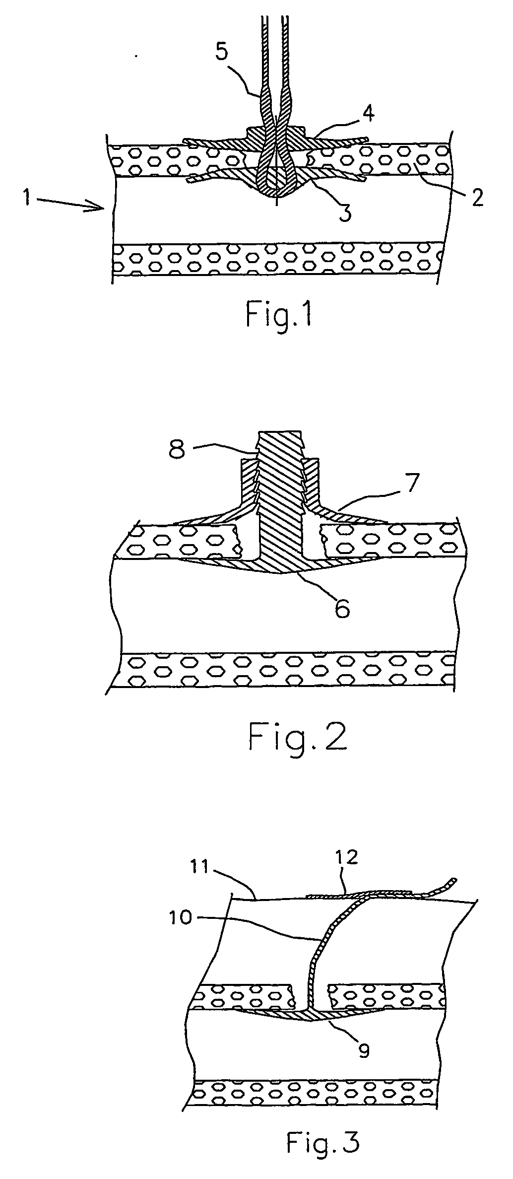
[0021] In FIG. 1 is shown a portion of a vessel 1 in a living body, whether a human or animal body, such as the femoral or radial artery. A puncture has been made through the vessel wall 2, thereby creating an opening, which has to be occluded after the treatment that made the puncture necessary. In FIG. 1, a wound closure device according to the present invention has been positioned to close the puncture wound. The wound closure device comprises a first sealing device 3, which is positioned against the inner surface of the vessel wall 2, and a second sealing device 4, which is positioned against the outer surface of the vessel wall 2. The wound closure device also comprises a fastener 5 in the form of a multifilament 5, which holds the first and second sealing devices 3, 4 together by means of friction locking.

second embodiment

[0022] a wound closure device according to the present invention is illustrated in FIG. 2. The wound closure device comprises a first sealing device 6, which is positioned against the inner surface of the vessel wall, and a second sealing device 7, which is positioned against the outer surface of the vessel wall. In this embodiment, the first and second sealing devices 6, 7 are held together by a saw-toothed guide 8, which extends integrally from the first sealing device 6.

third embodiment

[0023]FIG. 3 illustrates a wound closure device according to the present invention. The closure device shown in FIG. 3 comprises a sealing device 9, through which a suture 10 is threaded. In use, the sealing device 9 is urged toward the inner surface of the vessel wall by simply pulling the suture 10. In order to hold the sealing device 9 in place, the suture is then held and is secured in position on the patient's skin 11, such as by use of a strip of conventional tape 12.

[0024] As mentioned above, sealing devices of the types shown in FIG. 1 to FIG. 3 are conventionally made from materials that are described as being absorbable or biodegradable. Such sealing devices degrade by biological or chemical processes, in which covalent bonds in the molecules of the materials are broken. This can mean, for example, that a polymeric material is hydrolysed so that its molecular weight is reduced. Because of these relatively slow degradation processes, the corresponding degradation times may ...


PUM

PropertyMeasurementUnit
Timeaaaaaaaaaa
Timeaaaaaaaaaa
Timeaaaaaaaaaa

Abstract

The present invention provides a dissolvable medical sealing device (3, 4; 6, 7; 9) for closing a wound in vessel. A sealing device (3, 4, 6, 7, 9) according to the invention is made of a material that dissolves by means of physical processes, rather than by means of chemical or biological processes. Such a sealing device (3, 4; 6, 7; 9) can be made of polyethylene glycol, polypropylene glycol, copolymers containing ethylene glycol and propylene glycol, polyvinyl alcohol or polyvinyl pyrolidone, or any combinations thereof.

Description

FIELD OF THE INVENTION [0001] The present invention relates to a wound closure device for closing a wound in a vessel, comprising a first sealing device adapted to be positioned against the inner surface of the vessel wall and a fastener adapted to keep the first sealing device in place. BACKGROUND OF THE INVENTION [0002] During certain types of medical surgery or treatment, an introducer is used to access the vascular system of a patient. The introducer is inserted through the wall of a blood vessel in order to obtain access to the vascular system and may thereafter be used for guiding medical instruments such as catheters, guide wires and the like. [0003] After completion of the medical procedure, there will be an incision or a wound in the wall of the blood vessel corresponding to the size of the introducer. The bleeding from the wound, which is the result of such a surgical operation, can be stopped by applying direct pressure on the wound. However, applying direct pressure on t...

Claims


Application Information

Patent Timeline
IPC IPC(8): A61B17/00A61B17/12A61B17/03A61B17/064A61B17/08
CPCA61B17/0057A61L31/042A61B2017/00004A61B2017/00659A61B2017/0647A61L26/0009A61L17/10A61L26/0061A61L31/04A61L17/06A61B2017/00615A61B2017/00778A61L31/148A61K9/0024A61B17/0643
Inventor TENERZ, LARSAKERFELDT, DANMATHISEN, TORBJORN
Owner TERUMO MEDICAL CORP
Learn More