
Method for implanting flexible injection port

a flexible injection port technology, applied in the field of medicine, can solve the problems of uncomfortable or cosmetically objectionable protruding ports

Inactive Publication Date: 2005-06-16
ETHICON ENDO SURGERY INC
View PDF17 Cites 76 Cited by
  • Summary
  • Abstract
  • Description
  • Claims
  • Application Information

AI Technical Summary

Benefits of technology

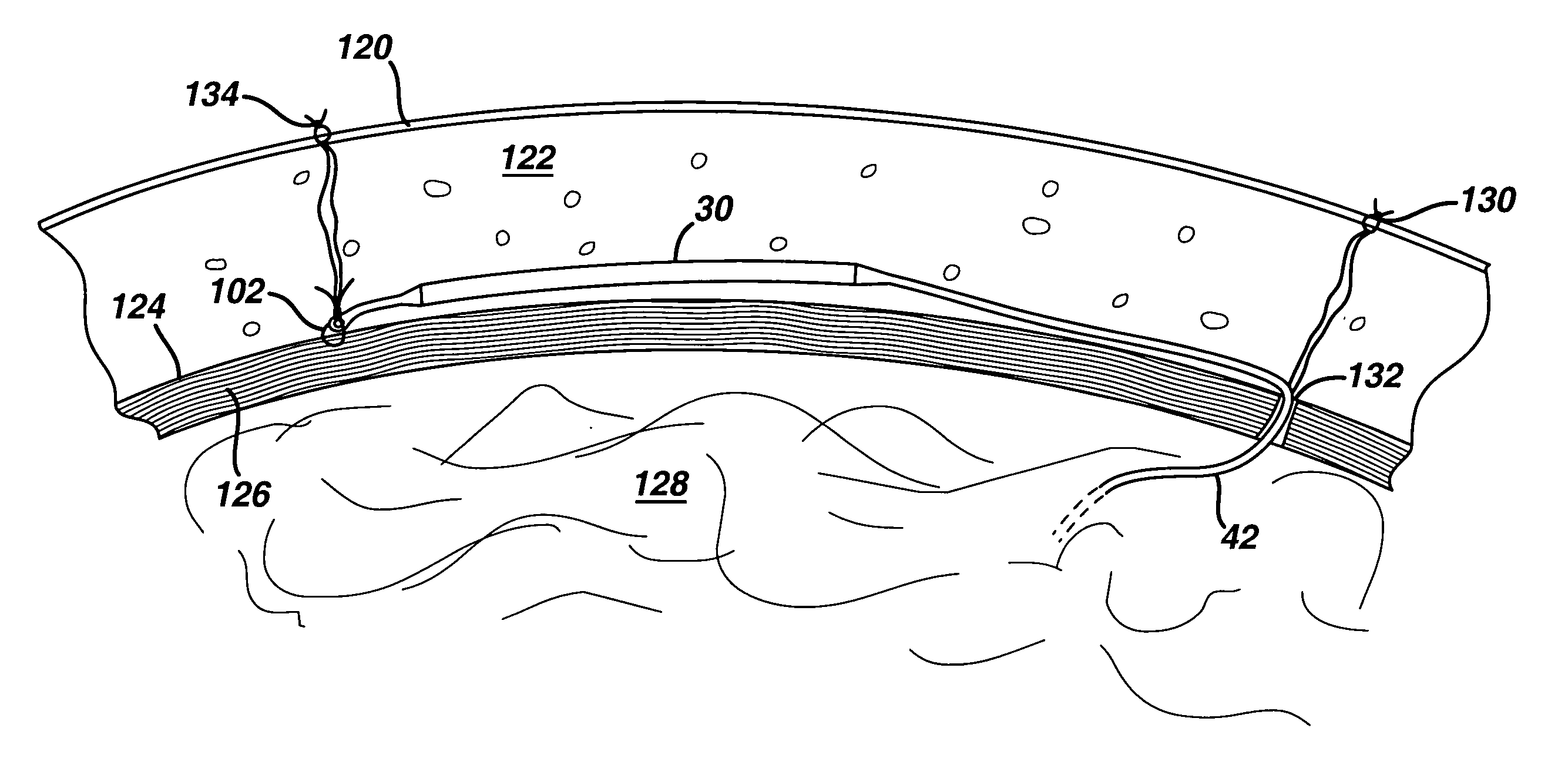
[0034] The present invention, a flexible injection port, as described in the preceding embodiments and their equivalents, has numerous advantages over the prior art injection ports. The flexible injection port may not require attachment to fascia, thus reducing the duration of the surgical procedure. The flexible injection port may require a smaller incision size and less tissue dissection for implantation, so that the patient has less pain, less scarring, a faster recovery, and less possibility of infection. Due to the integral construction of the flexible injection port and the catheter, the step of connecting the catheter to the injection port during the surgical procedure is not necessary, thus potentially reducing the number of surgical complications due to fluid leakage at the connection. Because the flexible injection port may be implanted in the fat layer near the skin surface, the surgeon or a trained clinician may use palpation to locate the injection port, and standard IV techniques to administer fluid, yet the implant is still cosmetically acceptable to the patient. In addition, shorter injection needles may be used to reduce patient anxiety during fluid administration. The flexible injection port may have no metallic parts, resulting in a flexible and lightweight implant for greater patient comfort and compatibility with magnetic resonance and fluoroscopic x-ray imaging. Finally, the injection portion of the flexible injection port is accessible with a hypodermic needle for most of the possible orientations of the flexible injection port within the subcutaneous fat layer of the patient.

Problems solved by technology

Although easy to locate with palpation, some patients regard the protruding port as uncomfortable or cosmetically objectionable.

Method used


Image

  • Method for implanting flexible injection port
  • Method for implanting flexible injection port
  • Method for implanting flexible injection port

Examples


first embodiment

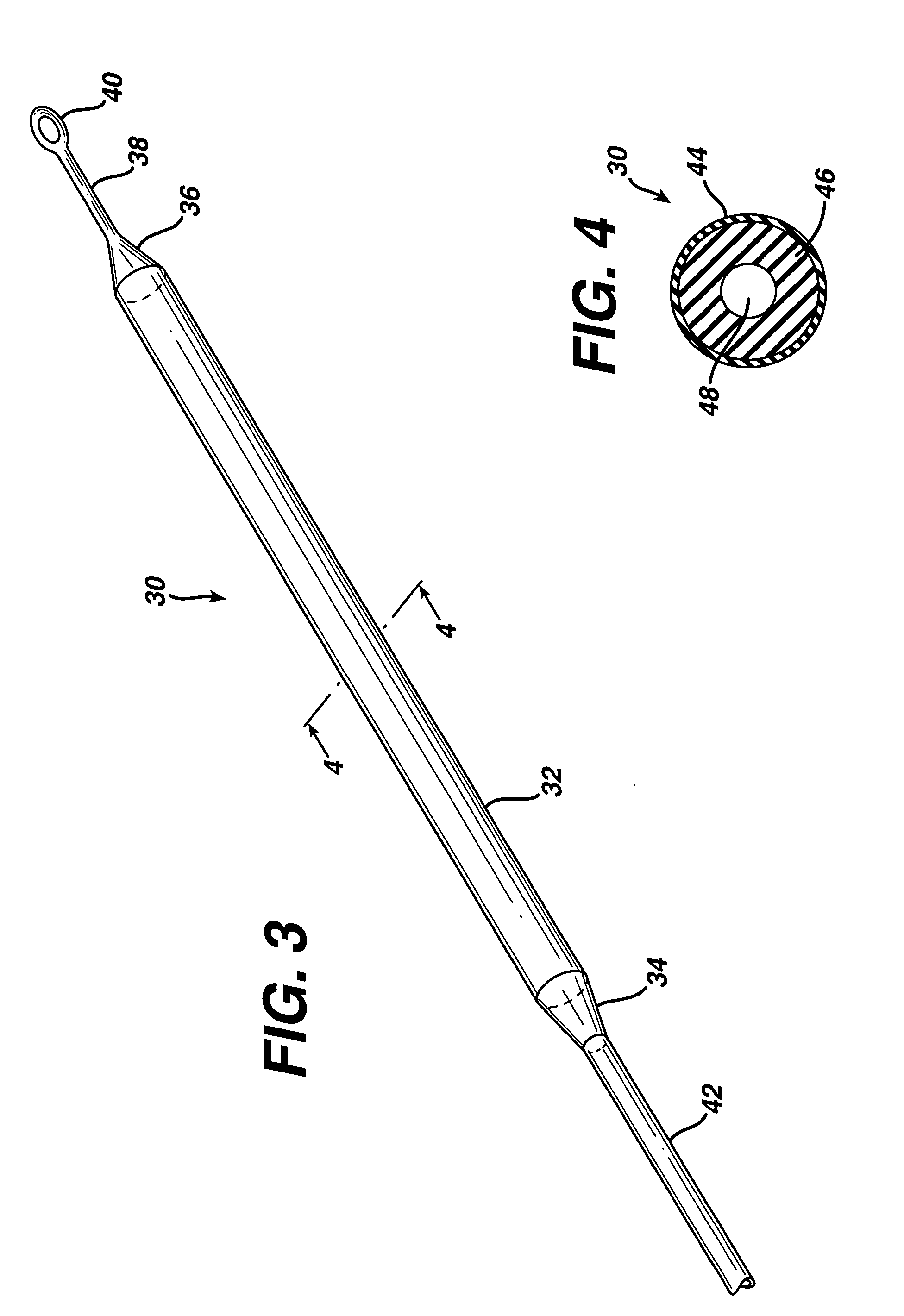
[0023]FIG. 3 is an isometric view of the present invention showing a flexible injection port or body 30, that generally comprises a first end 34, a second end 36, and a cylindrical injection portion 32 extending there between. A surgeon may use a hypodermic needle or the like to penetrate injection portion 32 and introduce a fluid such as a medication or saline solution into flexible injection port 30. Injection portion 32 self-seals when the surgeon removes the hypodermic needle. Injection portion 32 may have a length, but is not limited to, approximately 5-20 cm. Injection portion 32 may have a diameter, but is not limited to, approximately 5-12 mm. A catheter 42 attaches to first end 34 and distributes fluid injected into flexible injection port 30 to another portion of the patient's body. Catheter 42 is made from a silicone rubber or other biocompatible polymer such as known in the art for application to conventional injection ports, such as shown in FIGS. 1 and 2. A tether 38 h...

fourth embodiment

[0029]FIG. 8 is the present invention, a flexible injection port 80, comprising a first end 84 that attaches to a catheter 92, a second end 86 and an injection portion 82. Flexible injection port 80 further comprises a webbing 88 attached to and covering at least injection portion 82, and made of a thin, flexible, implantable material such as a polyester or polypropylene mesh, expanded PTFE, or the like. Webbing 88 provides broad margins for stapling or suturing to an underlying tissue such as fascia, as well as a large area for tissue in-growth, to enhance long-term stability and to substantially prevent migration of flexible injection port 80. FIG. 9 is a cross sectional view of flexible injection port 80, taken at line 9-9 of FIG. 8. Flexible injection port 80 comprises an outer tube 94 made of a heat shrinkable, PTFE material, and an inner tube 96 made of a silicone rubber having a durometer of approximately 20-40. Webbing 88 includes a pair of webbing layers, 91 and 93, that ma...


PUM


Abstract

In accordance with the present invention, there is provided a method for subcutaneously implanting an injection port for use with an implantable medical device. The method involves providing an injection port comprising an elongated flexible substantially non-rigid body having first and second ends and a wall therebetween, the wall is such that it will self seal after being punctured, the body further including and a fluid reservoir surrounded by the wall and a flexible elongated tubular catheter attached to the body which is in fluid communication with the reservoir. Thereafter, the method involves creating an incision within the patient, accessing the subcutaneous fat layer of the patient through the incision, creating a space in the subcutaneous fat layer and implanting the injection port within the subcutaneous fat layer such that the port can be found externally by palpitation.

Description

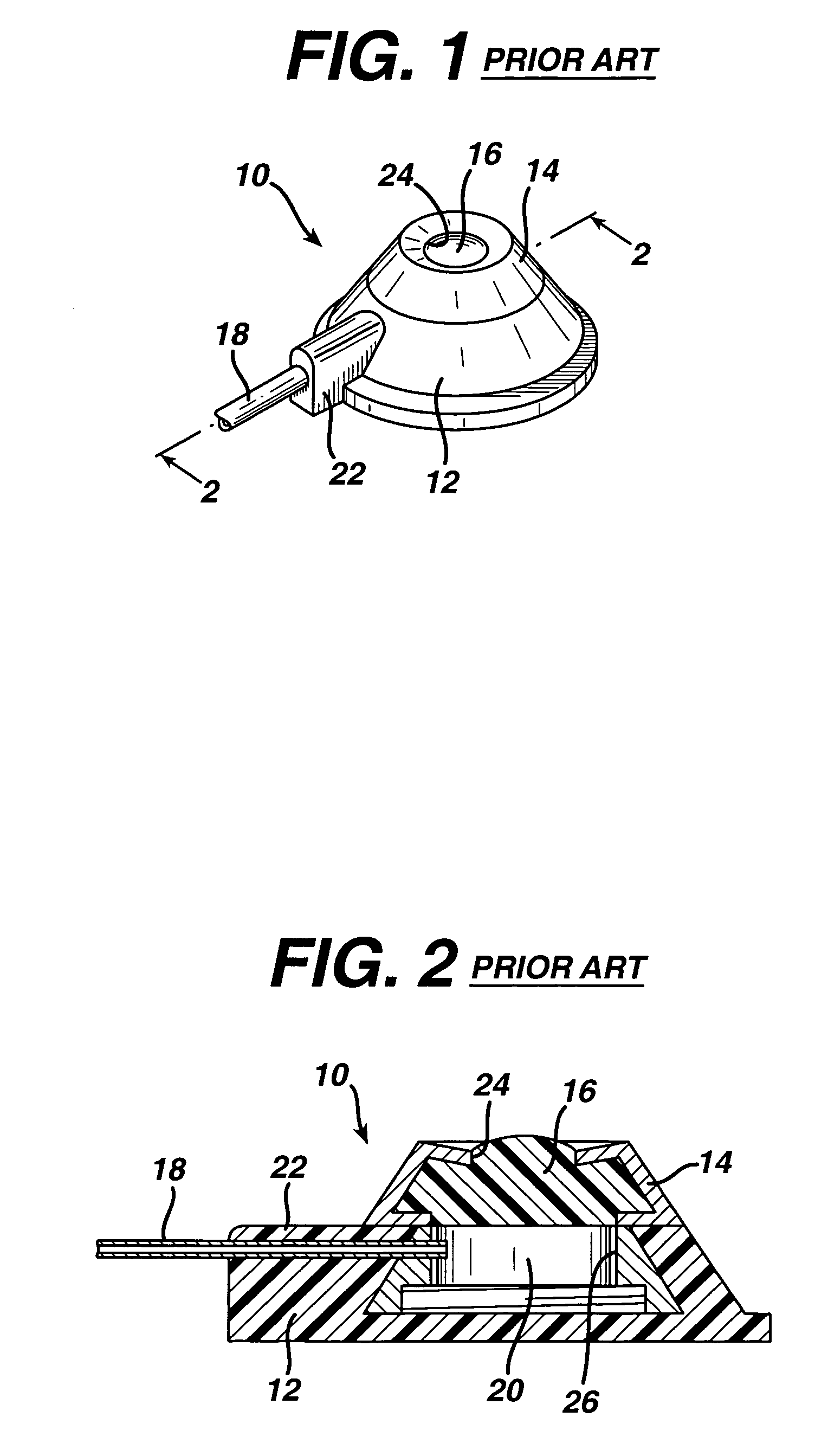
FIELD OF THE INVENTION [0001] This invention relates generally to the field of medicine, and more specifically to medical devices that are surgically implanted in a patient, and is particularly relevant to implantable injection or infusion ports such as used for chemotherapy and adjustable gastric band procedures. BACKGROUND [0002] Surgeons routinely implant subcutaneous injection ports in patients requiring long term, periodic fluid injections such as for chemotherapy and gastric band adjustments. The injection port connects to a flexible tube catheter to transport the fluid to the affected area (subclavian vein, etc.) or the gastric band. Current injection ports comprise a rigid metal or plastic housing, which is about 25 mm in diameter and 15 mm tall. A thick, silicone septum captured within the rigid housing covers an inner chamber that fluidly communicates with the catheter. The surgeon uses a hypodermic needle to inject fluid into the chamber through the silicone septum. [0003...

Claims


Application Information

Patent Timeline
IPC IPC(8): A61B17/00A61M5/00A61M37/00A61M25/00A61M31/00A61M39/00A61M39/02A61M39/04
CPCA61M39/0208A61M2039/022A61M2039/0072
Inventor CHEN, HOW-LUNCONLON, SEAN P.SCHULZE, DALE R.
Owner ETHICON ENDO SURGERY INC
Learn More