
Methods of isolation, expansion and differentiation of fetal stem cells from chorionic villus, amniotic fluid, and placenta and therapeutic uses thereof

Inactive Publication Date: 2005-06-09
CHILDRENS MEDICAL CENT CORP
View PDF34 Cites 225 Cited by
  • Summary
  • Abstract
  • Description
  • Claims
  • Application Information

AI Technical Summary

Benefits of technology

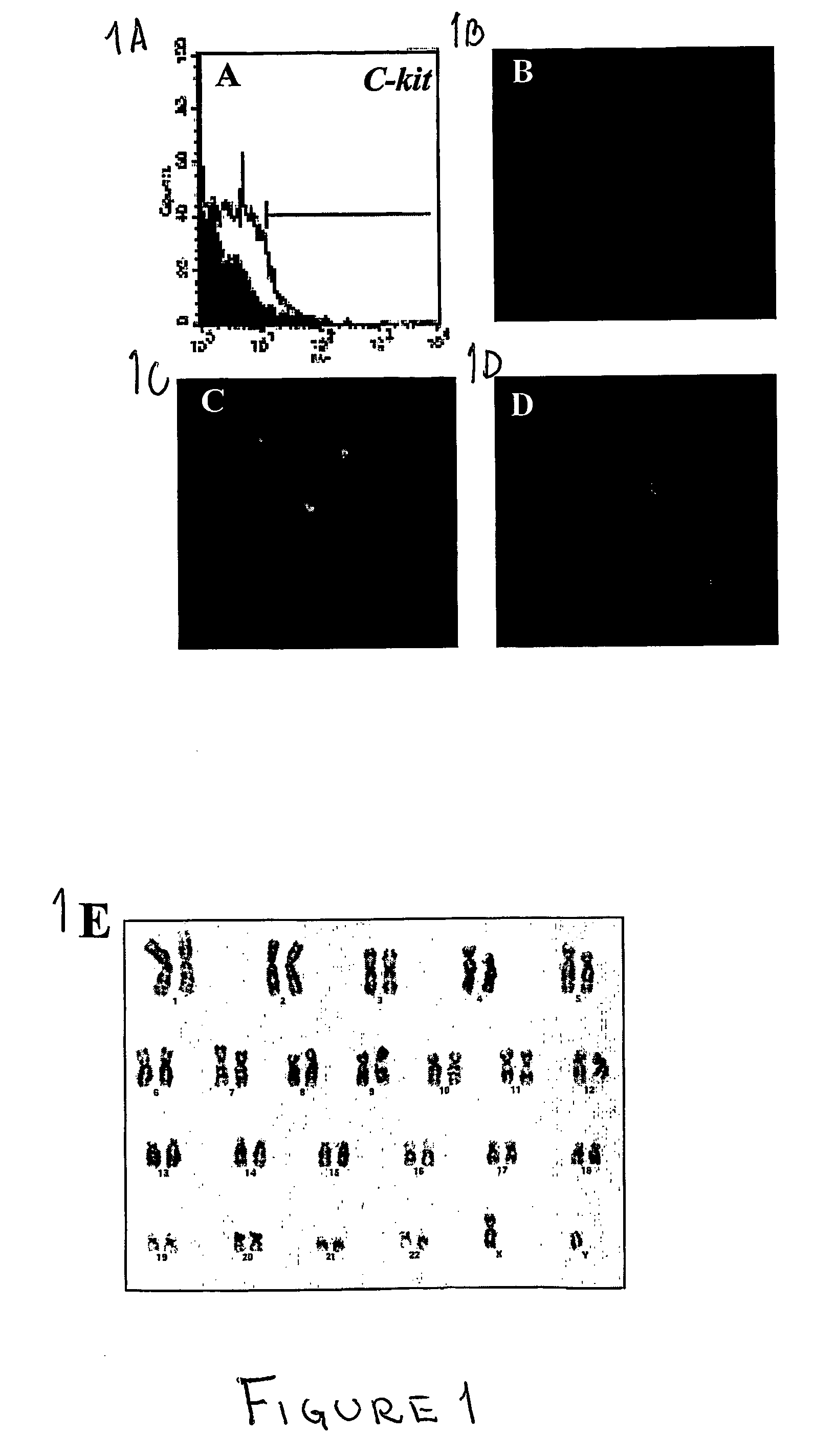
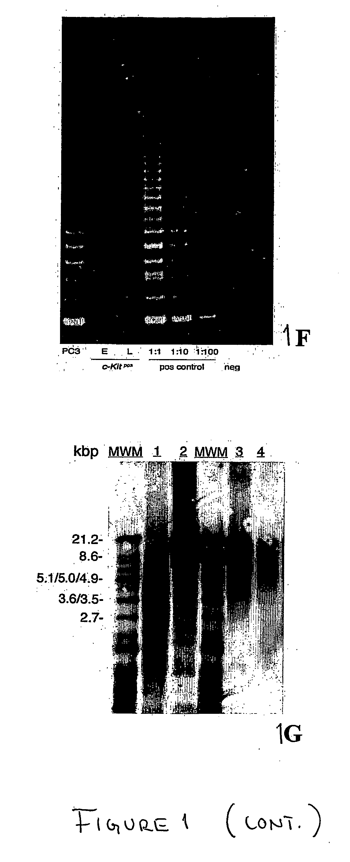
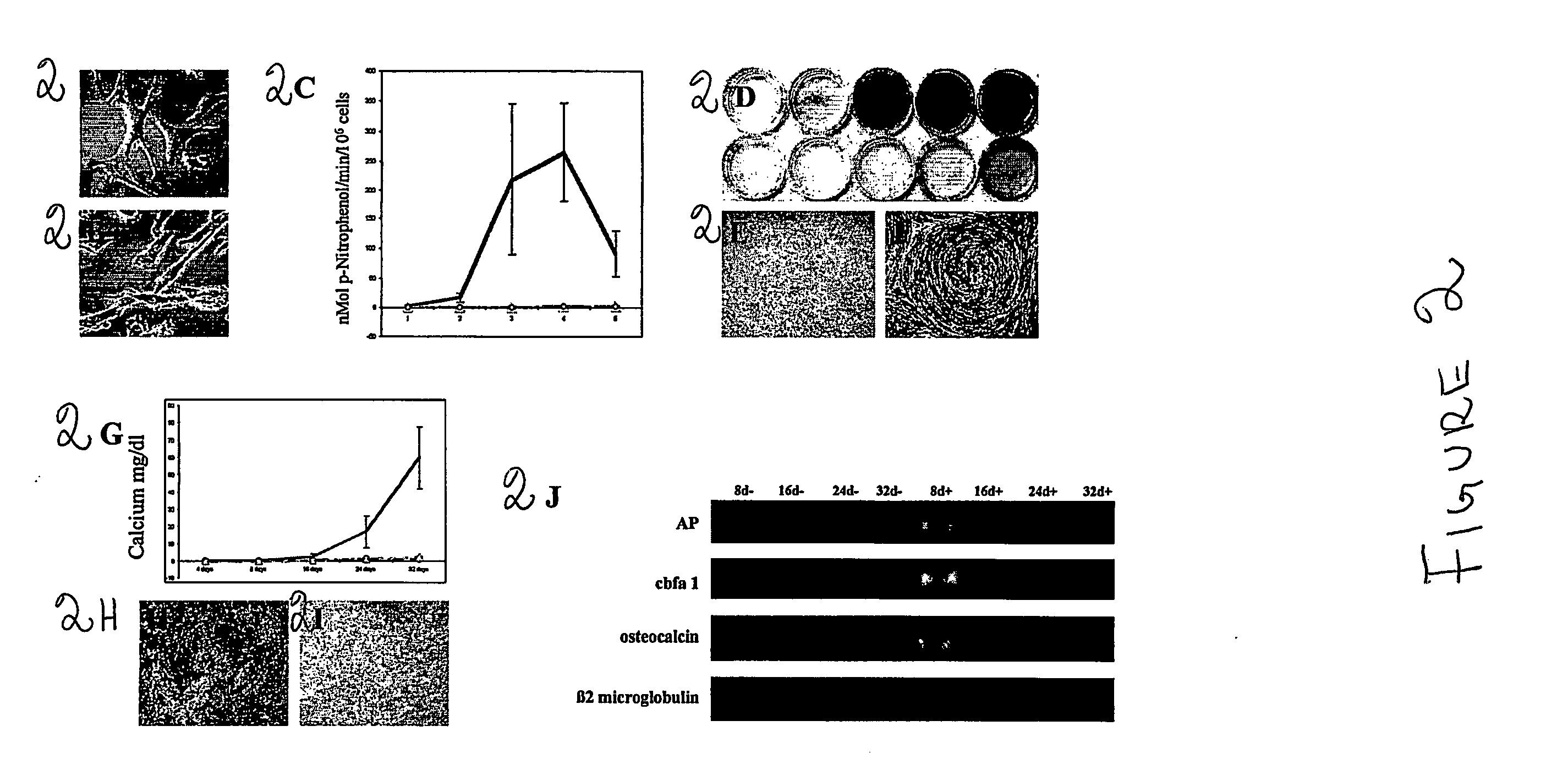
[0008] We have discovered that chorionic villus, amniotic fluid, and placenta provide an excellent source of pluripotent fetal stem cells for therapeutic applications. These fetal stem cells have a better potential for expansion than adult stem cells and avoid the current controversies associated with the use of human embryonic stem cells. The c-kitpos cells isolated from the chorionic villus, amniotic fluid and placenta samples differentiate into specific cell lineages, they do not need feeder layers to grow, and most importantly, the isolation of these cells does not require the sacrifice of human embryos for their isolation, thus avoiding the current controversies associated with the use of human embryonic stem cells.

Problems solved by technology

Although stem cells in adult tissues may be capable of developing into more cell types than originally thought, they have a limited cellular regeneration or turnover.
Adult stem cells have the disadvantage of not being immortal, and most of them lose their pluripotency after a defined number of passages in culture.
This short life-span may be a problem for clinical applications where a large amount of cells are needed.
ES cells tend to differentiate spontaneously into various types of tissues; however, specific growth induction conditions do not direct differentiation exclusively to specific cell types.
Although there is a great scientific interest in ES cell research, the destruction of embryos in order to harvest and experiment on ES cells still create unresolved ethical concerns.
However, fetal cell transplants are plagued by problems that are very difficult to overcome.
Fetal tissue can also be obtained from aborted embryos, but this resource is limited.
Beyond the ethical concerns regarding the use of cells from aborted fetuses or living fetuses, there are other issues which remain a challenge.

Method used


Image

  • Methods of isolation, expansion and differentiation of fetal stem cells from chorionic villus, amniotic fluid, and placenta and therapeutic uses thereof
  • Methods of isolation, expansion and differentiation of fetal stem cells from chorionic villus, amniotic fluid, and placenta and therapeutic uses thereof
  • Methods of isolation, expansion and differentiation of fetal stem cells from chorionic villus, amniotic fluid, and placenta and therapeutic uses thereof

Examples


examples

[0093] In this example the feasibility of isolating stem cells from human embryonic and fetal chorionic villi and amniotic fluid was investigated. Discarded cultures of chorionic villi cells and human amniotic fluid cells collected for prenatal diagnostic tests were obtained from more than 300 human pregnant females ranging from 23 to 42 years of age under an approved institutional Investigation Review Board protocol.

[0094] To establish the cultures, human amniotic fluid was obtained by transabdominal amniocentesis at 14 to 21 weeks of gestation, and human embryonic chorionic villus tissue specimens were obtained at 10 to 12 weeks of gestation through a transabdominal approach.

[0095] Amniotic fluid samples were centrifuged and the cell supernatant was resuspended in culture medium. Approximately 104 cells were seeded on 22×22 mm cover slips. Cultures were grown to confluence for about 3 to 4 weeks in 5% CO2 at 37° C. Fresh medium was applied after five days of culture and every th...


PUM

PropertyMeasurementUnit
Antigenicityaaaaaaaaaa

Abstract

The present invention is directed to pluripotent fetal stem cells derived from chorionic villus, amniotic fluid, and placenta and the methods for isolating, expanding and differentiating these cells, and their therapeutic uses such as manipulating the fetal stem cells by gene transfection and other means for therapeutic applications.

Description

FIELD OF THE INVENTION [0001] This invention relates to the isolation, expansion and differentiation of fetal stem cells from chorionic villus, amniotic fluid, and placenta and therapeutic uses thereof. BACKGROUND OF THE INVENTION [0002] Stem cells are unique cell populations with the ability to undergo both renewal and differentiation. This fate choice is highly regulated by intrinsic signals and the external microenvironment. They can be identified in many adult mammalian tissues, such as bone marrow, skeletal muscle, skin and adipose tissue, where they contribute to replenishment of cells lost through normal cellular senescence or injury. Although stem cells in adult tissues may be capable of developing into more cell types than originally thought, they have a limited cellular regeneration or turnover. [0003] Stem cells have been reported to exist during embryonic development and postnatally in bone marrow, skeletal muscle and skin. Embryonic stem (ES) cells are derived from the ...

Claims


Application Information

Patent Timeline
IPC IPC(8): C12N15/09A61K35/12A61K35/14A61K35/30A61K35/32A61K35/34A61K35/407A61P1/16A61P7/00A61P19/08A61P21/00A61P25/00A61P25/16A61P43/00C12N1/04C12N5/02C12N5/073C12N5/074G01N33/53G01N33/567
CPCA61K35/12C12N5/0607C12N5/0605A61P1/16A61P19/08A61P21/00A61P25/00A61P25/16A61P43/00A61P7/00
Inventor ATALA, ANTHONYDE COPPI, PAOLO
Owner CHILDRENS MEDICAL CENT CORP
Learn More