Method for stimulating wound healing
a wound healing and wound technology, applied in the direction of antibody medical ingredients, drug compositions, peptide/protein ingredients, etc., can solve the problems of skin erosion, ulceration, delay in healing process, etc., and achieve the effect of stimulating wound healing and stimulating wound healing
- Summary
- Abstract
- Description
- Claims
- Application Information
AI Technical Summary
Benefits of technology
Problems solved by technology
Method used
Image

Examples
example 1
Preparation of p43 Mutant Mouse
[0054] To prepare p43 mutant mice, p43 structural gene was allowed to be mutated using the gene trap method [Cecconi, F. & Meyer, B. I., FEBS Lett., 480:63-71, 2000]. The gene trap vector, VICTR20 (Lexicon Genetics, USA) was used to mutate the genomic DNA of SvEvBrd mice (Lexicon Genetics, USA) according to Zambrowicz's method [Zambrowicz, B. P. et al., Nature 392:608-611, 1998]. The mutated genomic DNA was introduced into the embryonic stem cells derived from 129 / SvEvBrd mice (Omnibank) to generate the mutant library. In the library, the clone containing p43 gene disrupted by the insertion of the gene trap vector was screened and called ‘OST58507’. The clone was used to generate the heterozygous C57 / BL6 mice (Samtako) by the standard method of Lexicon Genetics. The mating of the heterozygous mice generated 16 of wild type, 53 of heterozygous mutant mice and 28 of homozygous mutant mice. The ratio is close to the Mendelian segregation, indicating no s...
example 2
Characterization of p43 Mutant Mouse
2.1 Determination of the Gene Trap Vector's Site Inserted into p43 Allele
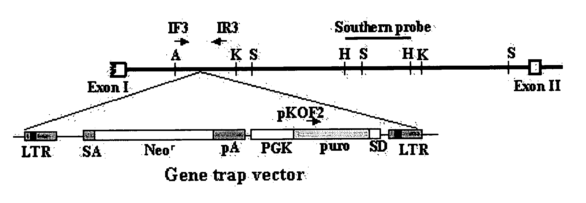
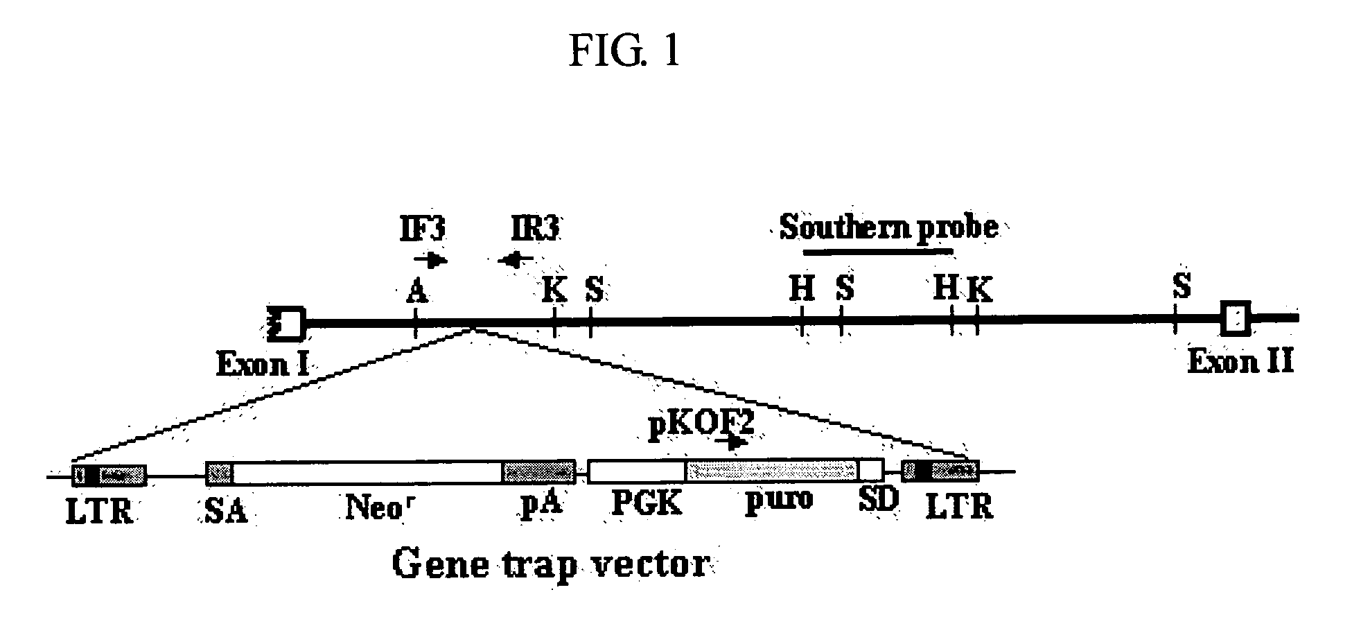
[0055] To determine the site where the gene trap vector was inserted into p43 allele, the p43 mutant allele was analyzed by sequencing. The sequencing was determined by Pangenomcis, a company for sequence analysis. As shown in FIG. 1, the first exon (Exon I) and second exon (Exon II) of p43 mutant gene were separated by the intron of about 7 kb. It was confirmed that the gene trap vector was inserted at about 1.5 kb downstream of the first exon.
2.2 Southern Blot
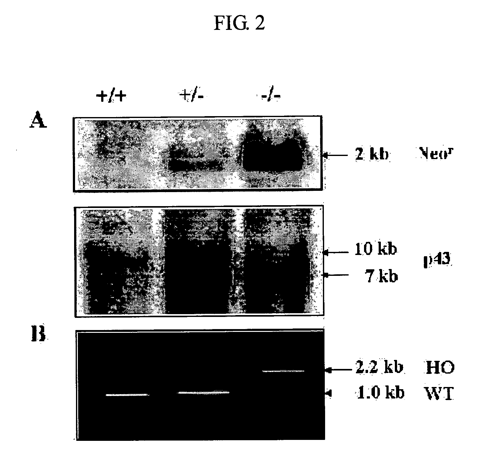
[0056] The genomic DNA was isolated from the tail of each mouse and digested with NcoI. The cleaved DNA fragments were separated by denaturing gel electrophoresis. Then, southern blot analysis was performed using the neomycin resistant gene (Neor)(SEQ ID NO: 2) of the gene trap vector or the HindIII fragment (SEQ ID NO: 3) of the p43 mutant gene as a prove, according to a conventional method [Southern, E. M., J. ...
example 3
Expression of p43 by p43 Mutant Mouse
3.1 Northern Blot
[0058] For Northern analysis, total cellular RNA was prepared from mouse embryonic fibroblast (MEF) cells using RNeasy Midi Kit (QIAGEN) according to the manufacturer's instruction. The isolated RNA was separated on denaturing gel, and then transferred to the Hybond-N+ membrane (Amersham). The RNAs on the membranes were hybridized with the specific probes for p43 set forth in SEQ ID NO: 7. As shown in FIG. 3A, p43 mRNA was detected in the wild type (+ / +) and the heterozygous mutant mice, but was not detected in the homozygous mutant mice (− / −). It indicates that p43 is not expressed in p43 mutant mice (− / −) of the present invention.
3.2 Western Blot
[0059] For Western blot analysis, the protein was isolated from the mouse organ according to Ziak et al. (Ziak M, et al., Biochem. Biophys. Res. Commun. 280:363-367, 2001). Western blotting was carried out with anti-p43 antibody as described previously [Park S. G., et al., J. Biol...
PUM
| Property | Measurement | Unit |
|---|---|---|
| diameter | aaaaa | aaaaa |
| temperature | aaaaa | aaaaa |
| temperature | aaaaa | aaaaa |
Abstract
Description
Claims
Application Information
 - R&D
- Intellectual Property
- Life Sciences
- Materials
- Tech Scout
- Unparalleled Data Quality
- Higher Quality Content
- 60% Fewer Hallucinations
Browse by: Latest US Patents, China's latest patents, Technical Efficacy Thesaurus, Application Domain, Technology Topic, Popular Technical Reports.
© 2025 PatSnap. All rights reserved.Legal|Privacy policy|Modern Slavery Act Transparency Statement|Sitemap|About US| Contact US: help@patsnap.com