System and method for generating digital images of a microscope slide

a technology of digital images and microscope slides, applied in the field of system and method for generating images of microscope slides, can solve the problems of insufficient information for the first sequence of images, the inability to capture an entire slide in focus using a fixed z-position, and the relatively slow start/stop acquisition system, as described abov

Inactive Publication Date: 2005-04-28
CARL ZEISS MICROIMAGING AIS
View PDF44 Cites 143 Cited by
  • Summary
  • Abstract
  • Description
  • Claims
  • Application Information

AI Technical Summary

Benefits of technology

[0030]FIG. 8 illustrates a speed curve that may be applied to control the motion of a microscope stage, in accordance with an embodiment of the invention; and

Problems solved by technology

Additionally, the surfaces of many tissues are uneven and contain local variations that make it difficult to capture an in-focus image of an entire slide using a fixed z-position.
In some cases, a first sequence of images does not provide sufficient information to determine a desired-focus position.
Start / stop acquisition systems, as described above, are relatively slow, because the microscope objective is often required to perform multiple focus-capture operations for each designated target point on the slide.
In addition, a high-power objective's field-of-view is limited; therefore, the number of points for which desired-focus information is directly obtained may be a relatively small portion of the entire slide.
First, as described above, the use of a high-power objective to obtain desired-focus data for a given target point is relatively slow.
Second, generating a focal map from a limited number of points on the slide can create inaccuracies in the resulting focal map.
Tissue on a slide often does not have a uniform, smooth surface.

Method used


Image

  • System and method for generating digital images of a microscope slide
  • System and method for generating digital images of a microscope slide
  • System and method for generating digital images of a microscope slide

Examples


Embodiment Construction

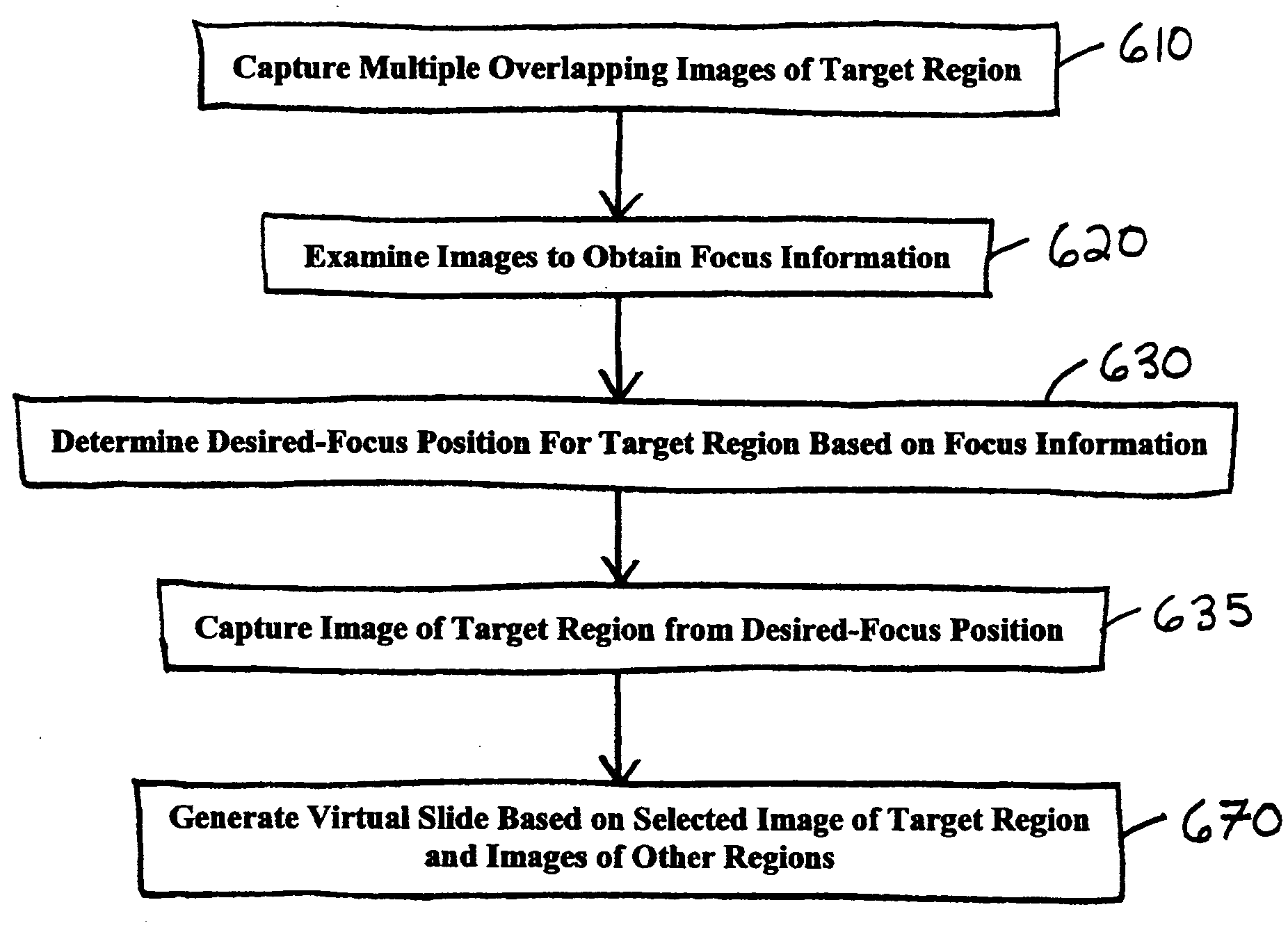
[0032] A virtual microscope slide typically comprises digital data representing a magnified image of all, or a portion of, a microscope slide. Because the virtual slide is in digital form, it can be stored on a medium, e.g., in a computer memory, and can be transmitted over a communication network, such as the Internet, an intranet, etc., to a viewer at a remote location.

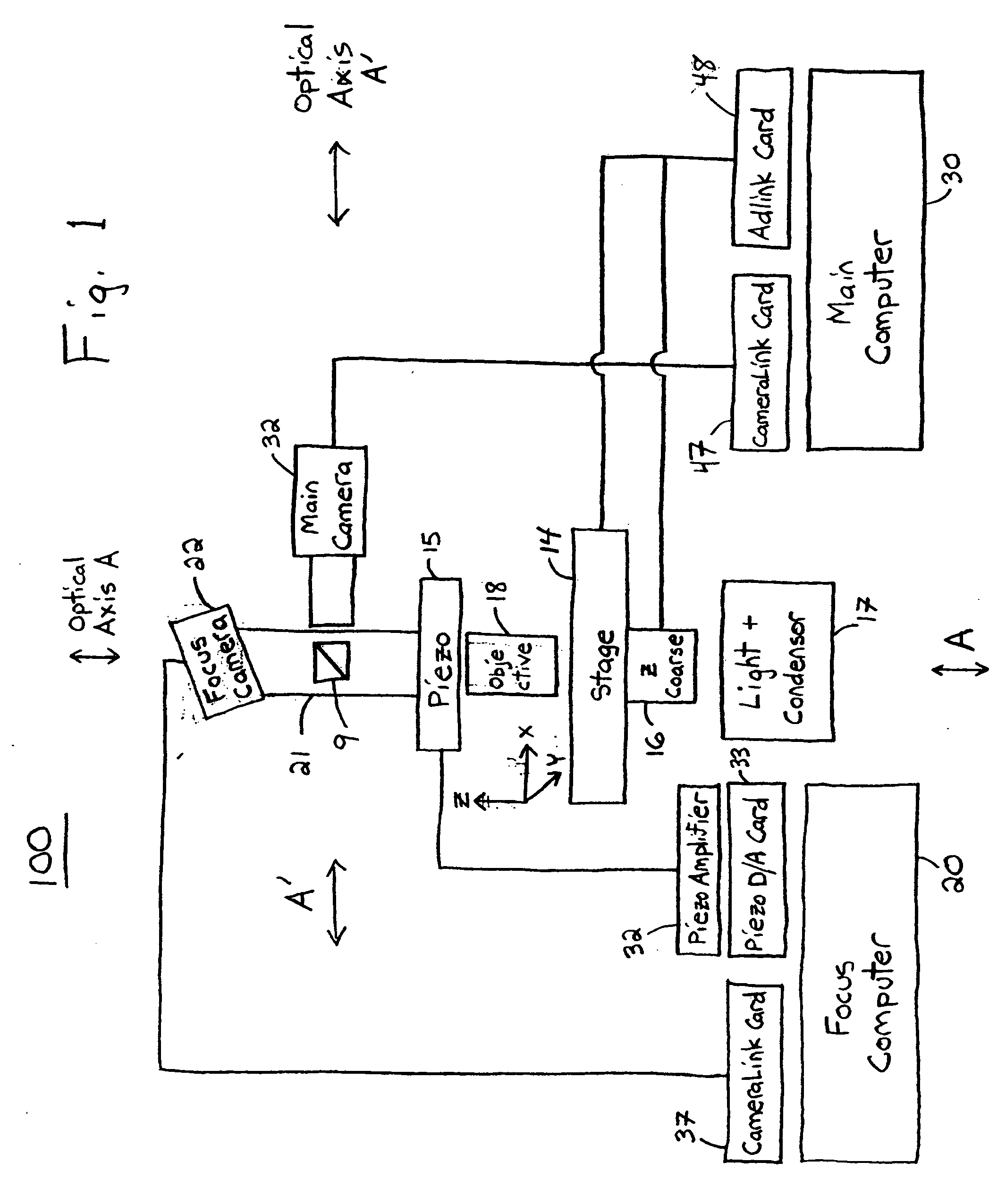
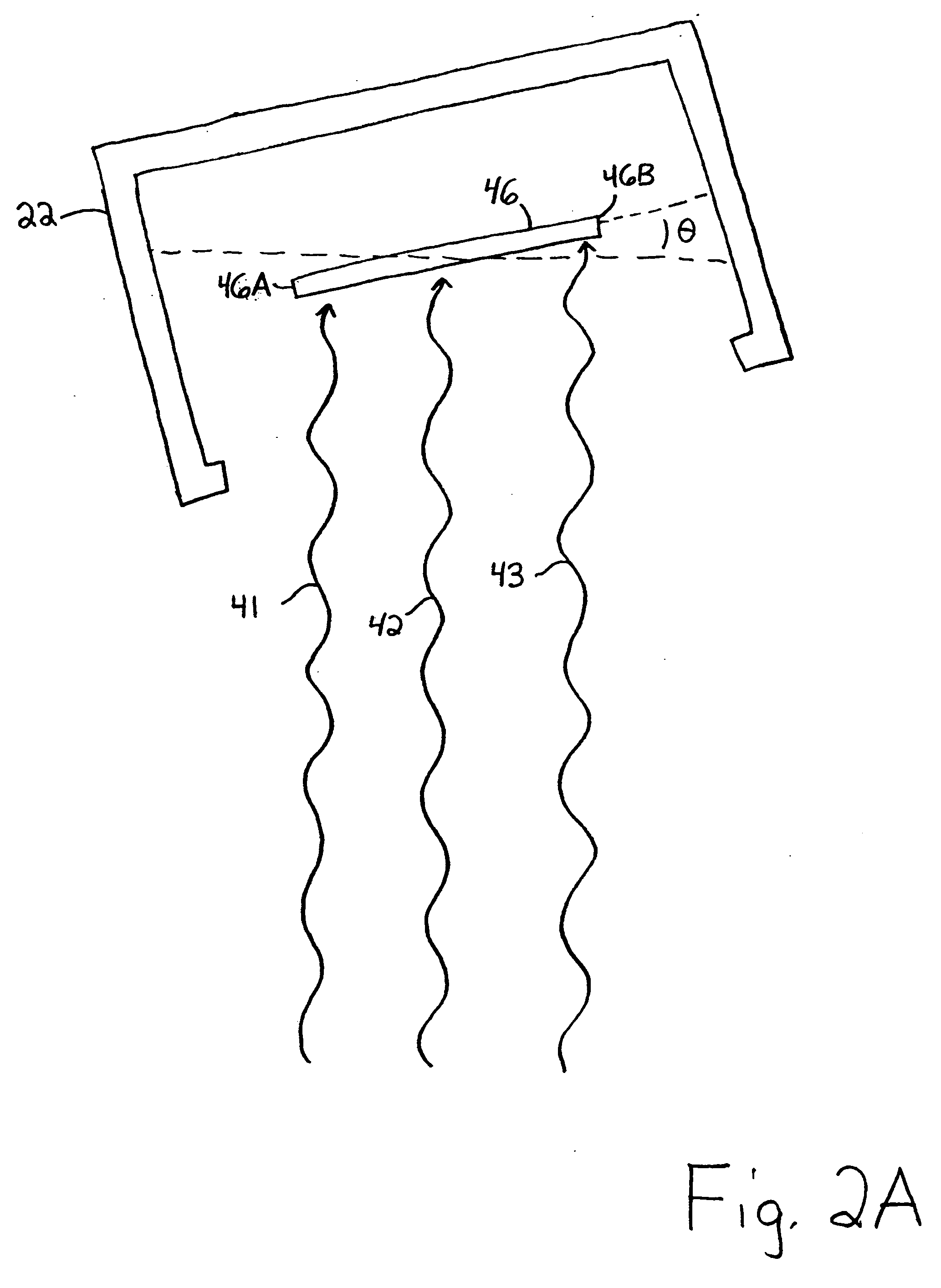
[0033] An improved system and method are provided for obtaining magnified images of a microscope slide for use in constructing a virtual slide. In an aspect of the invention, a focus camera captures a plurality of images of a target region. Each image covers a respective area that includes at least a portion of the target region. Additionally, each image contains information associated with multiple focal planes. In one embodiment, the sensor of the focus camera is positioned so that its focal plane is tilted relative to the focal plane of a main, scanning camera. In one example, the sensor in the focus camera is t...


PUM


Abstract

An improved system and method for obtaining images of a microscope slide are provided. In one embodiment, a focus camera includes an optical sensor that is tilted relative to the focal plane of a scanning camera. A scan of a target region is performed, and multiple overlapping images of the target region are captured from a plurality of x-y positions. Each image contains information associated with multiple focal planes. Focus information is obtained from the images, and a desired-focus position is determined for the target region based on the focus information. The scanning camera then captures an image of the target region from the desired-focus position. This procedure may be repeated for selected regions on the microscope slide and the resulting images of the respective regions are merged to create a virtual slide.

Description

[0001] This application claims the benefit of U.S. Application No. 60 / 489,769, filed on Jul. 22, 2003, assigned to the assignee of the present invention and incorporated by reference herein in its entirety.FIELD OF THE INVENTION [0002] The invention relates generally to a system and method for generating images of a microscope slide, and more particularly, to a system and method for obtaining focus information to be used in scanning a microscope slide. BACKGROUND OF THE INVENTION [0003] A virtual microscope slide typically comprises digital data representing a magnified image of a microscope slide. Because the virtual slide is in digital form, it can be stored on a medium, e.g., in a computer memory, and can be transmitted over a communication network, such as the Internet, an intranet, etc., to a viewer at a remote location. [0004] Virtual slides offer advantages over traditional microscope slides. In some cases, a virtual slide can enable a physician to render a diagnosis more qui...

Claims


Application Information

Patent Timeline
Patent Type & Authority Applications(United States)
IPC IPC(8): G02B21/24G02B21/36G06T3/40
CPCG02B21/241G02B21/361G06T3/4038G06K9/00134G02B21/367G06V20/693
Inventor DONG, RUI-TAORASHID, USMANZEINEH, JACK A.
Owner CARL ZEISS MICROIMAGING AIS
Why Eureka
Try Eureka