Light receiving and detection system for reading a radiographic image

a detection system and radiographic image technology, applied in the direction of optical radiation measurement, instruments, photometry, etc., can solve the problems of impending image interpretation and treatment based thereon, physicians on the ship may not have the necessary capability to review such radiographic images generated, and hospitals and physicians located in small towns do not have immediate availability, etc., to achieve excellent optical path and accurate reproduction of image pixel values on the substra

Inactive Publication Date: 2003-07-24
ERADLINK
View PDF8 Cites 4 Cited by
  • Summary
  • Abstract
  • Description
  • Claims
  • Application Information

AI Technical Summary

Benefits of technology

0033] The outputs of each Selfoc lens associated with each optical fiber are connected together so that there is, in effect, a collection of all of the information along the scan line, which is converted into an electrical analog signal. This analog signal from the scan line can then be digitized. In a sense, all of the photo-diodes thereupon become integrated into a type of optical integrating cylinder. At any given amplitude, the peak of the signal may vary, although, when graphically plotted on a clock time basis with clock signals on the clock track, there is a sampling of the light independently of the number of optical fibers emitting the light. The system provides an excellent optical path for recapture of most of the light from the light distribution bar, and the reading of the light through the substrate to obtain an accurate reproduction of pixel values of the image on that substrate.
0034] In the detection of the light, each scan line of the substrate includes usually five to ten pixels, and generally, no more than ten pixels. In effect, each individual scan operation thereby produces a light effectively as a point source of light. In contrast, any prior art scan usually will involve several hundred pixels.

Problems solved by technology

Very frequently, and as examples of the foregoing, hospitals and physicians located in small towns do not have the immediate availability of the services of a physician who has the capabilities of a radiologist, or other party having the capabilities of examining a particular type of radiographic image.
The use of a delivery service frequently involves days, and in some cases, interpretation of the image and treatment based thereon may be imminent.
In addition, with cruise ships and, for that matter, even military hospital ships, the physicians on those ships may not have the necessary capabilities of reviewing such radiographic images which are generated, and need to obtain input with respect to such radiographic images.
Unfortunately, the technology used in photocopiers does not easily translate into an efficient system for scanning of radiographic images.
Numerous problems which arise with the use of photocopier technology is due to the simple, yet critical, fact that photocopier technology is based on the use of a substrate which is typically opaque, and usually the image on only one side thereof is scanned, whereas in radiographic images, the substrate is generally transparent.
Prior art scanning systems, to the extent that they relied upon paper copier technology, were essentially rudimentary and not effective in generating high quality reproduction of images, and even more importantly, were not capable of generating those images without loss of detail.
This complex array of precision lenses and mirrors in the optical bench provided surfaces which contributed to veiling glare as a result of edge reflections and surface contamination.
They also relied upon a costly galvanometer to control raster scanning of the laser beam.
The costly gas laser also degraded over time, and required periodic calibration and eventual replacement by qualified technicians.
Consequently, the prior art laser scanning systems were not effective for generating true electrical representation of a radiographic image and transmitting same.
When the light passes through a radiographic film, there was a tendency for the light to scatter.
Otherwise, the quality of the image being detected for storage and transmission would be adversely affected.
Another problem with prior art detector systems was not only the need for constant readjustment and cleaning, but the fact that any substantial breakdown required personnel to service and repair that scanning apparatus.
Frequently, service personnel were not available in numerous locations, as for example, small towns, oceangoing vessels, and the like.

Method used


Image

  • Light receiving and detection system for reading a radiographic image
  • Light receiving and detection system for reading a radiographic image
  • Light receiving and detection system for reading a radiographic image

Examples


Embodiment Construction

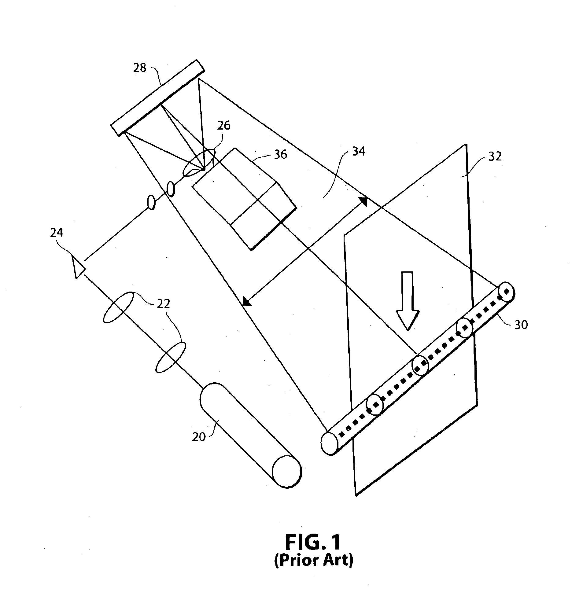
[0057] Referring now in more detail and by reference characters to the drawings, reference will initially be made to FIG. 1, which shows a typical prior art laser scanning system for scanning radiographic images. FIGS. 2-16 of the application more particularly relate to the apparatus and the method of the present invention.

[0058] Referring in more detail to FIG. 1, it can be observed that the prior art laser scanning systems generally rely upon a gas laser 20, including a plurality of condensing lenses 22, and a prism 24 directing light to a conical prism 26, and a folding mirror 28. That light from the mirror 28 is then directed to a photomultiplier detector 30. However, the light passes through the radiographic film substrate 32 and will thereupon provide a reading of the light and dark areas to the photomultiplier detector 30. The scanning beam generated at the folding mirror 28 is represented by the path 34. Moreover, a galvanometer 36 must be used in this arrangement to control...


PUM


Abstract

An apparatus and a method for receiving and detecting light from a plurality of light scanning operations on each scan line of a document, such as a radiographic image, which is being read. The light is detected by means of a photo-diode detector arrangement. Prior to detection, however, this light is received in an integrating arrangement. In effect, a progressive point of light from each optical fiber used for delivering of the light occurs from one end of a scan line to the other sequentially, from scan operation to scan operation. When the light emits from each of a plurality of optical fibers, the light then spreads somewhat into a cone. A reconcentrating and refocusing lens, such as a Selfoc lens, refocuses the spreading light into a point in the image plane of the radiographic film being scanned and essentially refocuses that light at that image plane. Detection of the light is made on a clock time basis so that the light content at each scan operation in a scan line so that recreation of the document is readily made.

Description

[0001] This application is based upon, succeeds to and claims the benefits of priority of my U.S. Provisional Patent Application Serial No. 60 / 351,210, filed Jan. 14, 2002, and entitled "Light Detection System for Receiving Radiograph Image Data."[0002] 1. Field of the Invention[0003] This invention relates in general to certain new and useful improvements in a system for receiving light, such as laser light, passed through a radiographic image film containing an image thereon and which processes the light to effectively represent each scan line of a radiographic film being read. More particularly, the invention relates to a generally sealed optical system which can be manufactured relatively inexpensively and eliminates the need for an optical bench and does not result in any veiling glare. As such, the system is highly effective for use in the examination and digitizing of a radiographic image.[0004] 2. Brief Description of Related Art[0005] In recent years, storage, retrieval and...

Claims


Application Information

Patent Timeline
Patent Type & Authority Applications(United States)
IPC IPC(8): H04N1/028
CPCH04N1/0281H04N1/02815H04N2201/0412H04N1/02895H04N1/02835
Inventor GERLACH, RICHARD
Owner ERADLINK
Why Eureka
Try Eureka