Method of testing squamous epithelial cells

a technology of epithelial cells and squamous cells, which is applied in the field of testing squamous epithelial cells, can solve the problems of oral squamous cell cancer, uncontrollable normal cell differentiation,

Inactive Publication Date: 2003-05-22
OKAMOTO TETABUJI
View PDF0 Cites 3 Cited by
  • Summary
  • Abstract
  • Description
  • Claims
  • Application Information

AI Technical Summary

Problems solved by technology

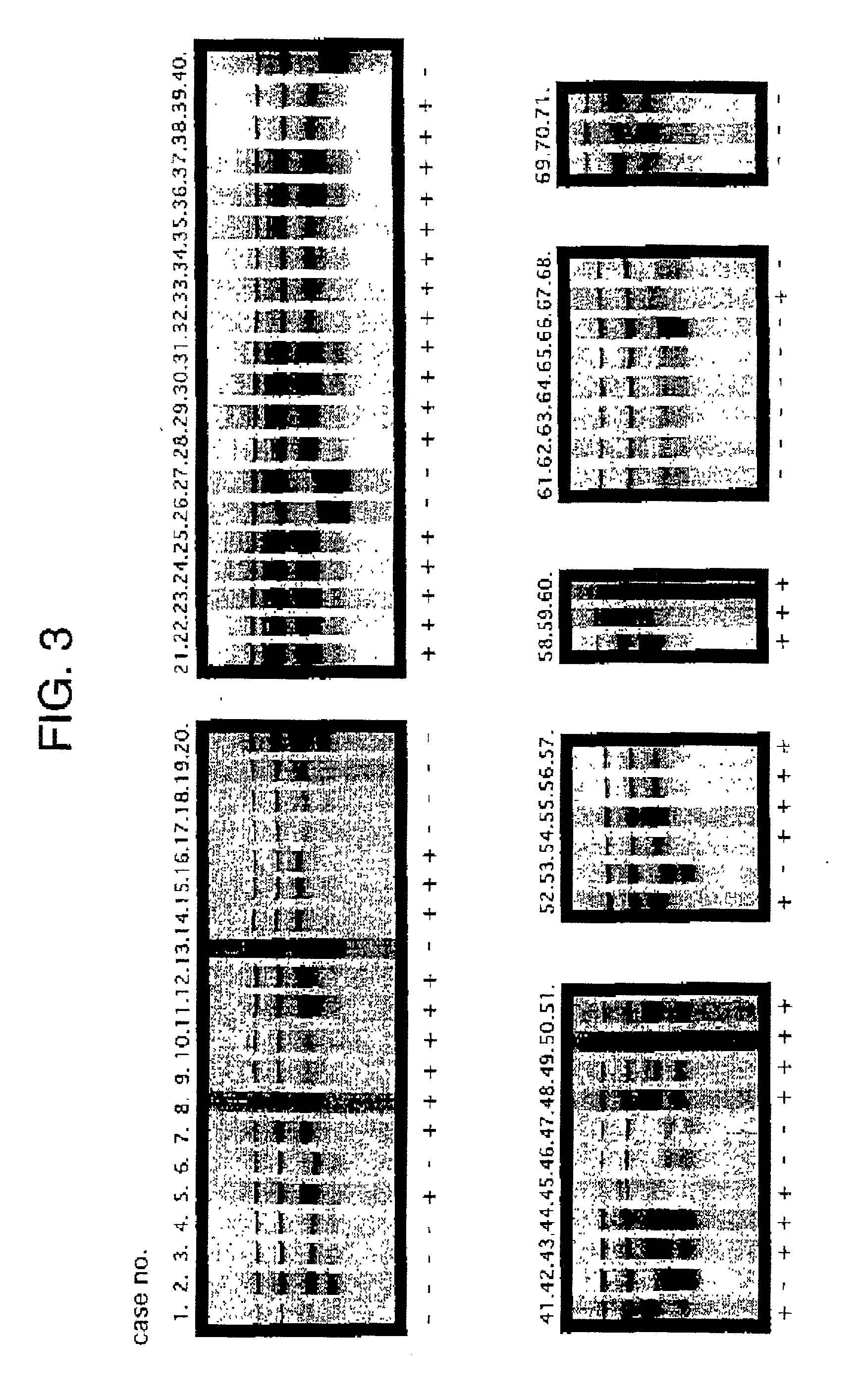
The result suggests that the mutation causes a ligand-independent permanent self-phosphorylation of FGFR3 and promotes signaling of FGFR3, rendering normal cell differentiation uncontrollable and, at the same time, negating growth inhibition by cell-to-cell contact, which results in the canceration of oral squamous cells.

Method used


Image

  • Method of testing squamous epithelial cells
  • Method of testing squamous epithelial cells
  • Method of testing squamous epithelial cells

Examples


example 2

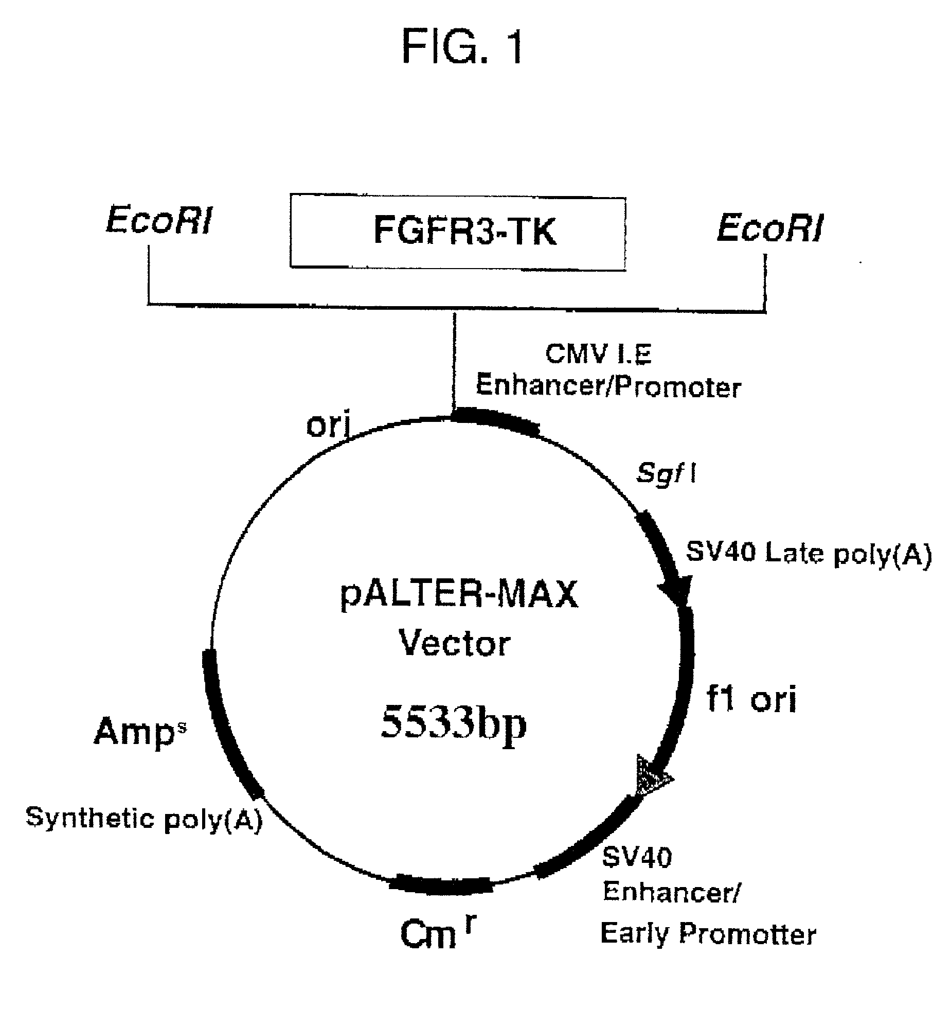
[0062] Example 2 Expression of FGFR3 protein in the tissues of oral squamous cell carcinoma and the normal epithelium

[0063] (1) The expression of FGFR3 protein was investigated using the Vectastatin ABC kit (Vector Laboratories, Inc.) by the immunoperoxidase staining method based on avidin-biotin-peroxidase complex method (ABC method).

[0064] Sections of 4 .mu.m in thickness were prepared on poly-L-lysine coated slides. After incubating at 37.degree. C. for 4 days, they were hydrated with the xylene and ethanol series, and the endogenous peroxidase was removed by MeOH / 0.3% H.sub.2O.sub.2 at room temperature for 30 minutes followed by treatment with 0.1% Triton X-100 / PBS on ice for 10 minutes, and then 0.05% Protenass-K / PBS at room temperature for 5 minutes. In order to prevent nonspecific reactions, blocking was performed by 10% goat serum / PBS at 37.degree. C. for 1 hour. Anti-FGFR3 rabbit polyclonal antibody (Santa Cruz Biotechnology, Inc.) as the primary antibody was allowed to rea...


PUM


Abstract

There is provided a method of testing squamous epithelial cells and a method of screening therapeutic agents for squamous cell carcinoma. That the base guanine at position 2128 (exon 17) of the FGFR3 gene is mutated to thymine in the cells of squamous cell carcinoma, or that the cells of squamous cell carcinoma produce an FGFR3 protein in which the amino acid glycine at position 697 is mutated to cysteine is utilized to facilitate the diagnosis and treatment of squamous cell carcinoma.

Description

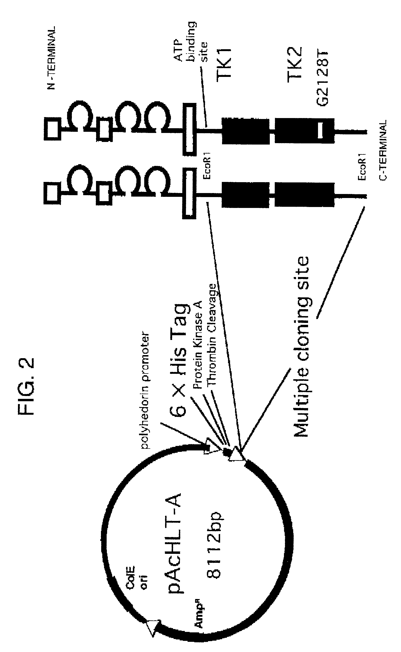
[0001] (1) Field of the Invention[0002] The present invention relates to a method of testing squamous epithelial cells. The present invention also relates to a method of screening therapeutic agents for squamous cell carcinoma.[0003] (2) Description of the Related Art[0004] Since the discovery of a basic fibroblast growth factor (FGF-2) as a growth factor for fibroblasts in the early 1970's.sup.11, the fibloblast growth factor (FGF) family that is encoded by 21 different genes has been elucidated up to now.sup.21. The activity of FGF is mediated by a high-affinity FGF receptor (FGFR) on the cell surface, and FGF has a structure composed of two or three extracellular immunoglobulin-like loop structures (1 g), a transmembrane domain, and, within the cell, a tyrosine kinase region divided into two parts.sup.31-51. It has been believed that FGFR, when bound to FGF in the presence of heparin or heparan sulfate proteoglycan, forms a dimer, which self-phosphorylates the tyrosine residue of...

Claims


Application Information

Patent Timeline
IPC IPC(8): A61K45/00A61P35/00G01N33/50C12N15/09C12Q1/68G01N33/15G01N33/574
CPCC12Q2600/136C12Q1/6886A61P35/00
Inventor OKAMOTO, TETSUJI
Owner OKAMOTO TETABUJI
Why Eureka
Try Eureka