Use of reinforced foam implants with enhanced integrity for soft tissue repair and regeneration
a technology of reinforced foam and soft tissue, which is applied in the direction of prosthesis, peptide/protein ingredients, antibody medical ingredients, etc., can solve the problems of osteoarthritis, strain or tear of the tendon itself, and difficult manufacture of synthetic materials, etc., and achieve the effect of sufficient structural integrity
- Summary
- Abstract
- Description
- Claims
- Application Information
AI Technical Summary
Problems solved by technology
Method used
Image
Examples
example 1
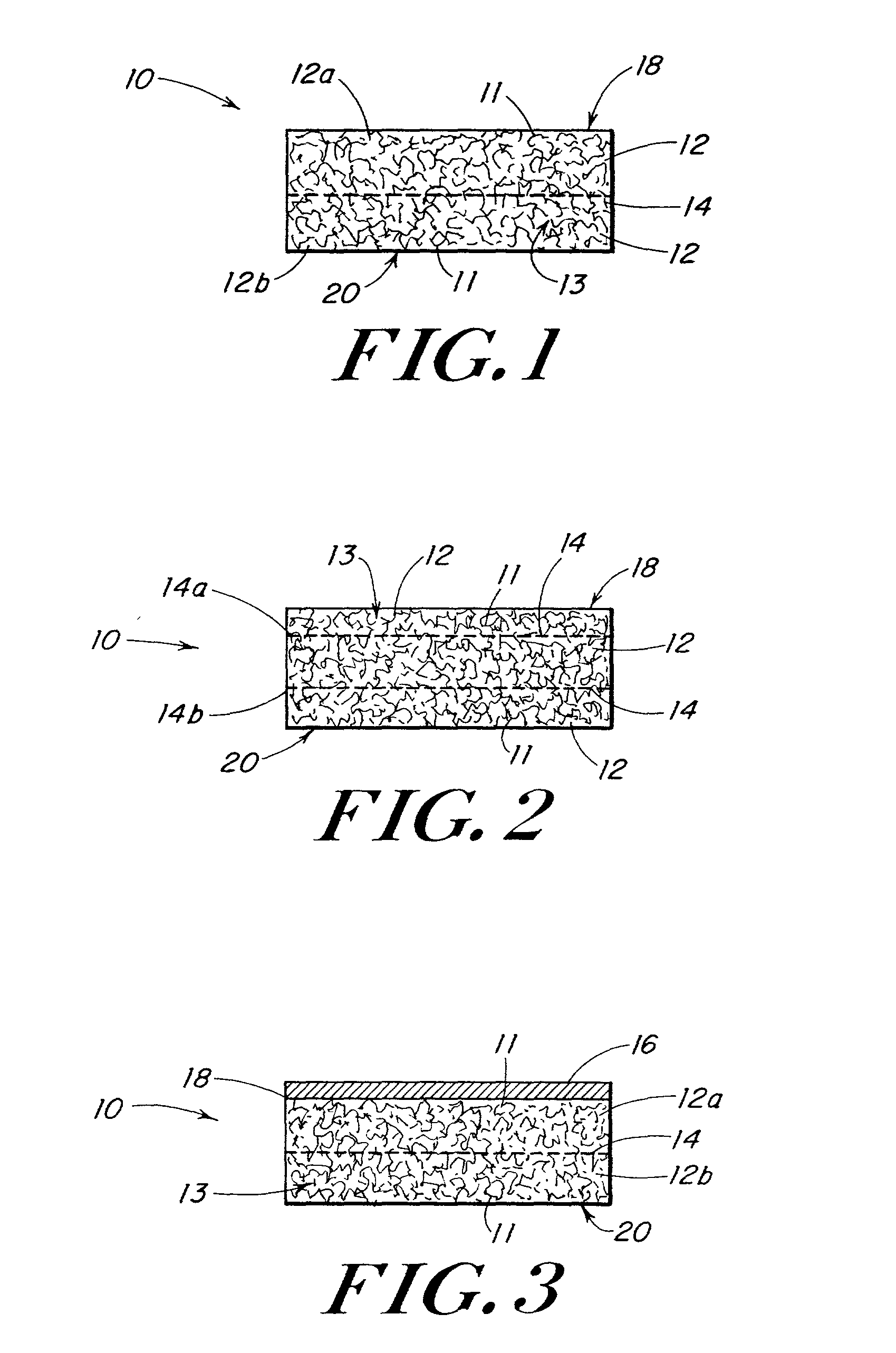
[0083] This example describes the preparation of three-dimensional elastomeric tissue implants with and without a reinforcement in the form of a biodegradable mesh.
[0084] A solution of the polymer to be lyophilized to form the foam component was prepared in a four step process. A 95 / 5 weight ratio solution of 1,4-dioxane / (40 / 60 PCL / PLA) was made and poured into a flask. The flask was placed in a water bath, stirring at 70.degree. C. for 5 hrs. The solution was filtered using an extraction thimble, extra coarse porosity, type ASTM 170-220 (EC) and stored in flasks.
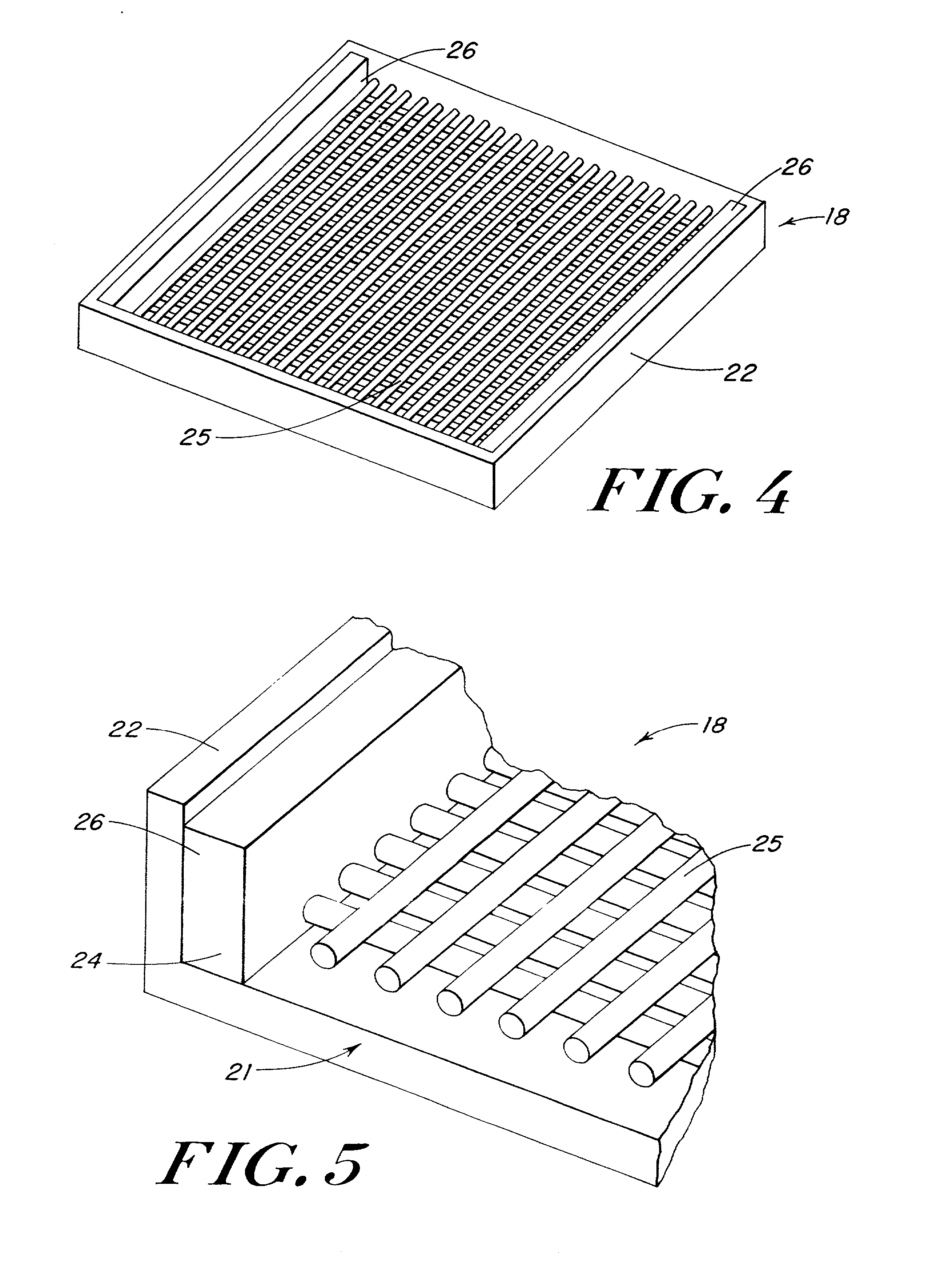
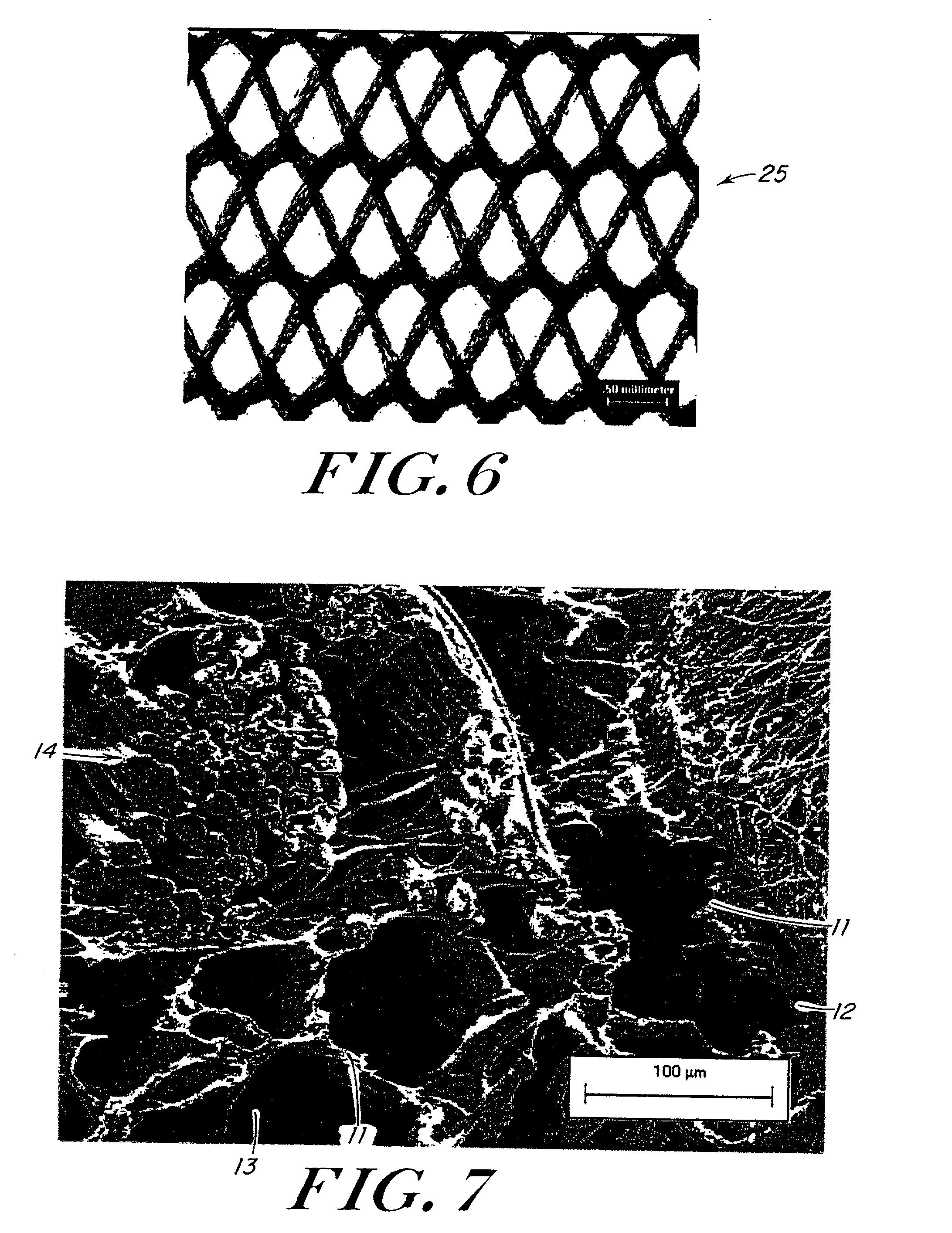
[0085] Reinforcing mesh materials formed of a 90 / 10 copolymer of polyglycolic / polylactic acid (PGA / PLA) knitted (Code VKM-M) and woven (Code VWM-M), both sold under the tradename VICRYL were rendered flat by ironing, using a compression molder at 80.degree. C. / 2 min. FIG. 6 is a scanning electron micrograph (SEM) of the knitted mesh. After preparing the meshes, 0.8-mm shims were placed at each end of a 15.3.times.15.3 cm al...
example 2
[0089] Lyophilized 40 / 60 polycaprolactone / polylactic acid, (PCL / PLA) foam, as well as the same foam reinforced with an embedded VICRYL knitted mesh, were fabricated as described in Example 1. These reinforced implants were tested for suture pull-out strength and burst strength and compared to both standard VICRYL mesh and non-reinforced foam prepared following the procedure of Example 1.
[0090] Specimens were tested both as fabricated, and after in vitro exposure. In vitro exposure was achieved by placing the implants in phosphate buffered saline (PBS) solutions held at 37.degree. C. in a temperature controlled waterbath.
[0091] For the suture pull-out strength test, the dimension of the specimens was approximately 5 cm.times.9 cm. Specimens were tested for pull-out strength in the wale direction of the mesh (knitting machine axis). A size 0 polypropylene monofilament suture (Code 8834H), sold under the tradename PROLENE (by Ethicon, Inc., Somerville, N.J.) was passed through the mesh...
example 3
[0095] Mesh reinforced foam implants were implanted in an animal study and compared to currently used pelvic floor repair materials. The purpose of this animal study was to evaluate the subcutaneous tissue reaction and absorption of various polymer scaffolds. The tissue reaction and absorption was assessed grossly and histologically at 14 and 28 days post-implantation in the dorsal subcutis. In addition, the effect of these scaffolds on the bursting strength of incisional wounds in the abdominal musculature was determined. Burst testing was done at 14 and 28 days on ventrally placed implants and the attached layer of abdominal muscle.
[0096] Lyophilized 40 / 60 polycaprolactone / polylactic acid (PCL / PLA) foam, as well as the same foam reinforced with an embedded VICRYL knitted mesh were fabricated as described in Example 1. The foam and mesh reinforced foam implant were packaged and sterilized with ethylene oxide gas following standard sterilization procedures. Controls for the study in...
PUM
| Property | Measurement | Unit |
|---|---|---|
| diameter | aaaaa | aaaaa |
| diameter | aaaaa | aaaaa |
| elongation | aaaaa | aaaaa |
Abstract
Description
Claims
Application Information
 - R&D
- Intellectual Property
- Life Sciences
- Materials
- Tech Scout
- Unparalleled Data Quality
- Higher Quality Content
- 60% Fewer Hallucinations
Browse by: Latest US Patents, China's latest patents, Technical Efficacy Thesaurus, Application Domain, Technology Topic, Popular Technical Reports.
© 2025 PatSnap. All rights reserved.Legal|Privacy policy|Modern Slavery Act Transparency Statement|Sitemap|About US| Contact US: help@patsnap.com


