
Vital stain

a vital stain and fluorescent dye technology, applied in the field of vital dye, can solve the problems of difficult endoscopy treatment, limited detection period of early cancer, and difficult to detect cancers of this size, so as to reduce the generation of ultraviolet rays in tissues, reduce the damage, and identify quickly

Active Publication Date: 2022-04-05
MIE UNIVERSITY
View PDF26 Cites 0 Cited by
  • Summary
  • Abstract
  • Description
  • Claims
  • Application Information

AI Technical Summary

Benefits of technology

The invention describes a method for identifying cancerous lesions on mucosal tissues using a vital stain that can reduce photodamage to cells and minimize DNA damage. The method utilizes the difference in dye affinities between cancer and normal cells to quickly and accurately diagnose the quality of mucosal lesions. The technique allows for cancer diagnosis to be performed using high-resolution fluorescent images and can easily and accurately remove detected cancer tissues. Overall, the invention provides a safer and more accurate approach for cancer diagnosis.

Problems solved by technology

It is extremely difficult to detect cancers of this size with existing endoscopes.
Because of the detection limit, however, with endoscopy it is only possible to discover early cancer in patients that have undergone medical examination during a period of 6 months before or after the 7th year.
The detectable period for early cancer is therefore limited.
In cases of advanced cancers that have exceeded this period, treatment by endoscope is difficult, and for advanced cancers in stage 2 or 3 it is necessary to rely on laparotomy by a surgeon for excision of the cancer.
However it is not possible to observe individual cells even by NBI, and biopsy is necessary for accurate diagnosis of lesions.
However, biopsy is an invasive procedure and also costly, while about 10 days are necessary to obtain diagnosis results.
Attempts to diagnose diseases from cellular images taken upon staining living cells have also been proposed for years, but the safety of the dyes or dye compounds used have been an issue.
The safety verification procedure for a new dye compound requires a minimum of 10 years and a cost of several billion yen per compound.
However, the images obtained under multiphoton laser microscopy are of low contrast.
In addition, because ultraviolet rays are emitted in the body upon irradiation of the multiphoton laser, it has not been approved for use in humans due to the risk of DNA damage.
Furthermore, the strength of the autologous fluorescence of cells differs significantly with different organs, and while epithelial cells of the gastrointestinal tract such as the large intestine, which have strong autologous fluorescence, allow visualization of cellular images using currently marketed multiphoton laser microscopes, ovarian epithelial cells or bladder epithelial cells, which have weak autologous fluorescence, are difficult to be visualized as cellular images using currently marketed multiphoton laser microscopes (Cruz, J., et al.

Method used


Image

  • Vital stain
  • Vital stain
  • Vital stain

Examples


example 1

1. Evaluation of Dye Compound Stainability

[0205]Drinking water containing 2% (w / v) dextran sodium sulfate (DSS) was given to 8-week-old C57B6 mice (male, approximately 20 g) for 7 days. After intraperitoneal injection of 0.2 ml of 5% chloral hydrate for anesthesia, the mouse abdominal wall was incised vertically to about 1 cm, and the gastrointestinal tract was raised over the abdominal wall to about 1 cm. Blood flow to the gastrointestinal tract was maintained during this time by the blood vessel flowing into the mesenteric attachment site. The gastrointestinal tract was incised vertically to about 1 cm on opposite side of the mesenteric attachment site, including the tunica muscularis and mucosa. The opposite side of the mesenteric attachment site was incised in order to prevent severance and damage of the blood vessel and minimize bleeding. When the incision site of the gastrointestinal tract was opened vertically, the mucosal surface of the gastrointestinal tract, as the food ch...

example 2

[0218]In vitro assay of MDCK-RasV12 cancerized cell small aggregates as a very early cancer model

(Materials)

[0219]MDCK normal cells; canine renal tubular epithelial cell-derived cell line.[0220]MDCK-GFP-RasV12 cells that express GFP-RasV12 which is the green fluorescent protein GFP fused to the constitutively activated oncogene product RasV12 (RasV12 being Ras with the 12th amino acid residue glycine replaced by valine, as a mutation found in 30% to 40% of colon cancers).[0221]DMEM (high glucose) with Phenol Red (Wako Pure Chemical Industries, Ltd.)[0222]DMEM (high glucose) without Phenol Red (Wako Pure Chemical Industries, Ltd.)[0223]Tetracycline System Approved Fetal bovine serum (Clontech)[0224]Penicillin / Streptomycin (×100) (Nacalai Tesque, Inc.)[0225]GlutaMax (×100) (Invitrogen)[0226]Trypsin (0.25%) (no phenol red, Invitrogen)+EDTA (3 mM) (Nacalai Tesque, Inc.)[0227]Zeocin (100 mg / ml, InvivoGen)[0228]Blasticidin (10 mg / ml, InvivoGen)[0229]Tetracycline (SIGMA) / 100% ethanol (100 ...

example 3

1. Evaluation of Dye Compound Stainability

[0242]Drinking water containing 2% (w / v) dextran sodium sulfate (DSS) was given to 8-week-old C57B6 mice (male, approximately 20 g) for 7 days. After intraperitoneal injection of 0.2 ml of 5% chloral hydrate for anesthesia, the mouse abdominal wall was incised vertically to about 1 cm, and the gastrointestinal tract was raised over the abdominal wall to about 1 cm. Blood flow to the gastrointestinal tract was maintained during this time by the blood vessel flowing into the mesenteric attachment site. The gastrointestinal tract was incised vertically to about 1 cm on the opposite side of the mesenteric attachment site, including the tunica muscularis and mucosa. The opposite side of the mesenteric attachment site was incised in order to prevent severance and damage of the blood vessel and minimize bleeding. When the incision site of the gastrointestinal tract was opened vertically, the mucosal surface of the gastrointestinal tract, as the foo...


PUM

PropertyMeasurementUnit
diameteraaaaaaaaaa
diameteraaaaaaaaaa
wavelengthaaaaaaaaaa

Abstract

A vital stain for observation under multiphoton laser microscopy, the vital stain comprising one or more edible dye compounds.

Description

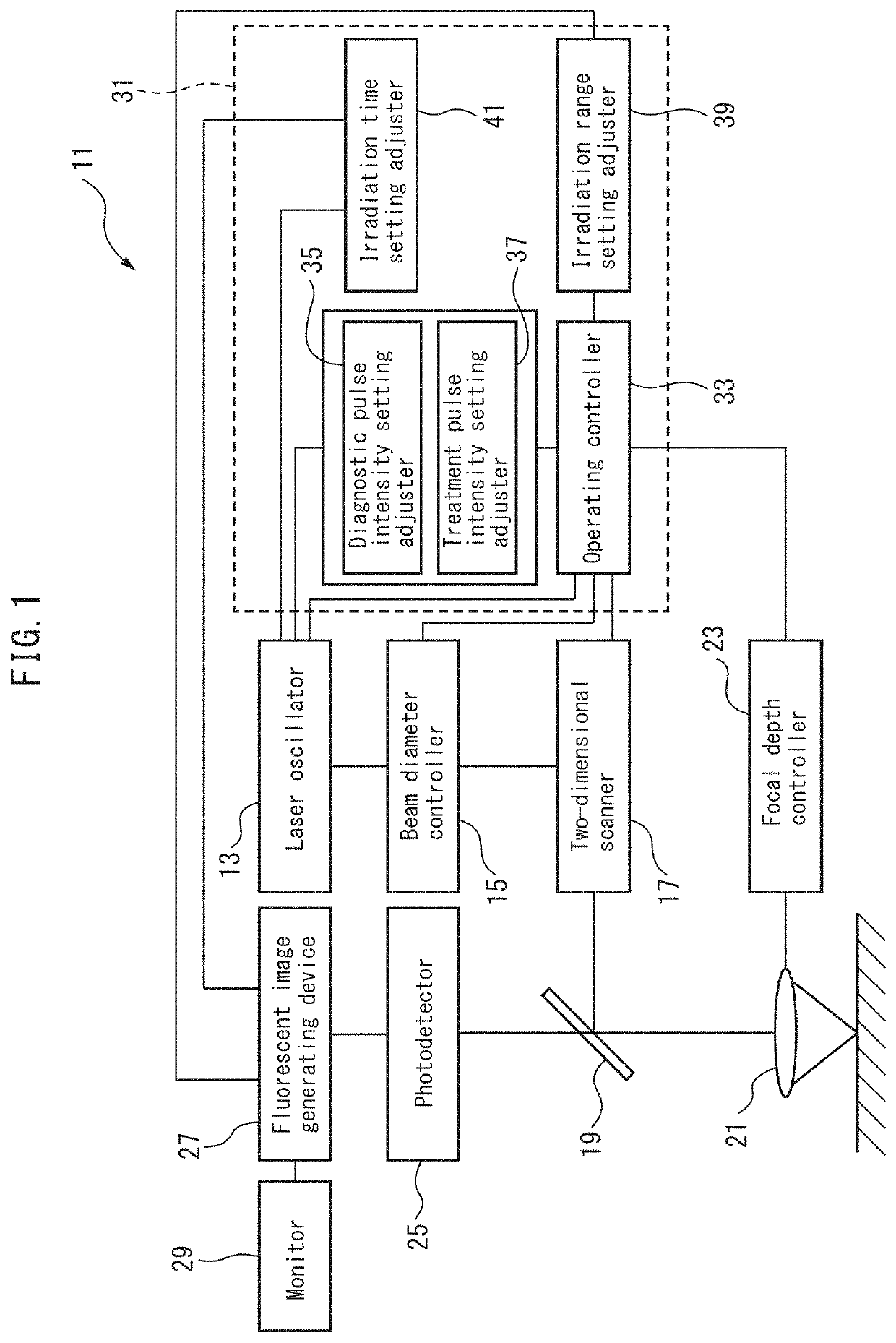
CROSS-REFERENCE TO RELATED APPLICATIONS[0001]This application is the U.S. National Phase of PCT / JP2014 / 059351, filed on Mar. 28, 2014, which is incorporated herein by reference it its entirety and which claims the benefit of JP Application No. 2013-074953, filed on Mar. 29, 2013, JP Application No. 2013-075150, filed on Mar. 29, 2013, and JP Application No. 2013-075256, filed on Mar. 29, 2013.TECHNICAL FIELD[0002]The present invention relates to a novel vital stain to be used in multiphoton laser microscopy and a method of observing cells using the stain, to a method of selecting and evaluating a novel vital stain to be used in multiphoton laser microscopy, to a novel cell stain selected by the method, to a novel tumor cell stain to be used in multiphoton laser microscopy and to a detection method for tumor cells using the stain, as well as to a multiphoton laser diagnosis and treatment apparatus for observation of patient tissues using the phenomenon of multiphoton absorption and f...

Claims


Application Information

Patent Timeline
Patent Type & Authority Patents(United States)
IPC IPC(8): G01N21/64A61B5/00G01N33/50C09B11/26C09B23/04G01N1/30
CPCG01N21/6486A61B5/0071C09B11/26C09B23/04G01N21/6458G01N33/50G01N1/30G01N2021/6439A61K49/006
Inventor MIZOGUCHI, AKIRAFUJIWARA, TAKESHITANAKA, KOJIWANG, SHUJIESAI, KOUSYOKUKIMURA, KAZUSHI
Owner MIE UNIVERSITY
Learn More